Keywords
osteotome, CAS-kit, indirect maxillary sinus lift, immediate implant placement, atrophic maxilla
This article is included in the Datta Meghe Institute of Higher Education and Research collection.
osteotome, CAS-kit, indirect maxillary sinus lift, immediate implant placement, atrophic maxilla
Dentistry is getting modernized with new equipment and techniques, such as implant rehabilitation.1 Dental implant is successful when it serves both functional and aesthetic purposes. Several studies have shown that for the success of an implant therapy there is a need for adequate bone density. Posterior maxillary rehabilitation is a challenge due to its anatomical variation. The presence of maxillary sinus makes implant surgery more complex due to the high chances of sinus membrane perforation. Sinus perforation is one of the most common complications. Overall, 50% of patients with atrophic maxilla who need implant rehabilitation undergo maxillary sinus lift treatment.2,3
There is abundant literature that states various techniques for sinus floor elevation with merits and demerits and their relevant indication, advantages, and disadvantages. Sinus lift technique is broadly classified into two major categories: the first one is the direct or lateral window technique.1 The drawback of this technique is that it requires a large flap for surgical access and is more technique-sensitive and time-consuming. Piezoelectric osteotomy is a technique in which piezoelectric handpiece is used to cut a bony window without damaging the sinus membrane. The advantage of piezoelectric tips is that when it comes in contact with non-mineralized tissue, the cutting action is terminated. Another method for sinus lift is an indirect approach, such as the osteotome technique given by Summer, and the crestal approach technique by Tatum, or the transalveolar approach, which are indicated in RBH of >6 mm.4
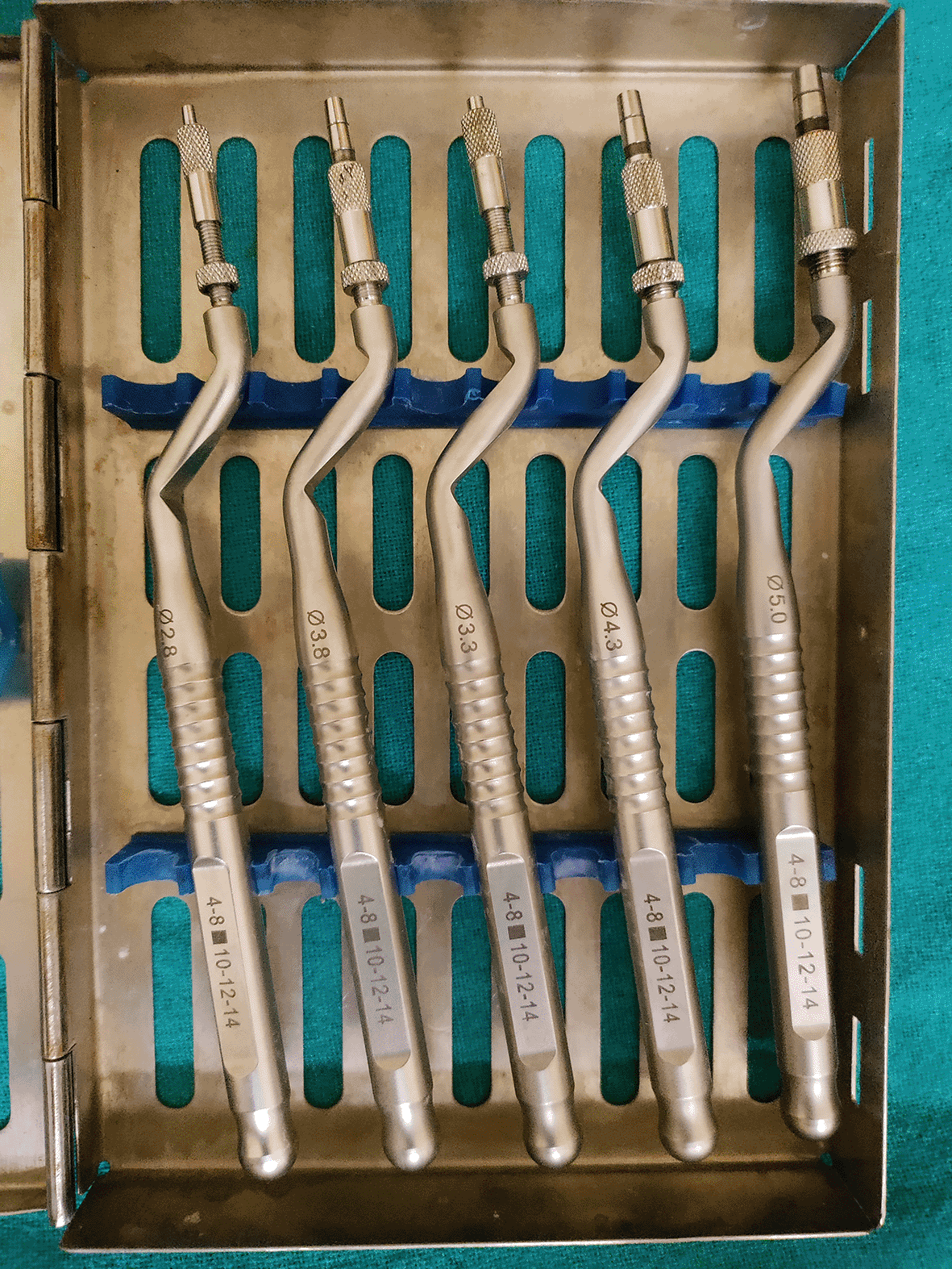
The osteotome technique is one of the oldest techniques, used since 1994, an efficient method with various modifications in the design using diverse materials like a platelet-rich fibrin (PRF) with or without bone graft.5 This technique is highly sensitive due to the increased risk of sinus floor fracture and elevation of the membrane. However, this technique uses a series of osteotomes tapped by malleate with the help of a chisel to create an osteotomy, which often causes patient discomfort. The advantage of this technique is that it increases the primary stability of the implant by increasing peri-implant bone volume, which ultimately results in compaction of bone instead of removing it.
Boyne reported the first maxillary sinus augmentation technique, which is a routinely performed procedure with various bone graft materials and has been practiced for PGST in the last 15 years.4 The literature has suggested that high success rate of implant survival in augmented sites. It has also been stated that implant placement with residual bone height (RBH) of a minimum 5 mm requires sinus augmentation for better primary stability, but in cases where RBH is less than 5 mm, it is better to go with a two-step approach.4
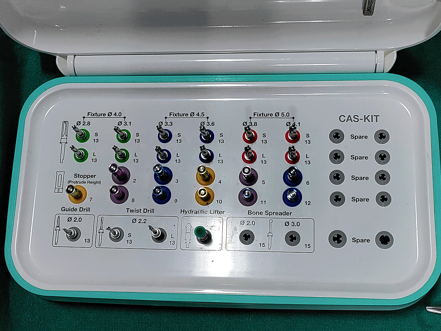
Using an osteotome is a risky procedure due to the high chances of sinus perforation; thus, to overcome this problem a Korean-based implant company (OSSTEM) has introduced a newer approach known as Crystal Approach System kit including a hydraulic lifter. The technique comprises a safe end-cutting drill with a vertical stopper. The drill is used to perform osteotomy in a conical shape with simultaneous fracture of bone and elevation of the sinus membrane with the help of hydraulic lifters. The advantage of this technique is that it provides good outcomes with low failure rates in implant placement and high bone regeneration around the implant.5 As bone is not removed in this technique; the stability is well maintained within a short period of time.
Though this technique has proven efficient, there is some uncertainty around whether the technique is suitable in SLE (sinus lift elevation) as only one study investigating the effectiveness of CAS kit has been published. It was found that no study has compared the two techniques for elevating sinus lift with minimal chance of membrane perforation and time consumption. Thus, the present trial was conducted to compare and evaluate clinical and radiological outcomes of conventional osteotome technique and CAS kit use for indirect sinus lifting and immediate implant placement in the atrophic posterior maxilla.
The present prospective, observational, case-control study included patients who came to the Department of Oral and Maxillofacial Surgery with edentulous posterior maxilla indicated for implant prosthesis in collaboration with the Department of Oral Radiology, Periodontics and Prosthodontics of a Sharad Pawar Dental College and Hospital, DMIHER, Sawangi (Meghe), Wardha for the duration of one and half year. Patients aged ≥18 years, requiring implant prosthesis in atrophic (RBH > 3 mm) posterior maxilla with a minimum width of 6 mm, and able to sign risk consent were included in the study. The study was performed in accordance with the Helsinki Declaration and its later amendments or comparable ethical standards and institutional ethical guidelines prescribed by the central ethics committee on human research (CECHR) of the institution. The study was approved by Datta Meghe Institute of Higher Education and Research [Ref No-DMIMS (DU)/IEC/2020-21/9421].
A detailed history of pre-existing illness was recorded, and the importance of the study was conveyed to the patients. After signing a written consent form, they were directed to Oral Radiology Department for CBCT evaluation. CBCT scans of the posterior maxillary area were performed to assess density, height, width, and neighbouring structures All patients were taken for surgery and prepared according to the standard surgical protocols. All patients were prophylactically given a single dose of antibiotic (625 mg amoxicillin and clavulanic acid combination or 600 mg clindamycin if allergic to penicillin) 1 hour prior to surgery. 0.2% chlorhexidine mouthwash was given to patients for oral mouth rinses. All patients underwent Lan A sensitivity test, xylocaine 2% with 1:100000 adrenaline was administered for regional block, and local infiltration was given. A Para crystal incision was made and full thickness mucoperiosteal flap was reflected at the implant recipient site and the bone was clinically inspected. The implant site was prepared. Patients were divided into two groups randomly by computerized allocation.
Group A: Patients undergoing a sinus lift with an osteotome (n=10)
Group B: Patients undergoing a sinus lift with CAS kit (n=10)
A total of 10 patients underwent osteotome procedure for sinus elevation, initiated with a pilot drill up to the depth of 1 to 2 mm coronal to sinus floor, followed by a 2 mm twist drill up to the same depth. Then, a sequential and suitable osteotome was selected to expand the alveolar bone to reach a suitable diameter. To fracture the sinus floor, an osteotome one size smaller diameter was used with increasing length, after which the same diameter osteotome was used to elevate the sinus membrane, and desired length was achieved.
The procedure was initiated with a 2 mm twist drill with a stopper set 1-2 mm inferior to the sinus floor. After the first drill, the diameter of the drill was increased up to 2.8 mm for soft bone and 3.1 mm for hard bone with 400 to 800 rpm. We checked the floor was intact and continued increasing the diameter with the same length for the respective implant size. After achieving the desired diameter, we increased the stopper length by 1-1 mm until complete erosion of the floor was achieved. Hydraulic lifter was used into the drilled hole; 1 to 3 mL of saline solution were gently injected and retracted back into the syringe, and the sinus membrane was elevated. After completion of osteotome and sinus lift procedures, sinus integrity was checked using the Valsalva manoeuvre.6 Perforation was managed by collagen sheet (4-5 mm) or left as it was (2-3 mm). Self-taping implant (Osteom) of selected diameter was placed in the prepared site by motor connection at the speed of 15rpm and 30 NCM torque to submerge the implant in the bone; if threads remained to be opened, then it was submerged by using a hand ratchet. The final torque was denoted as the primary stability of the implant which was checked using a hand ratchet. The cover screw was placed on the inserted implant and sutured with 3-0 vicryl (polyglactin910). After achieving haemostasis, a pressure pack and post-operative instructions were given.
Residual bone height (RBH): It is a distance measured from sinus floor to crestal bone evaluated from CBCT preoperatively.
Time required to perform the procedure: Time measured from starting the osteotomy procedure to the final implant insertion.
Sinus membrane perforation: Clinical assessment at the time of osteotome by Valsalva manoeuvre.
Primary stability: It is a biomechanical stability measured on implant insertion by hand ratchet.
Amount of bone regeneration: CBCT at three months and nine months after implant placement.
Marginal bone loss: mesial and distal bone level will be measured as a distance from the mesial and distal margin of the implant neck to the most coronal point where the bone appeared to be in contact with the implant. Measurements will be evaluated on CBCT at three months and nine months after implant placement. All pre-, intra-, and post-operative data was compiled in the master chart, and statistical analysis was performed.
Statistical analysis was done by using descriptive and inferential statistics. Qualitative data were analyzed using the Chi-square test. Dependent and independent variables were evaluated by the paired t-test and unpaired t-test respectively. The software used in the analysis was SPSS 27.0 and GraphPad Prism 7.0. p<0.05 was set as the level of significance.
A total of 20 subjects were recruited for the study.
The mean duration for implant placement in patients who underwent CAS group was 13.40±2.98 and 22.10±2.55 in the osteotome group (Table 1). By using Student’s unpaired t-test, astatistically significant difference was found in the duration of surgery in both groups (t=6.99, p=0.0001).
| Group | N | Mean | Standard deviation | Standard error of mean | t-value |
|---|---|---|---|---|---|
| CAS | 10 | 13.40 | 2.98 | 0.94 | 6.99 p=0.0001 |
| Osteotome | 10 | 22.10 | 2.55 | 0.80 |
The mean primary stability in patients in the CAS group was 44±6.14 and 35.50±7.61 in the osteotome group (Table 2). By using Student’s unpaired t-test, astatistically significant difference was found in primary stability between the groups (t=2.74, p=0.013).
| Group | N | Mean | Standard deviation | Standard error of mean | t-value |
|---|---|---|---|---|---|
| CAS | 10 | 44 | 6.14 | 1.94 | 2.74 p=0.013, S |
| Osteotome | 10 | 35.50 | 7.61 | 2.40 |
Mean marginal bone loss at three months in patients who underwent the CAS group was 0.02±0.04 and 0±0 in patients of the osteotome group (Table 3). By using Student’s unpaired t-test, no statistically significant difference was found in marginal bone loss between both groups (t=1.50, p=0.15).
NS: not significant.
| Group | N | Mean | 35.50±7.61 | 35.50±7.61 | t-value | |
|---|---|---|---|---|---|---|
| Three months | CAS | 10 | 0.02 | 0.04 | 0.01 | 1.50 p=0.15, NS |
| Osteotome | 10 | 0 | 0 | 0 | ||
| Nine months | CAS | 10 | 0.02 | 0.04 | 0.01 | 1.50 p=0.15, NS |
| Osteotome | 10 | 0 | 0 | 0 |
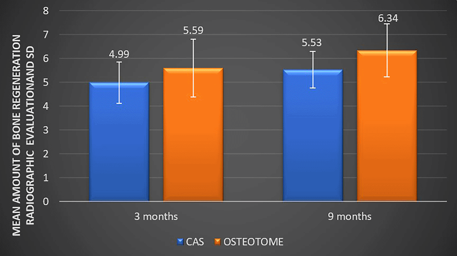
The mean amount of bone regeneration radiographic evaluation at three months in patients of the CAS group was 4.99±0.86 and 5.59±1.21 in the osteotome group. By using Student’s unpaired t-test, no statistically significant difference was found in bone regeneration from the radiographic between both groups (t=1.26, p=0.22). The mean amount of bone regeneration on radiographic evaluation at nine months in patients with CAS group and osteotome group was 5.53±0.77 and 6.34±1.11 respectively (Table 4). By using Student’s unpaired t-test no statistically significant difference was found in bone regeneration radiographic evaluation among both the groups (t=1.89, p=0.074).
| Group | N | Mean | 0±0 | 0±0 | t-value | |
|---|---|---|---|---|---|---|
| 3 months | CAS | 10 | 4.99 | 0.86 | 0.27 | 1.26 p=0.22, NS |
| Osteotome | 10 | 5.59 | 1.21 | 0.38 | ||
| 9 months | CAS | 10 | 5.53 | 0.77 | 0.24 | 1.89 p=0.074, NS |
| Osteotome | 10 | 6.34 | 1.11 | 0.35 |
Maxillary septa are the cortical bone barriers that separate the maxillary sinus into compartments, which are known as recesses. Septa are crucial for lifting the maxillary sinuses. Congenital septa develop during the middle face’s maturation. Primary septa and secondary septa are the two groups into which septa are divided. Secondary septa are those that are located above an edentulous ridge, whereas primary septa are those that are located above the maxillary sinus.4 According to a 2006 study by Ardekian et al., septa found in the maxillary sinus cavity induce sinus perforation. As a result, the maxillary cavity’s septa may also contribute to maxillary sinus perforation.7
To overcome this problem, Tatum (1970) developed a sinus augmentation technique which was later modified by Boyne and James in 1980. After a few years, a new technique was evolved by Summer in 1994. This technique mainly elevates the Schneiderian membrane, which ultimately increases the ridge height for implant placement. Today, two main procedures are performed for the sinus lift technique: the first is a one-stage method, i.e. osteotome sinus lift technique and the other is a two-stage technique using the lateral window technique.4
Our main focus in this study was to evaluate the efficacy of CAS kit over the osteotome technique during the sinus lift procedure. A total of 20 patients were included in both groups in which 60% of patients were male and 40% patients were female. The current study also included site-specific implant placement which showed that 40% of patients in the CAS Kit group and 50% of patients in the Osteotome group had an implant prosthesis in site 16 regions, while 20% of patients in the CAS group and 40% in the osteotome group had implants in site 26 regions and 20% of patients in CAS group and 10% in osteotome group in site 27 regions. Up till now, no study has demonstrated the site-specific implant insertion, thus this finding does not suggest any relevance with another study and was statistically insignificant.
The implant success depends upon the stability of an implant which is provided by peripheral bone. The present study found that mean RBH in the CAS group was 5.01±0.86 and 4.79±1.27 for patients of the osteotome group; statistically, there was no significant difference found in RBH between patients of the two groups. This is in accordance with the study by Rajkumar et al., in which the residual bone height was 4 to 7.5 mm for a sinus lift.8 A study performed by Lai et al., found RBH was 5.6±2.5 mm for the subjects without graft and 4.7±2.1 mm for graft subjects.9 However, the study performed by Movahedian Attar et al., included patients with higher range of residual bone in between 4.8 to 11 mm (a mean value of 7.9±1.27)10
Placing an implant in the atrophic maxilla is a time-challenging procedure as it has to be done meticulously without perforation of the sinus membrane. However, fewer studies have investigated time consumption with sinus lift procedure.
In the present study, the osteotome technique required more time for the sinus lift technique as compared to Cas kit, which was calculated from the beginning of an incision till sinus membrane elevation was performed. In both groups, there was a significant difference in the duration of surgery, which was 13.40±2.98 and 22.10±2.55 in CAS group and the osteotome group respectively; this contrasts with the animal study performed by Wu et al.,11 who evaluated the difference between Piezo surgery, CAS kit, and osteotome regarding the time required for completion of the procedure, and concluded that osteotome has the significantly shorter time required as compared with CAS kit and Piezo surgery.
The posterior atrophic maxilla is comprised of medullary bone which is less dense as compared with the mandible after extraction. Osteoclastic activity takes place at the alveolar ridge of the maxilla and eventually bone resorption.12 Thus, implant placement in the atrophic maxilla needs sinus membrane elevation with simultaneous bone regeneration to achieve implant stability. Bone regeneration can take place with or without graft placement.
In the current study, we evaluated bone regeneration/gain after graftless sinus elevation by radiographic means at three and nine months post-intervention in the CAS and osteotome groups. The mean bone regeneration in CAS and osteotome groups ranged 4-6 mm and 5.5-7 mm respectively, and was statistically not different. This was in accordance with the study performed by Chen et al.,13 in which sinus floor elevation followed by implant placement with a lateral trap-door window approach was done. The authors found that bone gain was 4.5 mm on average with a range of 3-9 mm. Another study performed by Thor et al.,14 measured a bone gain following the elevation of maxillary sinus floor, using lateral window corticotomy technique; the authors found that a mean bone gain of 6.51 mm (range 4-10 mm) after one year of follow-up period. Piero et al.,15 performed implant placement followed by angled osteotome and the window was prepared for sinus membrane elevation. The result evaluated bone gain, which was 8.2 mm and significantly higher without the use of a bone graft. In another study by Cricchio et al.,16 in which implant placement was done using lateral window sinus lift technique and simultaneous bone graft, it was found that bone gain was 4-7 mm after six years of follow-up period.
Another study performed by Gupta et al.,17 in which bone regeneration was evaluated using mesenchymal stem cell and blood coagulum, found bone regeneration to be 7.69+2.5 mm and 9.32+2 mm respectively for stem cell and blood coagulum after six months of the follow-up period. Nedir et al.18 in 2013 showed that the bone gain was higher in the graft group (4.6±1.4 mm) than in the non-graft group (3.3±1.5 mm). In a study by Crespi et al.,19 in which the authors evaluated a bone gain by performing sinus lift using the osteotome technique, bone gain was 4.08+1.25 mm after two years of follow-up period and was in contrast with our study.
The inner wall of the sinus is lined by the thin Schneiderian membrane, but each individual has a different thickness. Generally, the thickness varies from 0.3-0.8 mm and thus has a high chance of perforation during the sinus lift technique. Perforation of the sinus membrane does not only lead the graft material into the sinus but can also lead to sinusitis, infection, graft loss, and implant failure. Perforation of less than 0.5 does not affect the stability of an implant. During the placement of an implant, is there is sinus perforation less than 2 mm; on a radiograph, the sinus membrane will appear to have domelike shape. If perforation is more than 2 mm, it leads to implant exposure or loss of implant in the sinus.7
In the present study we found that 10% and 20% of patients had perforation in the CAS kit and Osteotome groups respectively, which was statistically insignificant and was similar to the study performed by Schwarz et al.20 Out of 407 sinus membrane elevations, 35 sinuses (8.6%) were perforated during the surgery and the author concluded that the presence of septa, habit of smoking, and RBH of less than 3.5 mm increased the risk of sinus membrane perforation. Wu et al.11 stated sinus perforation was lower in the CAS kit group (14.29, 21.43%) than in the Osteotome group (85.71%) using Piezo surgery.
Another study performed by Nolan et al.21 where sinus elevation was done using the lateral windows technique, showed very high sinus membrane perforation, i.e., 41% during elevation and 70.8% at augmentation. One of the studies by Xi et al.22 performed sinus elevation using OSFE and implant placement in which 42 patients were included and six of them presented with perforated sinuses; further, the study showed that there was a 100% of survival rate of implant in the perforated sinus. Therefore, the authors concluded that sinus perforation does not affect the implant survival rate, on implant placement in the posterior atrophic maxilla.
In the current study three patients underwent sinus perforation during sinus lift which was less than 2 mm in diameter; thus, it was sealed by a collagen sheet. Success of an implant depends upon the primary stability, which is driven by the quantity and quality of bone and surgical technique used for implant placement. After an implant placement, osseointegration takes place, which leads to primary stability. IT and IR are the parameters used to evaluate the primary stability of an implant.23
In the present study we found that the primary stability of the implant in patients with the CAS group was 44±6.14 NCM while patients with for the osteotome group it was 35.50±7.61 NCM, which was statistically significant. In contrast, a study done by Attanasio et al.23 found significantly higher primary stability (71.25±4.66 NCM) in osteotome-assisted implant placement. Alhayati et al.24 evaluated primary stability; sinus elevation was performed by using Versah burs for osseodensification and showed a primary stability of 69.85±9.74 NCM. Jelusic et al.25 studied stability of the implant in sinus lifts with and without using a graft material, which were 78.9±6.3 NCM and 78.7±6.1 NCM respectively. Ajanovic et al.26 in a pilot study measured mean implant stability with various diameters of the implant by creating a window by lateral approach, and sinus elevation was 77.73±2.93 NCM.
Marginal bone loss is a normal process that becomes more noticeable within the first year following implant placement. Average marginal bone loss is 0.1 mm in the first year, 0.07 mm per year from year 1 through 5, and 0.026 mm per year from 6 years through the end of follow-up.27 Marginal bone loss can be associated with patient-specific or implant-specific factors and can affect the stability of an implant.28
The current study evaluated marginal bone loss at three and nine months in CAS and osteotome groups, which was 0.02±0.04 mm and 0±0 mm respectively, and was statically insignificant. This contrasts with the study by Kim et al.29 who evaluated a marginal bone loss after sinus bone grafting and immediate implant placement at one year after prosthetic rehabilitation; after 20.8 months, bone loss was found to be 0.6 mm and 0.7 mm, respectively. Toledano et al. showed that marginal bone loss is greater in standard implant placement than in short implant placement.30
The present study evaluated all the essential parameter for the success of an implant. CAS kit is a new modality for implant placement in the atrophic maxilla due to its newer bur shape that prevents sinus perforation, and is a time-saving procedure for implant placement in the posterior atrophic maxilla. Use of Cas kit is an effective procedure for elevating sinus membrane in a posterior atrophic maxilla as it cuts intervention time and lowers the chance of sinus membrane perforation. However, osteotome is an older but gold standard procedure effective in bone regeneration. Multi-centric, split-mouth studies with a greater number of samples can help in evaluating the efficacy of both procedures.
Indirect sinus lift is a common employed procedure in cases where the RBH is less than 7 mm. The present study concluded that the CAS kit group had a shorter operative time, better primary stability, and less marginal bone loss. The osteotome group had better bone regeneration after three and nine months. CAS kit compared to osteotome was better in terms of operative time, primary stability, and marginal bone loss resulting in better modality to perform sinus lift in posterior atrophic maxilla as a recommended modality.
| Views | Downloads | |
|---|---|---|
| F1000Research | - | - |
|
PubMed Central
Data from PMC are received and updated monthly.
|
- | - |
Provide sufficient details of any financial or non-financial competing interests to enable users to assess whether your comments might lead a reasonable person to question your impartiality. Consider the following examples, but note that this is not an exhaustive list:
Sign up for content alerts and receive a weekly or monthly email with all newly published articles
Already registered? Sign in
The email address should be the one you originally registered with F1000.
You registered with F1000 via Google, so we cannot reset your password.
To sign in, please click here.
If you still need help with your Google account password, please click here.
You registered with F1000 via Facebook, so we cannot reset your password.
To sign in, please click here.
If you still need help with your Facebook account password, please click here.
If your email address is registered with us, we will email you instructions to reset your password.
If you think you should have received this email but it has not arrived, please check your spam filters and/or contact for further assistance.
Comments on this article Comments (0)