Keywords
giant bronchogenic cysts, surgical excision, complications, case report
giant bronchogenic cysts, surgical excision, complications, case report
Bronchogenic cysts (BCs) are defined as uncommon congenital anomalies.1 They account for 60% of mediastinal cyst lesions.1 Mediastinal BCs have often been described arising from an abnormality in tracheobronchial tree budding between the third and sixth weeks of gestation.2,3 In adult patients, BCs are frequently asymptomatic2,4–6 and are often randomly discovered during a chest X-ray.2 Whenever they become symptomatic, surgical resection becomes highly recommended. Typically, BCs are oval in shape, of diameter between 20 to 120 mm.2
The aim of our case, which was operated on in The Public Hospital of Abdarrahman Mami, Tunis, is to insist in the good timing of surgery of BCs in order to avoid compressive complications due to increasing size and to ensure easier operation.
A 49-year-old patient was referred to the Thoracic Surgery Department of Abderrahmane Mami Hospital in April 2021 with a dry cough, dyspnea and chest tightness progressing for six months. The patient had a medical history of diabetes mellitus, and arterial hypertension. She had no surgical history or family history of bronchogenic cysts.
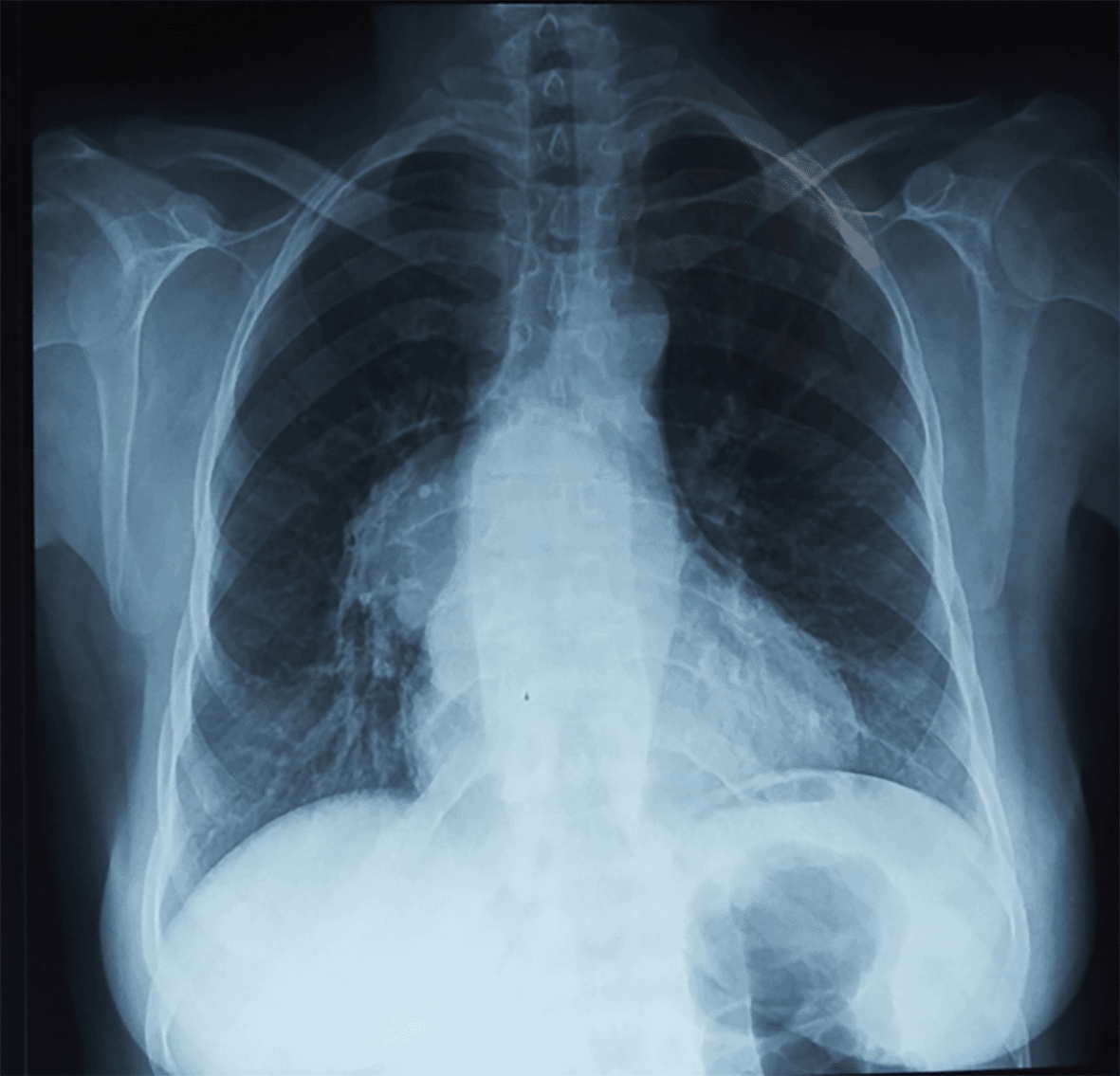
Chest radiography showed a mass overlapping the cardiac silhouette (Figure 1). A computed tomography (CT) scan of the chest revealed a round homogeneous cystic mass 96 mm * 80 mm in diameter, occupying the middle mediastinum between the aortic arch and right pulmonary artery in sub-carine of liquid density not enhanced after injection of a contrast product. Thoracic magnetic resonance imaging (MRI) showed a cystic formation of homogeneous signal with thin wall in T2 hypo signal, evoking a bronchogenic cyst (Figure 2).

Surgery was performed via a right posterolateral thoracotomy. Intraoperatively, there was a large cystic formation 100 mm*80 mm of the middle mediastinum in intimate contact with the origin of the right pulmonary artery, the lower right pulmonary vein as well as the right atrium exerting a compressive effect on the adjacent organs especially the pericardium (Figure 3A). After aspiration of a yellow liquid content and viscous consistency, the cystic was ‘en bloc’ resected in totality (Figure 3B).

The postoperative course was uncomplicated with removal of the drains on the fourth post-operative day. The patient was discharged from the hospital on May 16, 2021, on the fifth postoperative day. Histological examination confirmed diagnosis of a bronchogenic cyst. The patient was asymptomatic throughout the follow-up period.
BCs are relatively uncommon.1 They occur not only in pediatric patients, but also in adults.2 BCs are typically found near the large airways in the mediastinum, often just posterior to the carina, although they may be attached to the oesophagus or even inside the pericardium.3 BCs are thought to originate early in lung bud development, before bronchus formation.3 BCs contain clear fluid or less commonly, haemorrhagic secretions or air. Cases of intra-pericardial BCs are very rare in the literature.5 We present a distinctive case in which a giant BC was discovered compressing the pulmonary artery and pericardium in a very important way.
BCs are diagnosed incidentally because patients do not show symptoms.5–6 However, when BCs are huge, they sometimes bring about life-threatening complications including rupture, infections, an haemorrhage and compression of the surrounding structures.6–8 Those complications can be suggested by the presence of symptoms such as cough, fever, haemoptysis, dyspnea and chest pain. If the cyst is giant with invasion of the pericardium, there is an imminent risk of intra-pericardial rupture and pericardial effusion that can lead to cardiac tamponade and exacerbation of the initial symptoms.5
On radiography, BCs appear as a spherical opacity, non-calcified, with smooth outlines.2 A CT scan is very useful in preoperative diagnosis of BCs to ascertain the precise size and in order to show the relationship of the cyst to adjacent mediastinal structures.2–9 Thus, it shows homogeneous, smooth, solitary, round or ovoid masses usually located in middle mediastinum. The CT scan density of BCs can vary from typical water density to high density related to increased calcium content, anthracotic pigment, blood, or greater protein content of the fluid.9 MRI may provide specific diagnostic confirmation in regard to BCs and allows the limits within the pericardium and the large vessels to be specified.9 In Tunisia, hydatid cysts are common, and they are the first differential diagnosis to be suggested. In such contentious cases, only surgery associated with histological examination can affirm one of the two diagnoses.
Surgical resection is the most approved treatment option of BCs.6–8 The evolutionary possibilities of the cyst justify the legitimacy and the need of complete removal of this lesion.4 Despite the predictive diagnostic value of imaging, definitive diagnosis of BCs is confirmed by surgical excision and pathological examination.9 Different authors suggest various approaches such as CT guided or mediastinoscopic drainage, video-assisted thoracic surgery (VATS) or open thoracotomy. The choice of one or the other is according to the condition of the patient and characteristics of malformation (e.g., size, extension).1,6 Generally, considering the risk of serious complications and even malignancy degeneration, BCs require complete resection and should be performed every time when the diagnosis is suspected especially since the patient’s follow up is uncertain.1,5 Specifically, for giant BCs, surgical resection is recommended as early as possible in order to avoid serious complications due to the increasing size.5 In cases where there is a close relationship between the BC and the pericardium, it is important to consider emergency surgery in order to avoid a sudden rupture of the cyst in the pericardium that can lead to tamponade or acute pericarditis.10 The prognosis after complete excision is excellent.8 Incomplete excision will lead to a high recurrence rate and to the potential for more serious sequelae.8 Trans-tracheal and percutaneous drainage of the cyst are proposed by some authors as alternatives to surgery, but they are not widely accepted because of possible cyst recurrence.8
In our case, surgery was laborious because the cyst was firmly adhered to the pericardium, right pulmonary artery, and the lower right pulmonary vein with a very important compression of the bronchi. The surgical challenge was to do a radical excision without injuring the vascular and bronchial elements. We succeeded in performing this by dissecting the wall of the cyst step by step and we had the choice of approaching the patient with open thoracotomy.
Apart from the progressive malignant potential, BCs can increase in size becoming huge cysts and causes serious complications. Surgical excision is almost obligatory especially when the cyst is a certain size and in the case of giant BCs. The advent of VATS increasingly supports the idea of adhering to surgical treatment choice in the event of a bronchogenic cyst whatever the size. A careful analysis of the radiologic appearance allows us to better assess the limits of the BC and in order to prepare for sometimes laborious surgery. Incomplete surgical excision is associated with high late recurrence of the cyst.
| Views | Downloads | |
|---|---|---|
| F1000Research | - | - |
|
PubMed Central
Data from PMC are received and updated monthly.
|
- | - |
Provide sufficient details of any financial or non-financial competing interests to enable users to assess whether your comments might lead a reasonable person to question your impartiality. Consider the following examples, but note that this is not an exhaustive list:
Sign up for content alerts and receive a weekly or monthly email with all newly published articles
Already registered? Sign in
The email address should be the one you originally registered with F1000.
You registered with F1000 via Google, so we cannot reset your password.
To sign in, please click here.
If you still need help with your Google account password, please click here.
You registered with F1000 via Facebook, so we cannot reset your password.
To sign in, please click here.
If you still need help with your Facebook account password, please click here.
If your email address is registered with us, we will email you instructions to reset your password.
If you think you should have received this email but it has not arrived, please check your spam filters and/or contact for further assistance.
Comments on this article Comments (0)