Keywords
Spine, arteriovenous fistula, Peripheral Catheterization (source: MeSH).
Spinal dural arteriovenous fistulas (SDAVF) are a subtype of spinal arteriovenous malformation (AVM). Although rare, SDAVF are the most common spinal vascular malformations and have potential devastating neurologic consequences, sometimes irreversible. Originally, they were treated with surgery alone, however endovascular treatment of SDAVF has evolved recently with good results and the understanding of the anatomy plays a key role. The feeder vessel of the AV shunt is supplied by the radiculomedullary or radiculopial artery, the shunt is usually located within the root sleeve of the dura and the SDAVF is drained by the medullary vein via retrograde flow. Generally the endovascular approach uses the catheterization of the segmental arteries; however, aortic thoracolumbar aneurysms can render endovascular access difficult and risky. We present a case where an SDAVF in woman with aneurysmal dilation of the descending aorta and a mural thrombus was successfully treated through percutaneous catheterization of intercostal artery.
Spine, arteriovenous fistula, Peripheral Catheterization (source: MeSH).
Spinal dural arteriovenous fistulas (SDAVF) account for 70% of spinal vascular malformations. In SDAVFs, a direct shunt between a radiculomeningeal artery and a radicular vein results in increased venous pressure and decreased drainage of normal spinal veins, leading to venous congestion, intramedullary edema, chronic hypoxia, and myelopathy. In 90% of SDAVFs, the shunt is located in the lateral epidural space where the radicular vein passes through the dura at the dorsal surface of the dura root sleeve in the intervertebral foramen.1,2
An adult patient with no relevant medical history presented with intermittent upper and lower limb paresthesias and motor deficits, sometimes associated with cold sweats, nausea, and palpitations. She had no fever, recent trauma, or incontinence.
Physical examination revealed right lower-limb weakness and hyperalgesia.
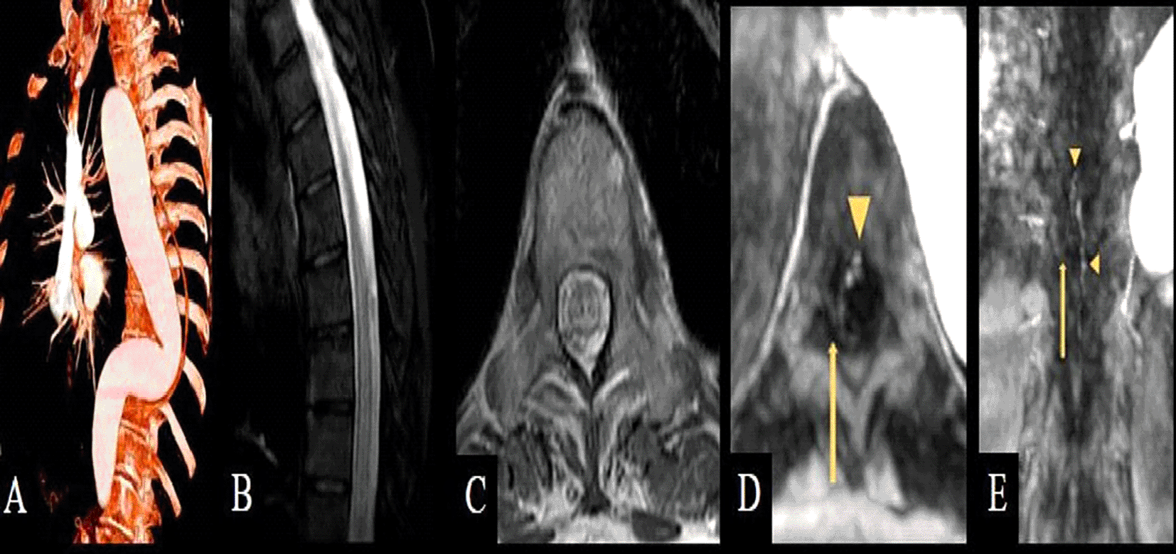
CTA showed aneurysmal dilation of the descending aorta with a mural thrombus. T2-weighted MRI sequences showed centromedullary edema over multiple spinal cord segments and dilated and coiled posterolateral perimedullary vessels. MRA done to locate the suspected SDAVF showed early venous filling with an arteriovenous shunt at the right T10 level (Figure 1).

[A] Volume rendering reconstruction from CTA shows the aneurysm and associated mural thrombus in the descending aorta; Sagittal [B] and axial [C] T2-weighted MRI show centromedullary edema and dilated perimedullary vessels. Axial [D] and coronal [E] first-pass contrast-enhanced MRA images show early venous filling (arrowheads) and the presumed shunt point (arrow) at the right T10 level.
DSA showed delayed venous return in the great anterior radiculomedullary artery originating from the right T11 segmental artery (not shown); however, the partially thrombosed aneurysmatic dilation of the aorta and the morphology and disposition of the thoracic segment precluded the catheterization of some lower thoracic segmental arteries.
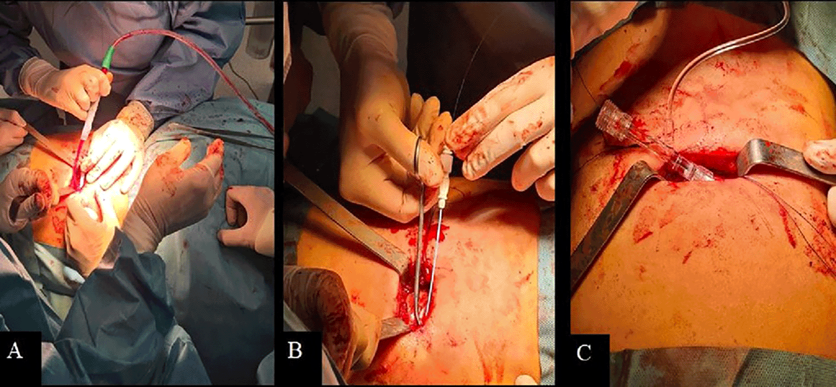
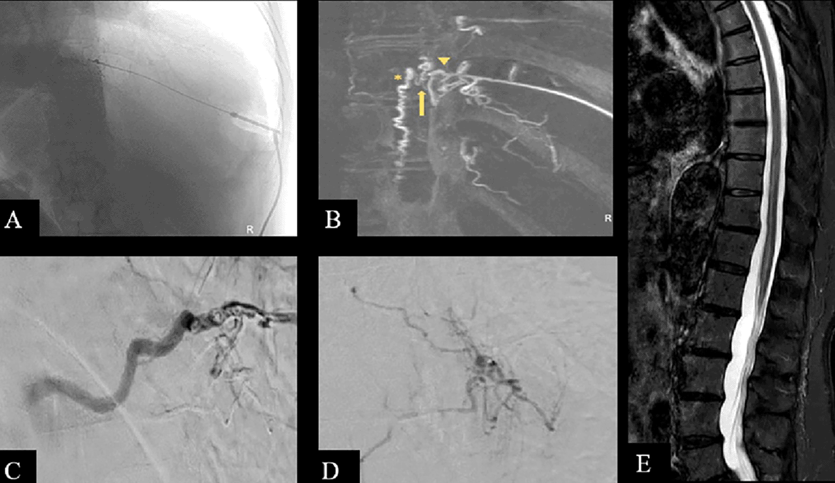
Surgical repair failed because the intradural vein receiving blood from the shunt could not be identified. The interdisciplinary team decided on a combined surgical and endovascular approach in which a minimal partial posterolateral rib resection with the patient in left lateral decubitus position exposed the ventral branch of the 10th right posterior intercostal artery (Figure 2), which was catheterized with a 4F introducer set (4F Micro-stick introducer set, Medcomp) and a 0.014 inch microwire. DSA revealed early venous filling and retrograde contrast uptake of the upper and lower radiculomedullary veins, confirming the diagnosis of SDAVF (Figure 3).

[A] Minimal partial posterolateral rib resection with the patient in left lateral decubitus to expose the ventral branch of the 10th right posterior intercostal artery. [B, C] Catheterization of the ventral branch of the 10th right posterior intercostal artery with a micropuncture set and a 0.014-inch microwire.

[A] Unsubstracted fluoroscopic image showing the intercostal access and opacification of the Spinal dural arteriovenous fistulas (SDAVF) at the T 10 level. Superselective injection and 3D rotational DSA with MIP reconstruction [B] show the feeding artery (arrowhead), the shunting zone (arrow), and the proximal draining vein (asterisk). [C, D] Exclusion of the SDAVF after embolization and coil placement in the segmentary artery. [E] Sagittal Short tau inversion recovery (STIR) MRI image 3-months after treatment showing less evident cord edema.
Injection from the segmental artery verified the absence of supply to the spinal cord from the pedicle feeding the SDAVF. After 3D-rotational DSA to better characterize the lesion, we injected approximately 0.5 cc of ethylene vinyl alcohol copolymer (SQUID-18, Emboflu; Gland, Switzerland, supplied by Balt) into the fistula with little pass to the draining vein. Finally, we placed a coil in the segmentary artery to mark the level of the fistula. Control images demonstrated the exclusion of the fistula and patency of the segmental artery (Figure 3).
The patient was discharged without complications after 2 days.
At follow-up, her lower limb strength had improved and on follow-up MRI three months after procedure (Figure 3) the prominent perimedullary vessels and cord edema were less evident. Even though there was not complete pass to the draining vein, it was enough to diminish the flow to the fistula and to get symptomatic relief.
Treatment for SDAVF aims to occlude the shunt. Surgical exclusion of the fistula by a classic approach or minimally invasive techniques yields excellent results,3,4 better than the endovascular approach.
Different results have been reported with endovascular treatment with different embolic materials.5–7 In our institution, endovascular treatment is considered for spinal arteriovenous shunts whenever safely possible. After superselective catheterization of the feeding artery, a liquid embolic agent is injected through the fistula to occlude the proximal segment of the draining vein to prevent subsequent intradural collateral filling of the fistula.6
In our case, MRI suggested SDAVF and MRA suggested the location, but the diagnosis could not be confirmed with conventional spinal DSA because the partially thrombosed thoracic aortic aneurysm and the morphology of the lower thoracic aorta made it impossible to catheterize the segmental arteries at the level of the suspected shunt. After surgical repair failed, a different approach was necessary.
The ventral branch of the posterior intercostal artery runs circumferentially under the rib; the dorsal branch has a spinal branch that enters the vertebral canal through the intervertebral foramen and gives rise to the radicular artery, among others.8
We used a novel technique to access the ventral branch of the posterior intercostal artery to catheterize the dorsal branch, confirm the SDAVF, and embolize the lesion after superselective catheterization.
All authors participated in design, writing, critical review and approved the final version of the manuscript.
The present study is a case report, so it does not need ethical approval. The authors received signed informed consent from the patient and are committed to respecting bioethical research principles as well as the Declaration of Helsinki.
We obtained the patient’s written informed consent to participate in this study. Also, the patient gave us written permission for the publication of images and data included in this case report.
All data underlying the results are available as part of the article and no additional source data are required.
| Views | Downloads | |
|---|---|---|
| F1000Research | - | - |
|
PubMed Central
Data from PMC are received and updated monthly.
|
- | - |
Provide sufficient details of any financial or non-financial competing interests to enable users to assess whether your comments might lead a reasonable person to question your impartiality. Consider the following examples, but note that this is not an exhaustive list:
Sign up for content alerts and receive a weekly or monthly email with all newly published articles
Already registered? Sign in
The email address should be the one you originally registered with F1000.
You registered with F1000 via Google, so we cannot reset your password.
To sign in, please click here.
If you still need help with your Google account password, please click here.
You registered with F1000 via Facebook, so we cannot reset your password.
To sign in, please click here.
If you still need help with your Facebook account password, please click here.
If your email address is registered with us, we will email you instructions to reset your password.
If you think you should have received this email but it has not arrived, please check your spam filters and/or contact for further assistance.
Comments on this article Comments (0)