Keywords
PAPVC, congenital heart diseases, open heart surgery, physiotherapy management, cardiac rehabilitation, case report
This article is included in the Datta Meghe Institute of Higher Education and Research collection.
PAPVC, congenital heart diseases, open heart surgery, physiotherapy management, cardiac rehabilitation, case report
The atypical drainage of pulmonary veins into the right atrium leads to a variety of congenital cardiac conditions known as partial anomalous pulmonary venous connections (PAPVC). This can occur by draining directly into the right atrium or through systemic veins.1 PAPVCs, which frequently coexist with atrial septal defect (ASD) and manifest as a left-to-right shunt, have negative consequences on cardio-pulmonary physiology.2
In 0.4-0.7% of autopsies, PAPVC are discovered. Most frequently, the right pulmonary veins that drain into the right atrium or superior vena cava (SVC) are involved in the aberrant venous connection. Less commonly, abnormal pulmonary veins flow into the coronary sinus, innominate vein, or inferior vena cava.2,3
Various transcathater or surgical approaches have been proven to improve the competency of the patient’s having anomalous venous connections, which includes balloon angioplasty and surgical repair of associated anomalies.4 To facilitate flow of blood from pulmonary veins to left atrium, the wall between the coronary sinus and left atrium is excised. The coronary sinus opening is then sealed off. ASD is also closed, if it is present.2 An anastomosis between the left atrium and the pulmonary venous confluence is made in infracardiac and supracardiac PAPVC. Unless there is severe pulmonary hypertension, the vertical vein is mostly ligated in the same surgical setting. In complicated and mixed type PAPVC, a number of other procedures, such as intra-atrial rerouting, have also been employed.2,5
Following heart surgery, there are a number of issues that call for specialised treatment, particularly in the respiratory system. These issues can extend a patient’s stay in the hospital, raising hospital expenses and playing a significant role in morbidity and death. Recent research has demonstrated that early mobilisation, such as getting out of bed and walking, improves the patient’s functional state and shortens the period of hospitalisation. Patients should receive physical treatment regularly to ensure their rehabilitation of their motor skills.6
These studies show that inspiratory muscle re-training (IMRT) is possibly beneficial in improving breathing mechanics and decreasing exercise-induced dyspnea. It is also likely useful in enhancing respiratory muscle strength and lowering airway closure (i.e., greater FVC). These mechanistic alterations reflect our developing understanding of the function of IMRT in paediatric patients and can be used to explain improvements in symptomology and clinical outcomes.7
A 10-year-old female patient presented to our hospital with a complaint of insidiously developing and increasingly worsening breathlessness which increases while walking uphill along with frequent episodes of fever. Following assessment she was admitted to the hospital. 2D Echo was done which reviled PAPVC along with 16 mm ASD. She underwent ASD closure along with pulmonary valvotomy.
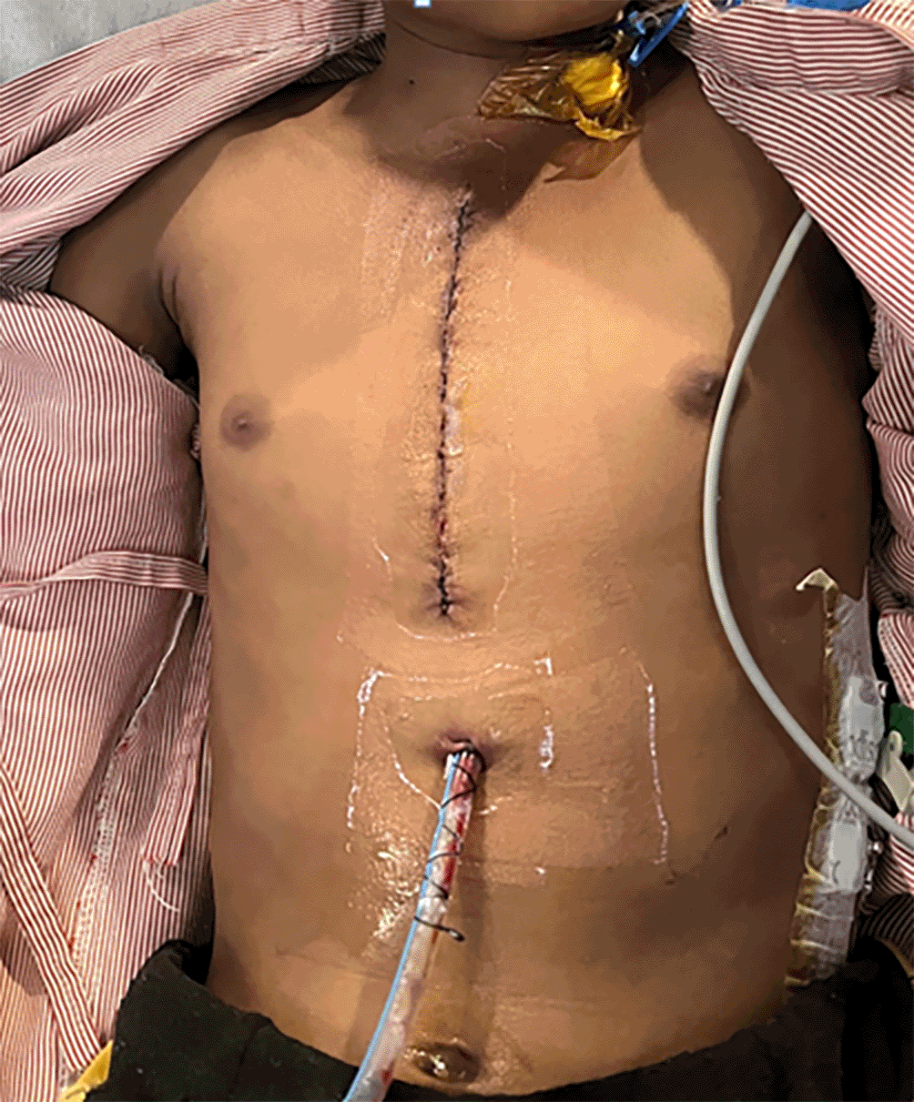
Prior to the examination, the patient’s caretaker provided informed permission, and the patient was then placed in a supine posture. The patient was conscious and following instructions. The patient’s BMI was 16.1 kg/m2, making her ectomorphic. Prior to surgery, the patient had gradual, insidious dyspnea that worsened as she walked uphill and subsided with rest. The patient is currently having discomfort at the suture site (Figure 1), which she rates 5/10 on a VAS (visual analogue scale). The pain was abrupt in start and dull aching in character. The pain aggravates on coughing and while inhaling deeply and she rated it 7/10 on VAS. Foley’s catheter, central line and drain were present in situ.
An examination of the cardiovascular and respiratory systems revealed a normal pericardium that was bilaterally symmetrical, had an apex beat at the level of the fifth intercostal space, and had sutures on the midsternal area. The heartbeat was regular and rhythmic at 89 beats per minute, and all of the peripheral pulsations were present. The middle and lower zones of the chest expanded less than usual, and the respiratory rate was 18 beats per minute.
Timeline: Table 1 included a timeline of the occurrences. It depicts series of incidence from the patient’s hospital admission until the commencement of the physiotherapy treatment.
Diagnostic assessment: 2D ECHO revealing 14×16 mm of Atrial septal defect along with PAPVC along with grade 1 pulmonary arterial hypertension with interstitial edema.
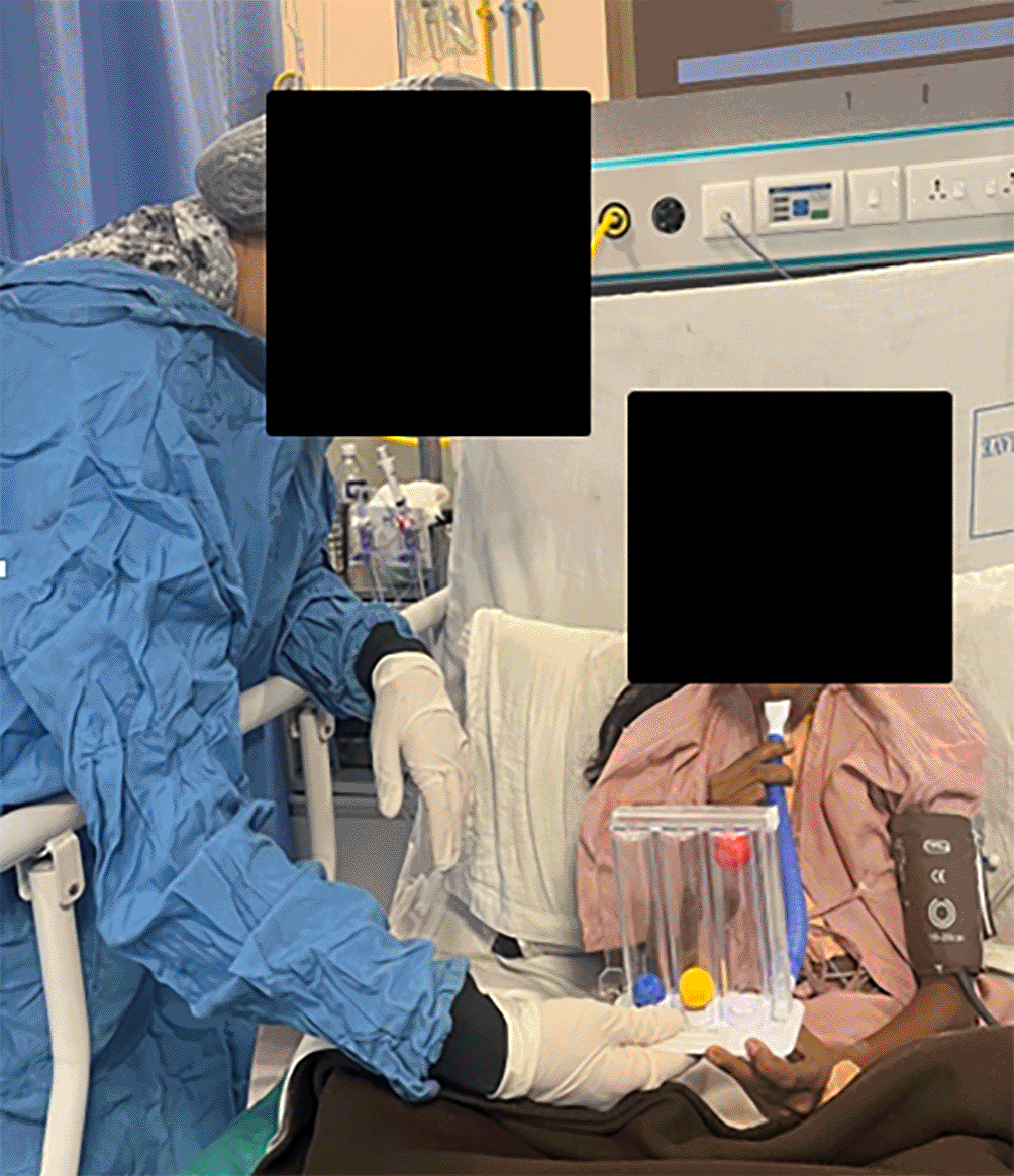
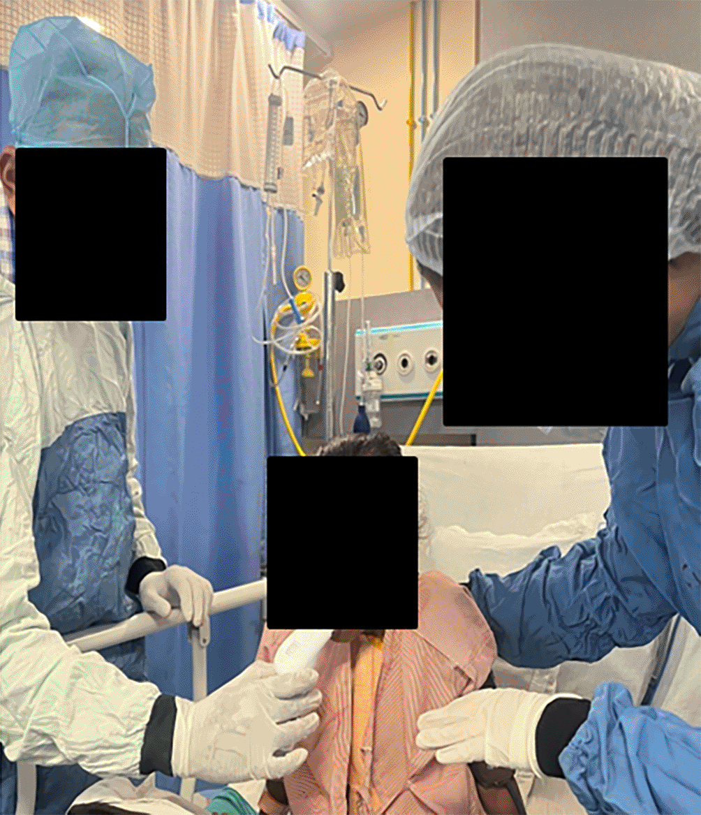
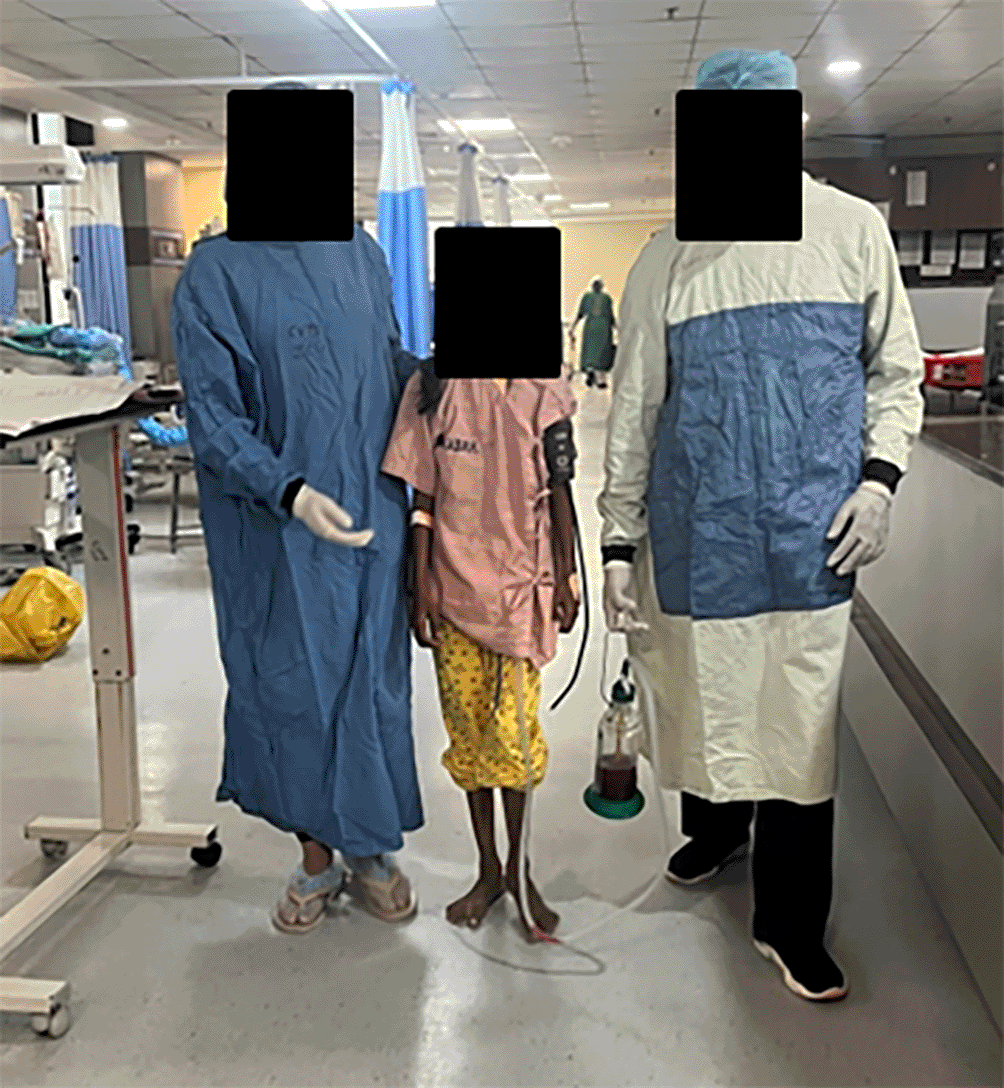
Therapeutic intervention: Table 2 represents a comprehensive rehabilitation protocol post cardiac surgeries in paediatric population. The protocol includes education of the patient’s care taker and family members about the condition and post-operative care. Pain control: using chest binder to support suture line and decrease pain. Promote bronchial hygiene: nebulization and initiating with active cycle of breathing technique for effective expectoration. Improving inspiratory hold and vital capacity by giving incentive spirometer (Figure 2) and diaphragmatic breathing exercises. Facilitation of normal chest movements by asking the patient to perform thoracic expansion exercises. Improving inspiratory muscle strength: making use of Powerbreathe medic plus device (Figure 3) which is exclusively used for re-training of inspiratory muscles. Hallway ambulation to improve functional capacity (Figure 4). Incorporating home exercise programme to promote functional independence and improving level of activities of daily living.
Follow-up and outcome measures: Preoperatively and on the first day of physical therapy rehabilitation, the outcome measures were evaluated. A follow-up was done after one week and two weeks of the physiotherapy intervention. The outcomes were listed in Table 3.
André Luiz Lisboa Cordeiro et al. in their studies have stated that Surgery for a coronary artery bypass graft (CABG) is linked to longer hospital stays, pulmonary problems, and reduced functional ability. Patients who are at a high risk for post-operative (PO) pulmonary problems prior to CABG experience these adverse effects to a greater degree. The healing phase for CABG patients has been demonstrated to be aided by inspiratory muscle training (IMT). However, there is insufficient evidence to support the use of IMT post-operatively to promote speedier recovery in high-risk patients.6 According to Tamires Daros Dos Santos et al. Patients undergoing CABG experienced additional advantages from short-term moderate-to-high intensity IMT combined with aerobic and resistance training in terms of exercise capacity, inspiratory muscle strength, QoL, and antioxidant profile.8 Studies conducted by Balbino Rivail Ventura Nepomuceno Jr et al. suggest that early implementation of inspiratory muscle training on hospitalised patients without established respiratory deficits may prevent adverse outcomes that are either directly or indirectly related to the loss of respiratory muscle mass inherent to a prolonged hospital stay. Training the respiratory muscles can prevent endotracheal intubation, muscular wasting, and death.9 According to Rhoia Neidenbach et al. Respiratory training is known to improve exercise capacity and cardiopulmonary function showing significant improvement in oxygen saturation and maximum workload capacity.10 In this current case we have found significant improvement in PIMax and functional capacity.
| Views | Downloads | |
|---|---|---|
| F1000Research | - | - |
|
PubMed Central
Data from PMC are received and updated monthly.
|
- | - |
Provide sufficient details of any financial or non-financial competing interests to enable users to assess whether your comments might lead a reasonable person to question your impartiality. Consider the following examples, but note that this is not an exhaustive list:
Sign up for content alerts and receive a weekly or monthly email with all newly published articles
Already registered? Sign in
The email address should be the one you originally registered with F1000.
You registered with F1000 via Google, so we cannot reset your password.
To sign in, please click here.
If you still need help with your Google account password, please click here.
You registered with F1000 via Facebook, so we cannot reset your password.
To sign in, please click here.
If you still need help with your Facebook account password, please click here.
If your email address is registered with us, we will email you instructions to reset your password.
If you think you should have received this email but it has not arrived, please check your spam filters and/or contact for further assistance.
Comments on this article Comments (0)