Keywords
Case Report, Congenital Diaphragmatic Hernia, Ex utero Intrapartum Technique Procedures, Hernia Repair, Twins
Case Report, Congenital Diaphragmatic Hernia, Ex utero Intrapartum Technique Procedures, Hernia Repair, Twins
Congenital diaphragmatic hernia occurs when abdominal contents herniate into the thoracic cavity, causing lung hypoplasia and pulmonary vascular alteration. Hernias are classified into different types based on the defects’ location in the diaphragm which are: (a) The most frequent type, Bochdalek hernias (occur in the posterolateral part of the diaphragm) which mostly occurs on the left side rather than the right side, (b) Morgagni hernias (20% to 25%), and (c) central hernias, accounting for 2% to 5% of all cases.1,2
Cases of CDH in one twin have rarely been reported. Studies reporting CDH in twin pregnancies are still rare and reported as cases of CDH in general. A retrospective study from 2010 to 2016 reported only 11 twin pregnancies with CDH in one of the twins.3 Infants with CDH have a higher mortality rate without prompt management, and they also have a wide range of long-term issues that last beyond infancy, such as complications of the respiratory system, gastrointestinal system, neurobehavioral disorders, and orthopaedic deformities. Hernia recurrence is another risk that can develop later, especially in newborns with significant defects. It has been established that each of these clinical issues has an impact on the quality of life of affected children. Thus, prenatal diagnosis especially with ultrasound holds a pivotal role to prevent further fetal morbidity and mortality.4,5
The majority of CDH cases are identified prenatally between 18–24 weeks of pregnancy during a standard abnormality scan. CDH is distinguished by the visualization of thorax filled with abdominal contents and the mediastinal shifting from the defect to the contralateral side in prenatal imaging. Milder cases, on the other hand, might not be discovered until later due to minor defects that lead on to cause many complications.6,7 The prognosis of CDH is influenced by the extent of lung hypoplasia, the location of the liver, and co-existing abnormalities.
Fetal lung volume is measured by the lung-to-head ratio (LHR) which is obtained from the contralateral lung area divided by the fetal head circumference. Fetal LHR measurements have been used to predict neonatal pulmonary hypoplasia. Between 12 and 32 weeks of gestation, there is an 18-fold increase in lung area and a four-fold increase in head circumference. Thus, LHR on the left or right-side increases exponentially with gestational age because the measurements to determine LHR differ depending on gestational age. The value of the ratio is very important, in the study by Metkus, all neonates died if the ratio reached 0.6, on the contrary, they would survive better with a survival rate of 100% if the ratio reached 1.35, while the ratio between 0.6 to 1.35 had a survival rate of 61%.8 Advances in prenatal diagnosis have made it possible to identify a number of birth anomalies that may obstruct the fetus’s airway or impede ventilation at birth. Following the confirmation of CDH diagnosis, close monitoring to check fetal well-being is strongly advised.6,8,9
Selective centers in developed countries are providing fetal therapy Tracheal Occlusion To Accelerate Lung Growth Trial (TOTAL) and Fetoscopic Endoluminal Tracheal Occlusion or FETO as management for qualified patients in moderate and severe cases of CDH.10 The delivery of infants with CDH is recommended until term gestation. It is also strongly recommended that the infants are delivered at a tertiary center with expertise in CDH management and have that access to extracorporeal membrane oxygenation (ECMO) therapy.11,12 In cases of prenatally diagnosed CDH, it is advised to place a nasogastric tube shortly after delivery for stomach and intestines decompressions. Thus, in twin pregnancy, ultrasound guidance plays a critical role in determining which twin suffered from CDH. Ex utero intrapartum treatment procedures could be performed on the neonates after the baby with CDH was delivered. A variety of fetal airway obstructions have been successfully treated with EXIT procedures, enabling control of the airway at birth. The EXIT procedure can be performed on babies with CDH at birth, however, babies with CDH still need postnatal surgical repair.11–13
Here, we present a case report of prenatally diagnosed CDH in one of the twins who underwent EXIT procedure without previous antepartum fetal therapy. This case is very rare since diagnosing one of the twins with CDH during the intrapartum period to determine which one will undergo EXIT procedure is quite challenging. There have been no case reports published regarding prenatally diagnosed CDH in one of the twins followed by EXIT procedure, especially in developing countries. Therefore, the aim of this study was to report a rare case as well as to analyze the pearls and difficulties in diagnosing and managing these patients.
This following case is described according to the CARE checklist.14 A 32-year-old multiparous Indonesian woman with no occupation only a housewife, came to the Maternal Fetal Clinic, in a tertiary hospital in Indonesia, as referred by an obstetrician with multiple pregnancy and suspected CDH on the 1st baby. The mother had a history of twin babies in her family. She had no comorbidities and no previous history of surgery. The patient was compos mentis. Her vital signs, physical examination results, and laboratory profiles were within normal limits. The patient underwent further investigation with ultrasound and it revealed multiple pregnancies with placenta inserted at the posterior, positive septum confirmed monochorionic-diamniotic multiple pregnancies with discordance of 18%.
The first fetus was alive with a cephalic lie equal to 32–33 weeks of pregnancy, and an estimated fetal weight of 1,504 grams. In the thoracal region, the four-chamber view diagnostic planes showed a dextroposition with narrow left ventricle; the measured cardiothoracic area (CTAR) was 20%, and the axis was 29.13 degree. A mass (stomach) was found in this thoracal region, which was 1.98 cm × 1.20 cm in size, located as high as the four-chamber view, and the right ventricle was pushed forward anteriorly as shown in Figure 1 and Figure 2 (Video of Babies’ Ultrasound). LHR was 0.14 and observed per expected LHR (o/e LHR) was 4.79%. Ultrasound assessment showed urinary bladder filled with fluid which shows normal kidney function, shape and appearance of the fetal kidney within normal limits, also umbilical artery within normal limits. The single deepest pocket (SDP) was 5.44 cm. Further examination with Doppler velocimetry revealed middle cerebral artery pulsatile index (MCA-PI) and umbilical pulsatile index (umbilical PI) were within normal limit with normal flow of ductus venosus. No abnormality was found in other regions. These findings suggested that the first fetus had a severe left-side CDH with no lung hypoplasia, polyhydramnios, hyperexpanded lungs, and anatomic compressions were found.
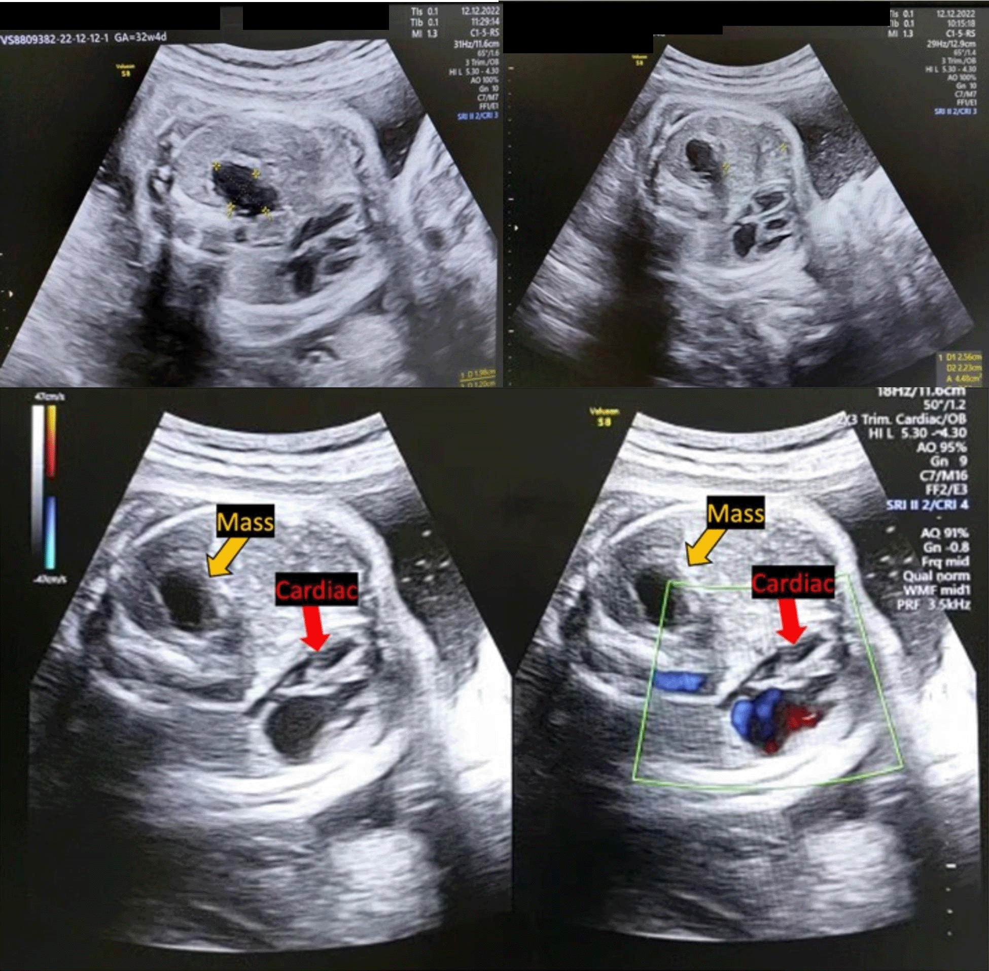
The four-chamber view diagnostic planes showed a mass (stomach) (yellow arrow) was found in this thoracal region, was 1.98 cm × 1.20 cm in size. The right ventricle is pushed forward anteriorly (red arrow). LHR was 0.14 and o/e LHR was 4.79%. Lung hypoplasia prediction was also within normal limit.
This is the video recording of the four-chamber view diagnostic planes which showed a mass (stomach) that was found in this thoracal region. It was 1.98 cm × 1.20 cm in size. The right ventricle is pushed forward anteriorly. LHR was 0.14 and o/e LHR was 4.79%. Lung hypoplasia prediction was also within normal limit.
The second fetus was alive with transverse lie equal to 31–32 weeks of pregnancy and an estimated fetal weight of 1,824 grams. No abnormality was found in the thoracal and abdominal regions. Further examination with Doppler velocimetry revealed MCA-PI and umbilical pulsatile index (umbilical PI) were within normal limits with normal flow of ductus venosus. The single deepest pocket (SDP) was 5.44 cm.
Three weeks later, she was admitted to the obstetric ward and had undergone expectant management until term pregnancies. Caesarean delivery was planned for the patients in our case due to an obstetric indication (transverse lie of the second fetus), which was also accompanied by CDH of the first fetus. Following the confirmation of the CDH and the completion of the imaging workup, a multidisciplinary team of obstetricians, anesthesiologists, neonatologists, and pediatric surgeons was assembled. An intervention aimed at establishing and maintaining airway patency immediately after cesarean delivery was considered mandatory, thus direct laryngoscopy intubation as the first approach in EXIT-to-airway procedure was planned.
The first baby was a male born weighing 1,600 grams with a 1st minute APGAR score of 3 and the 5th minute APGAR score was 5, while the second baby was also a male; born weighing 2,000 grams, with a 1st minute APGAR score of 6, while the 5th minute APGAR score was 8. Following informed consent, the group in charge carried out the EXIT-to-airway procedure planned previously. The babies were admitted to the neonatal intensive care unit (NICU) for general condition improvement and adaptation of pulmonary vasculature before definite surgical repair for CDH. The mother was given supportive treatments in obstetric wards. Four days later after stabilization, the first baby underwent primary repair surgery for his CDH. Intraoperative findings demonstrated left-sided Bochdalek type of CDH with a large defect size of 7.5 cm × 6 cm in size. Unfortunately, the baby died the next day due to some complications from previous CDH primary repair.
CDH in one twin is rarely reported. As the pearl of this case, advances in prenatal diagnosis played an important role because this method allows the detection of a number of birth anomalies that can obstruct the fetal airway or obstruct ventilation at birth. Therefore, this condition should be referred to a tertiary center with standard neonatal management in the NICU and optimal pediatric surgery services which will improve outcomes.15 More than half of all CDH cases are discovered prenatally in the second trimester. However, in this case, the patient was diagnosed in the third trimester in tertiary care, because it was not diagnosed earlier by a specialist or perhaps because it had an initial minor defect that was not identified until the third trimester.6,7,16 Thus, accurate prediction of outcome is essential because prenatal interventions, which currently consist of FETO, may be beneficial for the fetus with the worst prognosis. The location of the defect, the estimated lung size, and the presence of liver herniation, predicted or measured by ultrasound and MRI, are used to make predictions and case-specific prognoses.
In prenatal imaging, CDH is distinguished by the visualization of thorax filled with abdominal contents and the mediastinal shifting from the defect to the contralateral side.6 The size of the heart is tiny (CTAR 20%), especially the left ventricle due to mass pressure from the left. In this case, the stomach pressure had a 100% impact on the small size of the heart. Pressure by other organs on the intrathoracic side affect the effectiveness of the heart’s workload, causing an urgent effect on the heart when it beats. The heart does not have enough space during diastole because of pressure from that organ. In the end the amount of blood pumped by the heart is limited because of the limitations of the heart expanding during diastole.7 Metkus et al.8 first described the lung-to-head ratio (LHR). LHR provides an indirect estimation of a lung size contralateral to the hernia. It is commonly measured using two-dimensional ultrasound at the four-chamber view by dividing the contralateral lung area divided by the fetal head circumference. In this case the LHR ratio was only 0.14. When referring to the Metkus study, postpartum babies have a poor prognosis and are unlikely to survive because the LHR ratio is below 0.6.8 The most precise techniques for measuring the lung include tracing its contours or applying the long axis method. Severe lung hypoplasia is considered when the o/e LHR in left-sided CDH (LCDH) is <25%. However, if LHR 25% with o/e LHR 35% regardless of liver position, or LHR 35% with o/e LHR 45% with “up” type of liver position, this case is considered a “moderate” level even though the survival rate is limited to only 50–60% and survivors have significant morbidity. An o/e LHR emerges as a better predictor of fetal survival.6–8 The patient in this case was diagnosed prenatally with CDH on the first fetus with an o/e LHR of 4.79%, thus this condition was considered a severe CDH.
It is highly recommended to closely monitor and check the well-being of the fetus after confirming the diagnosis of CDH.6,8,9 Eligible patients can be offered intrauterine management such as TOTAL and FETO especially in cases of moderate or severe CDH. However, because both of these invasive procedures were performed intrauterine, the potential for preterm labor due to rupture of the membranes or the emergence of early labor must also be considered. However, studies have shown that the group of fetuses that underwent this procedure had a higher survival rate than the control group.16 One advantage of the EXIT procedure is that it allows intubation and ventilation during labor. However, the patient and family refused prenatal intervention in this case after informed consent was obtained, with the consideration that the family refused because it could worsen the prognosis of the first fetus if there was an earlier delivery.
The delivery of infants with CDH is recommended until term gestation. Therefore, patients underwent expectative management until term pregnancy in this case.17 As another pearl, diagnosing one of the twins with CDH during intrapartum period to determine which one will undergo EXIT procedure is quite challenging. Therefore, many aspects are required to be considered in evaluating the diagnosis and therapy. Following patient consent after delivery, the assembled group carried out the EXIT-to-airway procedure planned. With an overall survival rate of 83%, the EXIT-to-airway procedure reduced mortality of neonates. Nonetheless, maternal and fetal complications persist. The fetus is at risk of barotrauma and inadequate ventilation during airway securing, hence appropriate tube placement must be confirmed before cord clamping.18
All infants undergoing intrauterine fetal surgery still require postnatal CDH repair and the success rate depends on many factors.11–13,15 So far, CDH has been managed as a disorder requiring emergency surgery for many years, but even so, if surgical therapy is successful, there are still other pathological conditions, namely a relatively frequent threat of death, often from pulmonary hypertension (PH). Previous studies have challenged the practice of sending newborns directly from the delivery room to the operating room and have found no appreciable therapeutic benefit in the presence of pulmonary hypertension. There is currently advice for surgical correction to be delayed for at least 48 to 72 hours after delivery to allow time for the pulmonary vessels to adapt. Surgery is delayed longer in children with severe PH until the condition is effectively controlled. If ECMO is required, the survival rate of newborns with CDH drops to approximately 50%.18
Historically, CDH was treated surgically through a subcostal abdominal incision. Nowadays, thoracoscopic surgery is used to conduct many CDH repairs on stable children since minimally invasive surgery has become increasingly popular over time.19 The use of minimally invasive procedures may reduce postoperative discomfort and prevent thoracotomy and laparotomy-related complications. When the diaphragmatic defect is significant, a primary repair may be impractical or impossible. Based on the defect size in this patient, the patient underwent a primary repair. To fill the defect, one may employ an abdominal or thoracic muscle flap, a biologic patch, or a prosthetic patch.20 However, all of these treatments carry the risk of future surgical failure and CDH recurrence. In this case, the baby died the next day due to some complications from previous CDH primary repair using synthetic patches. Synthetic patches were chosen as widely used patches of defect closure, but have been implicated as a significant contributor to morbidity and further complications in recent studies.20,21 This condition might be also affected by technical rather than intrinsic to the patch. Thus, considering the risk and benefit of surgical method based on the defect size and patches or mesh used become important to determine patient’s survival expectations.
Although CDH in one of the twins is rarely reported, it has a high morbidity and mortality rate. The diagnosis and management are quite challenging since they involve several specialists as well as the adoption of particular uncommon procedures in clinical practices. In consequences, proper prenatal diagnosis using ultrasound since the second trimester, close and regular maternal-fetal monitoring, prenatal intervention, intrapartum ultrasound guidance, and post-natal procedures hold a pivotal role. Accurate preoperative planning with a multidisciplinary approach, followed by all necessary precautions and procedures are crucial for a successful EXIT-to-airway procedure. The pearls and pitfalls previously described become considerations for further provision and guidelines for CDH.
| Views | Downloads | |
|---|---|---|
| F1000Research | - | - |
|
PubMed Central
Data from PMC are received and updated monthly.
|
- | - |
Provide sufficient details of any financial or non-financial competing interests to enable users to assess whether your comments might lead a reasonable person to question your impartiality. Consider the following examples, but note that this is not an exhaustive list:
Sign up for content alerts and receive a weekly or monthly email with all newly published articles
Already registered? Sign in
The email address should be the one you originally registered with F1000.
You registered with F1000 via Google, so we cannot reset your password.
To sign in, please click here.
If you still need help with your Google account password, please click here.
You registered with F1000 via Facebook, so we cannot reset your password.
To sign in, please click here.
If you still need help with your Facebook account password, please click here.
If your email address is registered with us, we will email you instructions to reset your password.
If you think you should have received this email but it has not arrived, please check your spam filters and/or contact for further assistance.
Thank you for submitting your case report on a rare and complex presentation of congenital diaphragmatic hernia (CDH) in one twin with EXIT procedure. The ... Continue reading Dear Dr. Pribadi and Co-authors,
Thank you for submitting your case report on a rare and complex presentation of congenital diaphragmatic hernia (CDH) in one twin with EXIT procedure. The topic is clinically important, particularly in showcasing the challenges of fetal diagnosis and surgical coordination in a developing country context. However, to make your manuscript worthy of publication in a peer-reviewed medical journal, several major areas must be addressed to meet professional and academic standards.
�� MAJOR ISSUES TO FIX
1. ENGLISH LANGUAGE & GRAMMAR
- The manuscript requires substantial English editing to correct grammar, improve sentence structure, and reduce awkward phrasing.
- Many sentences are repetitive, overly long, or unclear. This affects reader comprehension and the scientific impact of your findings.
�� Recommendation: Have the entire manuscript professionally proofread by a native English speaker or editing service.2. STRUCTURE AND STYLE
- The abstract lacks clear structure (background, case description, conclusion) and includes unnecessary narrative elements. It should concisely reflect the content and key findings.
- Figures and videos are poorly integrated, with captions repeated or mismatched.
- There are redundant explanations (e.g., LHR and o/e LHR described multiple times across sections with the same phrasing).
- The main discussion lacks focus at times and wanders into generic textbook material rather than emphasizing what was unique about your case.
�� Recommendation: Streamline the content. Focus more on critical insights from your case and how it advances understanding of CDH in twins and EXIT procedures in low-resource settings.3. REFERENCES
- The citation format is inconsistent. Several citations are incomplete or out of place, and many PubMed links are pasted without being properly referenced.
- References need to follow a uniform style with journal abbreviation, year, volume, and page numbers.
�� Recommendation: Reformat all references according to the target journal’s style guide (e.g., Vancouver style or AMA).4. ETHICS AND REPORTING GUIDELINES
- There is no explicit statement of IRB approval or confirmation that the case followed the Declaration of Helsinki or institutional ethical standards.
- While CARE checklist use is mentioned, this section is underemphasized.
�� Recommendation: Add a formal Ethical Approval section in the methods or end of the manuscript, even if local guidelines do not require approval for single case reports.5. CLINICAL DEPTH AND FOLLOW-UP
- The report would be strengthened by:
- Postmortem insights (if available) explaining the complications leading to death.
- A clearer justification for choosing EXIT without prenatal intervention, considering an o/e LHR of only 4.79% (extremely poor prognosis).
- Discussion of the technical aspects of EXIT and surgery—what could have been done differently?
�� Recommendation: Provide deeper critical analysis of the decision-making process and outcomes. Expand on limitations and what clinicians in similar settings should take away.✅ STRONG POINTS
- The case is original and rare.
- Prenatal imaging is well-documented.
- Ethical patient consent is addressed.
- It gives valuable insight into fetal surgery in developing countries.
�� BOTTOM LINEYour case is important, but it is not yet ready for indexing in its current form. With substantial improvements in language, structure, reference formatting, ethical transparency, and focused clinical insight, it could become a valuable contribution to fetal medicine literature.
Please consider revising carefully with these points in mind. I would encourage resubmission once these issues are addressed.
Respectfully,
Wiku Andonotopo, MD, PhD, HDGO, FICS.
Department of Obstetrics & Gynecology, Women Health Center, Fetomaternal Clinic, Eka Hospital Serpong, Tangerang, Banten, INDONESIA