Keywords
intestinal obstruction, metachronous, small intestinal adenocarcinoma, case report
intestinal obstruction, metachronous, small intestinal adenocarcinoma, case report
Small intestinal adenocarcinomas, comprising less than 5% of digestive cancers, present a unique and increasingly recognized medical challenge.1,2 Their rarity and association with predisposing pathologies contribute to delayed diagnoses, often at advanced stages.1 Individuals undergoing treatment for colorectal cancer face a heightened likelihood of developing another primary malignant disease.1,3
Small bowel intussusception is a frequently encountered condition in childhood, primarily of unknown origin. Nevertheless, it can occur in adults as well, albeit rarely, with approximately 90% of cases attributed to an underlying organic disease.1
This case illuminates the complex journey of a 73-year-old male, previously treated for adenocarcinoma of the right colon, now facing acute intestinal obstruction due to a metastatic lesion. The patient’s intricate medical history, including metachronous liver metastasis and a prior colectomy, underscores the multifaceted nature of these malignancies. Admitted for intestinal obstruction, the patient underwent surgery, including small intestine resection and stoma creation, leading to an uneventful postoperative course and subsequent close oncological follow-up.
This report aims to dissect the complexities of small intestinal adenocarcinomas, exploring diagnostic challenges, treatment strategies, and postoperative care nuances within the context of a unique clinical scenario.
The 73-year-old male, with a medical history notable for adenocarcinoma (ADK) of the right colon, underwent a curative right colectomy in 2021. A histopathological examination confirmed the presence of low-grade mucinous adenocarcinoma of the right colon, with infiltration extending through all layers and perforation of the visceral peritoneum, categorized as pT4pN0. Additionally, the presence of a K-RAS gene mutation was identified, leading to the completion of chemotherapy using capecitabine.
Subsequently, he developed metachronous liver metastasis one year later, successfully managed through a combination of chemotherapy and radiofrequency therapy. The patient presented to our department due to an episode of acute intestinal obstruction related to a previously treated flange three months ago.
Upon current admission, the patient reported symptoms of acute intestinal obstruction, including abdominal pain, abdominal distension, and a cessation of bowel movements and gas for a duration of two days. A thorough physical examination revealed the patient to be hemodynamically stable, with a markedly distended abdomen exhibiting a 25 cm median incisional hernia. Importantly, there were no signs of peritoneal irritation, such as defense or contracture. Additionally, a digital rectal examination did not detect any palpable masses.
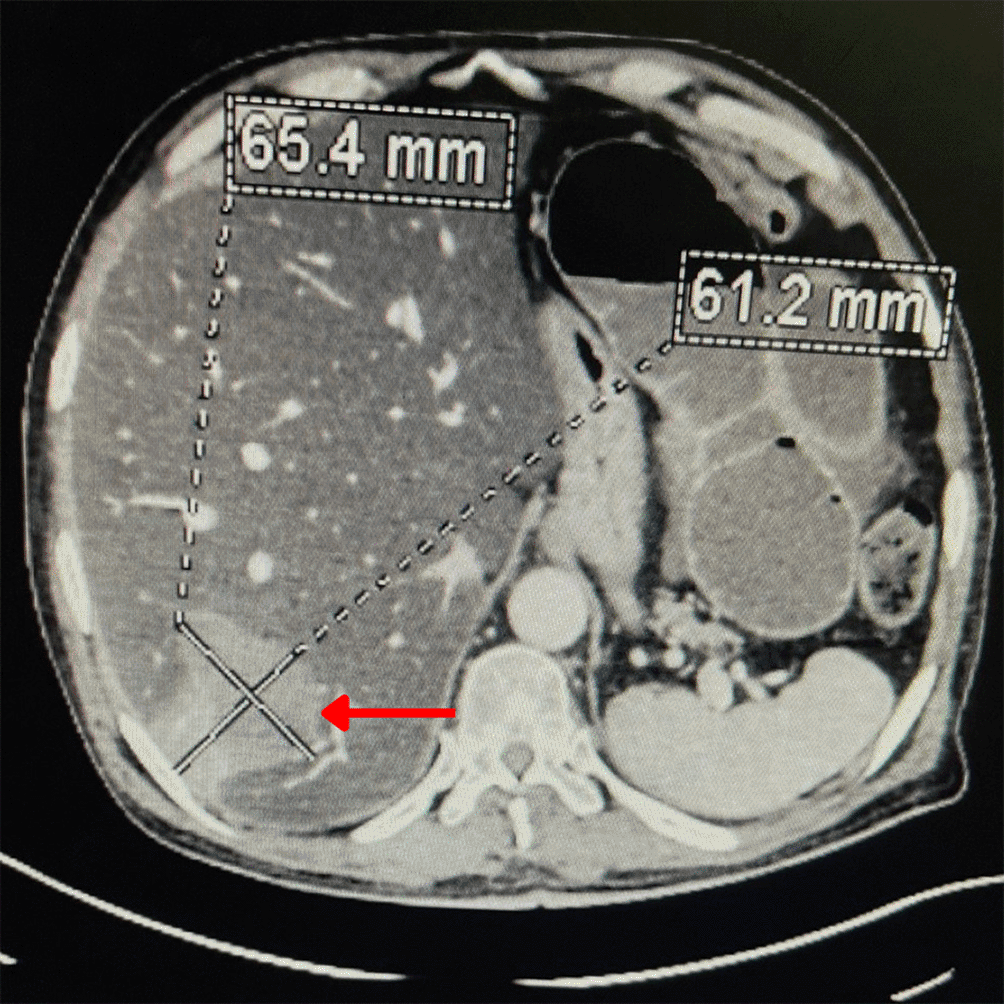
Abdominal X-ray examination demonstrated features consistent with intestinal obstruction, characterized by multiple gas-liquid levels. Laboratory tests yielded normal results, indicating no immediate systemic abnormalities. Further assessment through abdominal computed tomography (CT) revealed evidence of acute mechanical intestinal obstruction affecting the small intestine proximal to a likely flange, situated at the level of the right flank (Figure 1). The maximum distension measures approximately 60 mm without radiological signs of intestinal distress. Additionally, a sizable hepatic metastatic lesion has been identified spanning segments V and VI, measuring 6.5 cm in greatest diameter (Figure 2).

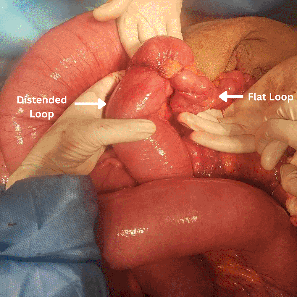
Initially, a conservative approach was pursued with medical management. However, in the absence of clinical improvement, a decision was made to proceed with surgical intervention. Intraoperatively, a moderate serous effusion was noted along with distension of the small bowel loops upstream of a transitional level. A distended loop, flattened loop configuration was identified, indicative of a tumor involving the inverted small intestine and adherent to the parietal peritoneum (Figure 3).
The surgical procedure entailed the resection of the small intestine on both sides, maintaining a 5 cm margin from the tumor, and the creation of a double-barrel stoma to address the surgical requirements. The histopathological examination of the operative specimen confirmed the presence of adenocarcinoma of the small intestine with clear margins. This tumoral lesion is responsible for an upstream intestinal intussusception. It is classified as pT3 N2. The postoperative course was uneventful, and the patient is currently under close follow-up in the oncological department, emphasizing the comprehensive and multidisciplinary nature of his ongoing care. This approach ensures continuous monitoring, addressing potential oncological considerations, and optimizing the patient’s overall well-being. The patient is on the verge of initiating supplementary chemotherapy treatment.
Small intestinal adenocarcinomas, constituting less than 5% of digestive cancers, are emerging as rare malignancies with an increasing incidence.1 Unlike their more prevalent counterparts in the colon, these tumors are characterized by their association with predisposing diseases, making their early diagnosis challenging.1,4 The duodenum stands out as the primary site for these tumors (46–82% of the cases), with the jejunum (11–31%) and ileum (7–21%) following in frequency.1,2,5
Given their scarcity and subtle symptomatology, small intestinal adenocarcinomas often elude early detection, resulting in diagnoses at advanced stages.1,6 Abdominal pain, gastrointestinal bleeding, and intestinal obstruction are common but nonspecific symptoms.7 The delayed diagnosis contributes to the presence of lymph node metastases in a considerable percentage of cases at the time of identification.3,7
Notably, the difference in incidence between small bowel tumors and colorectal adenocarcinomas implies distinct environmental exposures and underlying molecular mechanisms.3,5 The molecular abnormalities observed in small intestinal adenocarcinomas share similarities with colonic adenocarcinomas, but their varying frequencies suggest unique carcinogenic pathways.3,7
These rare tumors are intriguingly linked to predisposing pathologies and genetic syndromes.4,7 Lynch syndrome, familial adenomatous polyposis (FAP), and Peutz-Jeghers syndrome, associated with STK11 gene mutations, are identified as contributing factors, significantly elevating the risk of developing small bowel adenocarcinoma.4,7 This genetic predisposition underscores the importance of considering familial and hereditary factors in the context of small intestinal malignancies.1,6,7
Diagnostic tools, such as multi-detector helical CT scans, play a pivotal role in the identification and characterization of small bowel diseases.7 In the presented case, CT imaging revealed acute mechanical intestinal obstruction, which initially posed a diagnostic challenge due to the rarity of small bowel tumors, compounded by the patient’s history of colonic surgery. This scenario highlights the need for heightened clinical suspicion and advanced imaging techniques for accurate and timely diagnosis. The comprehensive performance metrics of multidetector row helical computed tomography (CT) enteroclysis for small bowel disease indicate a sensitivity, specificity, and accuracy of 100%, 95%, and 97%, respectively.1 In contrast, magnetic resonance (MR) enteroclysis demonstrates sensitivity, specificity, and accuracy of 86%, 98%, and 97%, respectively.4,7 Video capsule enteroscopy (VCE), introduced in 2001, achieves a diagnostic yield of approximately 50–60% for small bowel lesions.1,7 Despite this, there is an average delay in diagnosis reported at 8.2 months, primarily attributed to physicians failing to order the appropriate tests, and a delay of 12 months due to radiologists failing to confirm the diagnosis.1,4
Surgical resection remains the cornerstone of treatment, with the specific approach tailored to factors such as tumor location, invasion extent, and its relationship with surrounding organs.5 In the case presented, the patient underwent small intestinal resection and creation of a double stoma without any additional resection procedure due to the absence of invasion of neighboring organs.
The role of adjuvant chemotherapy in small intestinal adenocarcinomas has been a subject of debate.2,3,7 While some retrospective studies have not shown a clear benefit, an American registry study suggests a significant improvement in overall survival, particularly in stage III cases.3 However, the level of evidence supporting adjuvant chemotherapy remains relatively low.3 The National Thesaurus of Digestive Oncology TNCD recommends considering adjuvant chemotherapy, specifically a combination of fluoropyrimidine and oxaliplatin, after curative resection of stage III or in the case of stage IIB.8
Prognosis varies across stages, with the 5-year overall survival rates ranging from 50-60% for stage I, 40-55% for stage II, 10-40% for stage III, and 3-5% for stage IV.3,8 This places small intestinal adenocarcinomas in an intermediate prognostic category between colon and stomach cancers.3,8 Despite the lack of a standardized monitoring protocol after resection, the TNCD recommends regular clinical examinations and imaging studies, underscoring the importance of postoperative follow-up for patients capable of withstanding reoperation or chemotherapy.8
In conclusion, small intestinal adenocarcinomas present a complex clinical scenario, characterized by their rarity, association with predisposing pathologies, challenges in early diagnosis, and varied treatment outcomes.1,3 Ongoing research and advancements in diagnostic techniques contribute to a deeper understanding of these malignancies, guiding improved therapeutic strategies and postoperative management.1,6
This case highlights the intricate nature of diagnosing and treating metachronous complicated small bowel adenocarcinomas. Surgical interventions, customized based on tumor location, played a critical role in achieving a positive outcome.2 The ongoing discussion regarding adjuvant chemotherapy and the importance of vigilant postoperative monitoring are emphasized, illustrating the evolving dynamics of rare malignancies.2 As we navigate through this complex medical landscape, continuous research and multidisciplinary approaches are indispensable for enhancing our comprehension and enhancing outcomes in small intestinal adenocarcinomas.1,3
| Views | Downloads | |
|---|---|---|
| F1000Research | - | - |
|
PubMed Central
Data from PMC are received and updated monthly.
|
- | - |
Provide sufficient details of any financial or non-financial competing interests to enable users to assess whether your comments might lead a reasonable person to question your impartiality. Consider the following examples, but note that this is not an exhaustive list:
Sign up for content alerts and receive a weekly or monthly email with all newly published articles
Already registered? Sign in
The email address should be the one you originally registered with F1000.
You registered with F1000 via Google, so we cannot reset your password.
To sign in, please click here.
If you still need help with your Google account password, please click here.
You registered with F1000 via Facebook, so we cannot reset your password.
To sign in, please click here.
If you still need help with your Facebook account password, please click here.
If your email address is registered with us, we will email you instructions to reset your password.
If you think you should have received this email but it has not arrived, please check your spam filters and/or contact for further assistance.
Comments on this article Comments (0)