Keywords
Case report, Atypical meningioma, Histopathology, Radiosurgery, Magnetic resonance imaging, MR spectroscopy.
This article is included in the Datta Meghe Institute of Higher Education and Research collection.
Case report, Atypical meningioma, Histopathology, Radiosurgery, Magnetic resonance imaging, MR spectroscopy.
Meningiomas originate from arachnoid cells on the dura’s inner surface and the latter originate from mesodermal and neuronal crest-derived meningeal progenitor cells. Grade I Benign meningioma is seen in 90% of cases, Grade II atypical meningioma is seen in 5-7% of cases, and Grade III malignant meningioma is seen in 1-3% of cases.1 When compared to WHO grade I meningiomas, grade II meningiomas have a higher re- occurrence and mortality rate. Atypical meningioma has two histological variants:- clear cell meningioma, and choroid meningioma, which is a more aggressive form of meningioma and signals a WHO grade 2 tumor. The risk of brain tumors increases with age, overweight, hormones, obesity, any previous radiotherapy treatments and in certain syndromes such as Li–Fraumeni, Gorlin, Cowden and von Hippel–Lindau syndromes, multiple endocrine neoplasia type 1 and neurofibromatosis type 2 (NF2) can determine the development of meningiomas. The tumors associated with these conditions are often multiple and occur mostly in children. Patients with atypical meningiomas have a survival rate of 63.8% which is 5-year survival. It is difficult to differentiate between grade I and II meningiomas based on radiological criteria; therefore histopathology is required. The Histopathologic features, such as mitoses between 4 and 19 per high-power field, brain invasion, or three of five particular histologies, including spontaneous necrosis, sheeting conspicuous nucleoli, high cellularity, and small cells, are used to make the diagnosis.1 The Ki-67 proliferation index is inversely proportional to the apparent diffusion coefficient (ADC) value, which helps distinguish low-grade and aggressive meningiomas. Histologically, mitotic activity, necrosis, increased nucleus-to-cytoplasm ratio and tumor cells of atypical meningiomas result in decreased water diffusion rates, thus leading to lower ADC values.2 Therefore, early detection of atypical meningioma helps in determining the location, size, and structures that are affected by the growing tumor and helps in early surgical excision of the tumor. Complete surgical resection is frequently curative, reduces peritumoral edema and improves neurological symptoms. However, radiotherapy may be used in cases of incompletely resected or recurring cancers that have not previously received radiation. There are different ways of delivering radiotherapy, including traditional external beam irradiation, Lekshell gamma knife, linear accelerator, and Cyberknife radiosurgery.1,3,4 Radiotherapy for aggressive cancers can prevent re-occurrence. Immunochemotherapy may be considered if the meningioma is incurable or when all previous treatments (surgery or radiation) fail. While cyclophosphamide, adriamycin, vincristine, ifosfamide/mesna, and adriamycin/dacarbazine have been used to treat patients with aggressive or malignant meningiomas, hydroxyurea, interferon-alpha, tamoxifen, and mifepristone have only modest success in treating patients with recurrent meningiomas.1
Patient information – A 40-year-old female presented to the general medicine department complaining of dizziness, fatigue, loss of balance, nausea, reduced sense of smell, and holo cranial headache for eight months. The patient also had weakness and a tingling sensation in the right upper limb, which made her unable to lift or grip objects. There was no head trauma, seizures/convulsions, fever, loss of consciousness, ear-nose-tongue bleeding, or history of migraine. There were no known comorbidities such as Diabetes Mellitus, Hypertension, Bronchial Asthma or Tuberculosis.
History of past operative, trauma, or history of any other malignancy.
Family history is significant.
Clinical findings
I) General examination – Built-average, decubitus – supine, nutritional status – normal, temperature – afebrile, pulse – 88 beats/mints, B.P – 114/88 mmHg.
II) Systemic examination:
A) Central nervous system (CNS) examination: A complete neurological examination was performed that included the following assessments:
1) Glasgow coma scale (GCS) [We have obtained a copywrite license for this assessment tool] – Eye opening response was given a score of 3, verbal response was given a score of 5 and motor response was given a score of 5 = Total 13/15.
2) Mental status – The patient was well oriented to the time, place, and person.
3) Motor function and balance – Muscle appearance and tone were normal in all four limbs. The muscle power was reduced in the right upper limb (3/5), and the rest of the limbs showed normal power. The reflexes were sluggish in the right upper and lower limbs, the patient had ataxia, and on performing cerebellar signs-Romberg sign-patient was asked to walk in a straight line with closed eyes. She could not walk straight, so the Romberg sign was positive. The remaining cerebellar signs were negative.
4) The patient was unable to recognize a light touch in the right upper limb. The remaining evaluations of temperature, position sense, vibration, and discriminative sensations were normal.
5) Cranial nerve assessment examining the olfactory nerve(1st cranial nerve): There was a reduced sense of smell in both nostrils. Examination of the trigeminal cranial nerve (5th CN) revealed- reduced sensation on the right side of the face, and the rest of the nerve examination was normal.
B) Respiratory, C) cardiovascular, and D) gastrointestinal systems appear to be normal.
Investigations:
CBC investigation: Hb – 10.2 grams/deciliter (g/dl); total Red Blood Cell count – 4.4 million cells, total WBC count – 10,200 cells/cubic millimeter, total platelet count – 2.5. All values were within the normal ranges.
Cerebrospinal fluid examination (CSF) was – clear and colorless in nature, PH – 7.2, protein content – 10 g/L, lactate dehydrogenase (LDH) – 62 units/liter, Glucose – 79 milligram/deciliter (mg/dl), no RBC, no pus cells, no organism seen, and negative for acid – fast bacilli.
Histopathological examination: Excisional biopsy of the tumor showed a grade II atypical meningioma (WHO classification).
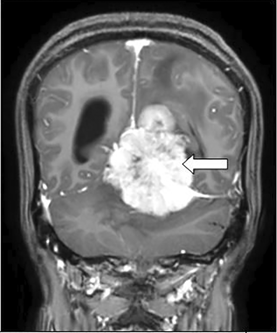
4) Radiological investigation – The patient complained of dizziness and headache. A magnetic resonance imaging (MRI) was performed in which atypical meningioma was diagnosed, which showed a well-defined lesion that was arose from the left tentorial leaflet appearing heterogeneously iso to hypointense on T1 image shown in Figure 1 causing a mass effect in the form of effacement of adjacent sulcogyral spaces and compression of the 4th ventricle shown by the lower arrow in Figure 2 with mild dilatation of the 3rd ventricle as shown by the upper arrow in Figure 2, heterogeneously iso to hyperintense with surrounding vasogenic edema on T2 Image as shown in Figures 3 and 4, heterogeneously enhancing with few non enhancing necrotic areas within the tumor on T1 contrast images as shown in Figures 5 and 6. The lesion shows restriction on the diffusion-weighted image (DWI), shown by the arrow in Figure 7 and the apparent diffusion coefficient (ADC) shows low signal intensity, as shown by the arrow in Figure 8.

A well defined lesion that arose from the left tentorial leaflet appearing heterogeneously iso to hypointense on T1 image.
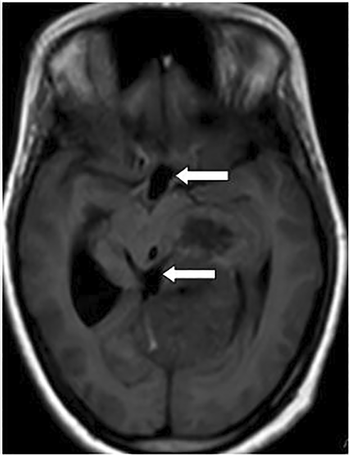
A well-defined lesion that was arose from the left tentorial leaflet causing mass effect in the form of effacement of adjacent sulcogyral spaces and compression of the 4th ventricle shown by the white arrow with mild dilatation of the 3rd ventricle as shown by the white arrow.

A well-defined lesion that was arose from the left tentorial leaflet is a well-defined lesion that was arising from the left tentorial heterogeneously iso to hyperintense with surrounding vasogenic edema causing mass effect in the form of effacement of adjacent sulco-gyral spaces shown by the white arrow.

A well-defined lesion that was arising from the left tentorial heterogeneously iso to hyperintense with surrounding vasogenic edema causing mass effect in the form of effacement of adjacent sulco-gyral spaces shown by the white arrow.
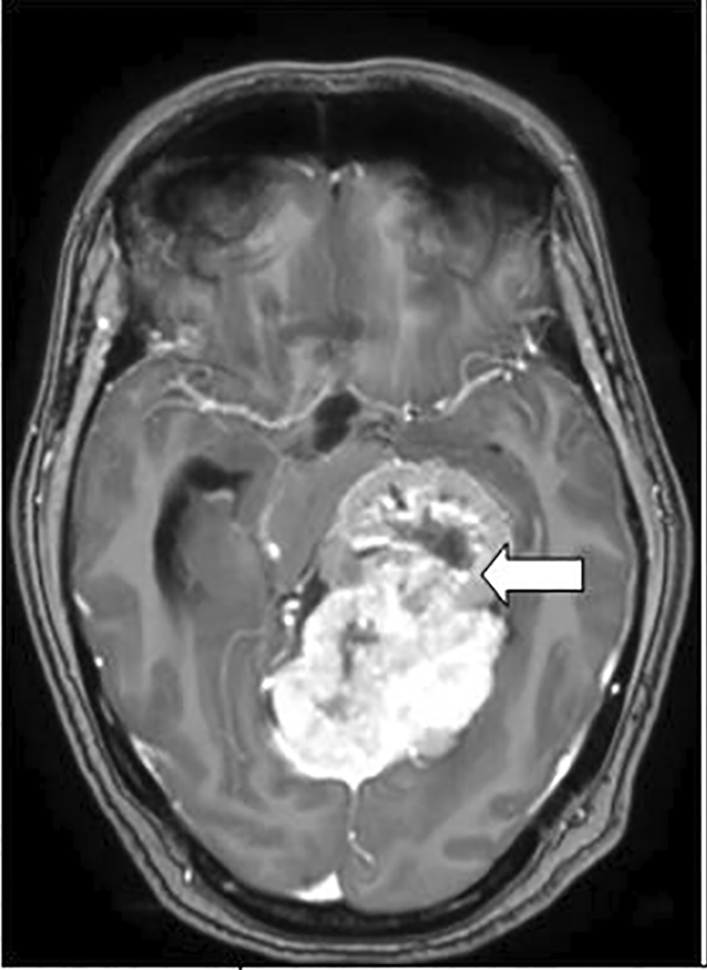
A well defined heterogeneously enhancing mass lesion with few non enhancing necrotic areas within the tumor shown by the white arrow.
A well defined heterogeneously enhancing mass lesion with few non enhancing necrotic areas within the tumor shown by the white arrow.

The lesion shows restriction on the diffusion-weighted image shown by the white arrow.

The lesion shows low signal intensity on apparent diffusion coefficient (ADC)as shown by the white arrow.
There is dilatation of the bilateral lateral ventricles are shown by the arrow in Figure 9.

There is dilation of the bilateral lateral ventricles shown by the white arrow.
MR spectroscopy showed increased choline values and reduced N-acetyl aspartate values, indicating showing increased choline/NAA ratio suggesting that the lesion was malignant in Figure 10.

The lesion shows increased choline values and reduced N-acetyl aspartate values, indicating increased choline/NAA ration suggesting that the lesion was malignant.
Medical management
The patient was managed conservatively and was administered ibuprofen 200 mg tablet once a day for headache, betahistine 16 mg twice daily (histamine agonists) for dizziness, and ondansetron 8 mg sos-if required for nausea and vomiting.
Surgical management
A basal brain tumor sub-total excision of atypical meningioma was performed along with ventriculo-peritoneal shunting for non-communicating hydrocephalus. Post operative adjuvant radiotherapy was started to reduce the size of the tumor and to prevent reoccurrence. A Post operative contrast-enhanced CT scan was performed which showed a lesion that was heterogeneously enhanced on post contrast with surrounding perilesional edema few areas of calcific foci and a central area of hypodensity mostly due to necrosis noted arising from the left tentorial leaflet suggestive of the residual lesion shown in Figure 11A with right-sided ventriculoperitoneal shunt shown by the arrow in Figure 11B.

There is a heterogeneously enhancing mass lesion on post contrast with surrounding perilesional edema few areas of calcific foci and a central area of hypodensity mostly due to necrosis noted arising from the left tentorial leaflet suggestive of the residual lesion shown by the white arrow in (A) with a right-sided ventriculoperitoneal shunt traversing througfh the right parietal lobe with its tip into the body of right lateral ventricle shown by the white arrow in (B).
Meningiomas are common intracranial tumors that can be identified based on their radiological and histological characteristics. However, meningiomas can also mimic other brain tumors. Females are more likely to have meningiomas, and the incidence rate increases with age. Meningioma development has been linked to specific molecular changes and ionizing radiation. Meningiomas are divided into 15 subtypes and are categorized into three classes, with survival and re-occurrence rates getting worse as the grade rises.5 Atypical meningioma (WHO Grade II) is a more aggressive form of meningioma with a higher five-year re-occurrence rate (41%), than grade I (benign) meningiomas (12%), and the diagnosis is mainly on histopathologic findings on biopsy as it is difficult to differentiate between grade I and II meningioma based on radiologic criteria. The Symptoms include headache, seizures, change in personality or behavior, development of a localized neurologic impairment, drowsiness, confusion, loss of hearing or tinnitus, fatigue on exertion, projectile vomiting and visual disturbances. If a meningioma is left untreated, it can cause problems such as loss of neurological function, weakness/numbness, hearing or vision loss, and balance problems. Sometimes there is worsening of the signs of persistent headache, the origin of new episodes of seizures, or increased intracranial pressure (e.g., vomiting, swelling of the 2nd cranial nerve), initially a neurological evaluation should be performed followed by radiological studies if necessary initial diagnoses are made using contrast-enhanced CT or MRI. A wait-and-see strategy is used for tiny, asymptomatic tumors, but total surgical removal is the best course of action for symptomatic meningiomas.5 Intraoperative MRI helps to obtain tissue samples and remove the tumor during surgery. The biochemical profile and nature of the tumor can be better visualized using on MR spectroscopy. In adults, prognosis is mostly age-dependent. In general, early diagnosis has a better prognosis. If the entire tumor is operable, the outcome is better. Tumor removal is the main goal of surgery, however the patient’s neurological capabilities must be preserved or improved first.6 Preoperative embolization of the tumor is widely performed for patient safety throughout the surgical process. In patients when the total tumor is removed, there is a comorbidity risk (any side effect that can cause deterioration in the quality of life) and it may be preferable to leave some of the tumors in place and monitor future growth with routine imaging examinations.7 In the case of a GRADE II atypical meningioma, an MRI scan should be performed every 6-12 months. After 5 years, MRI is performed every 2 years.8
Sophisticated imaging techniques can be used to diagnose meningioma. The main radiological investigations performed for diagnostic purposes are magnetic resonance imaging (MRI) and computed tomography. Intraoperative magnetic resonance imaging (MRI) was performed to guide tissue samples and tumor removal during surgery. Magnetic resonance spectroscopy (MRS) can be used to investigate the biochemical profile of a tumor and determine the nature of the lesions observed on the MRI. An atypical meningioma was diagnosed in this case, which appeared as a well-defined lesion arising from the left tentorial leaflet with surrounding perilesional vasogenic edema appearing heterogeneously isointense to hypointense on T1 Weighted Image and heterogeneously isointense to hyperintense on T2 Weighted Image and showing heterogeneous enhancement with few areas of necrosis within the contrast images. No evidence of calvarial destruction or intra-osseous enhancement was observed. The combination of necrosis and invasion of the brain is generally associated with an increased risk of re-occurrence of tumor and such a combination shows a radio-resistance nature. MR spectroscopy showed increased choline values and reduced N-acetylaspartate values, thereby showing an increased choline/NAA ratio suggesting a malignant lesion in the case of atypical meningioma. A basal brain tumor excision of atypical meningioma was done along with ventriculoperitoneal shunting for non-communicating hydrocephalus was the surgical management carried out in this case. Post operative adjuvant radiotherapy was started to reduce the size of the remaining tumor as there was incomplete resection due to involvement of crucial structures of the brain and to prevent chances of reoccurrence. Follow-up was advised every 6 months to determine the prognosis of the surgery and re-occurrence of the tumor.
The patient complained of dizziness, fatigue, loss of balance, nausea, reduced sense of smell, and holocranial headache for 8 months. The patient also had weakness and a tingling sensation in the right upper limb, which made her unable to lift or grip objects. She was given medical treatment for the relief of the above-mentioned symptoms, and after receiving the medication she experienced some relief in her condition. Furthermore, she was advised to undergo an MRI brain contrast scan to rule out the cause of her neurological symptoms which showed an atypical meningioma at the base of the skull. After the radiological diagnosis, an excisional biopsy of the tumor was planned so that the histological diagnosis could be correlated with the radiological findings. The histopathological findings were consistent with the radiological findings. After getting an idea about the nature of the tumor, she planned to undergo surgery for tumor removal. It took a few weeks to recover from the surgery, but as time passed, her symptoms started improving. She is thankful to all the doctors who took part in her diagnosis and management, kept her motivated, and gave her positive hope throughout the journey.
Written informed consent for publication of their clinical details and images was obtained from the patient, who volunteered to participate in this study and gave permission for this study. She was explained the possible risks and benefits of this study and provided adequate information concerning the study in her language.
| Views | Downloads | |
|---|---|---|
| F1000Research | - | - |
|
PubMed Central
Data from PMC are received and updated monthly.
|
- | - |
Provide sufficient details of any financial or non-financial competing interests to enable users to assess whether your comments might lead a reasonable person to question your impartiality. Consider the following examples, but note that this is not an exhaustive list:
Sign up for content alerts and receive a weekly or monthly email with all newly published articles
Already registered? Sign in
The email address should be the one you originally registered with F1000.
You registered with F1000 via Google, so we cannot reset your password.
To sign in, please click here.
If you still need help with your Google account password, please click here.
You registered with F1000 via Facebook, so we cannot reset your password.
To sign in, please click here.
If you still need help with your Facebook account password, please click here.
If your email address is registered with us, we will email you instructions to reset your password.
If you think you should have received this email but it has not arrived, please check your spam filters and/or contact for further assistance.
Comments on this article Comments (0)