Keywords
Pediatric maxillofacial trauma
This article is included in the Datta Meghe Institute of Higher Education and Research collection.
Pediatric maxillofacial trauma
Pediatric facial fractures are rare and make up 5–15% of all facial fractures. The low incidence of facial fractures among children is due to physiological and environmental factors such as greater resilience of the pediatric skeleton, higher bone to tooth ratio, direct parental supervision and limited outdoor activity.1 In children of 5–10 years of age nasal fractures are by far the most frequent (58.6%), followed by mandibular fractures (21.5%). Orbital (9.5%), frontal (5.1%), and midfacial (3.8%) fractures are next in frequency.2 Le Fort fractures are very rare in children, and there is a paucity of literature presenting their frequency and characteristics. In the mandible condylar fracture is the most common (38.9%) followed by angle (20.6%), parasymphysis (18.3%), body (15.3%) and symphysis (5.3%).3 Facial fractures are more common in boys, though this varies greatly from 1.1:1 to 8.5:1.4
The majority of facial fractures in children result from falls (7.8–48%) and sports-related injuries (4.4–42%).5
In primary or mixed dentition, the placement of internal fixation hardware is challenging due to the risk of injuring unerupted permanent tooth follicles. The use of resorbable plates and screws offer a potential solution to the growing pediatric facial bone as the standard titanium fixation systems carry the risk of translocation, growth restriction, and dental injury. Dental splinting or suspension wires can also be used in place of direct plating. Fortunately, mild malocclusion at this age has the potential to be improved through bony remodeling, the eruption of permanent dentition, compensation of the mastication mechanism, and posttraumatic orthodontia.6
Accordingly, the goal of treatment is to restore the underlying bony architecture to its preinjury position in a stable fashion with minimal residual functional and esthetic impairment.
Here we present a case of a 5-year-old male child of Indian origin who reported to the Department of Oral and Maxillofacial Surgery with a chief complaint of pain in lower and upper jaws since the past 2 days due to fall from stairs. There was a positive history of oral bleed, bilateral nasal bleed, three episodes of vomiting, and avulsion of 51, 61 and 62 respectively.
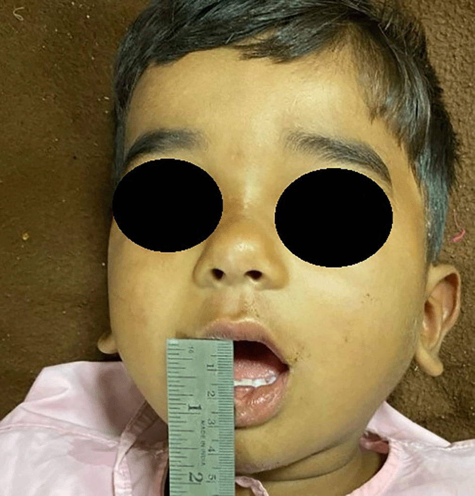
Extraoral examination revealed gross asymmetry due to the presence of diffuse swelling over the face along with the presence of a sutured contused lacerated wound over the chin.
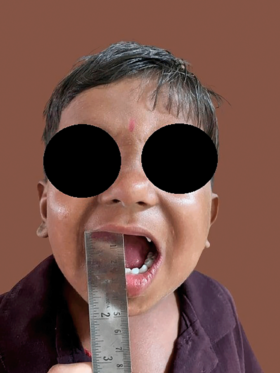
Bilateral temporomandibular joint movements were restricted due to pain and the mouth opening was reduced (12mm) (Figure 1).
On maxillofacial examination step and tenderness over the symphysis region and tenderness over bilateral maxilla and pre-auricular region was noted.
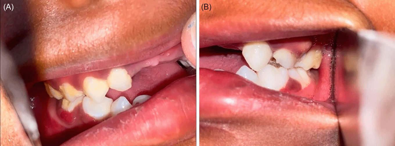
Intraoral examination revealed luxated right maxillary central incisors, left maxillary central and lateral incisors, intersegmental mobility in between the mandibular primary central incisors and anterior open bite (Figure 2). Maxilla was mobile at Le-fort I level bilaterally and ecchymosis was present over right maxillary buccal vestibule.

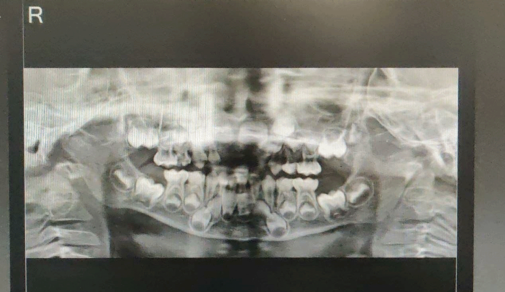
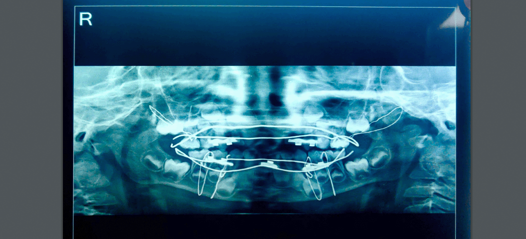
An orthopantomogram (OPG) revealed the presence of a fracture line in the mandibular symphysis region along with bilateral condylar fracture (Figure 3).
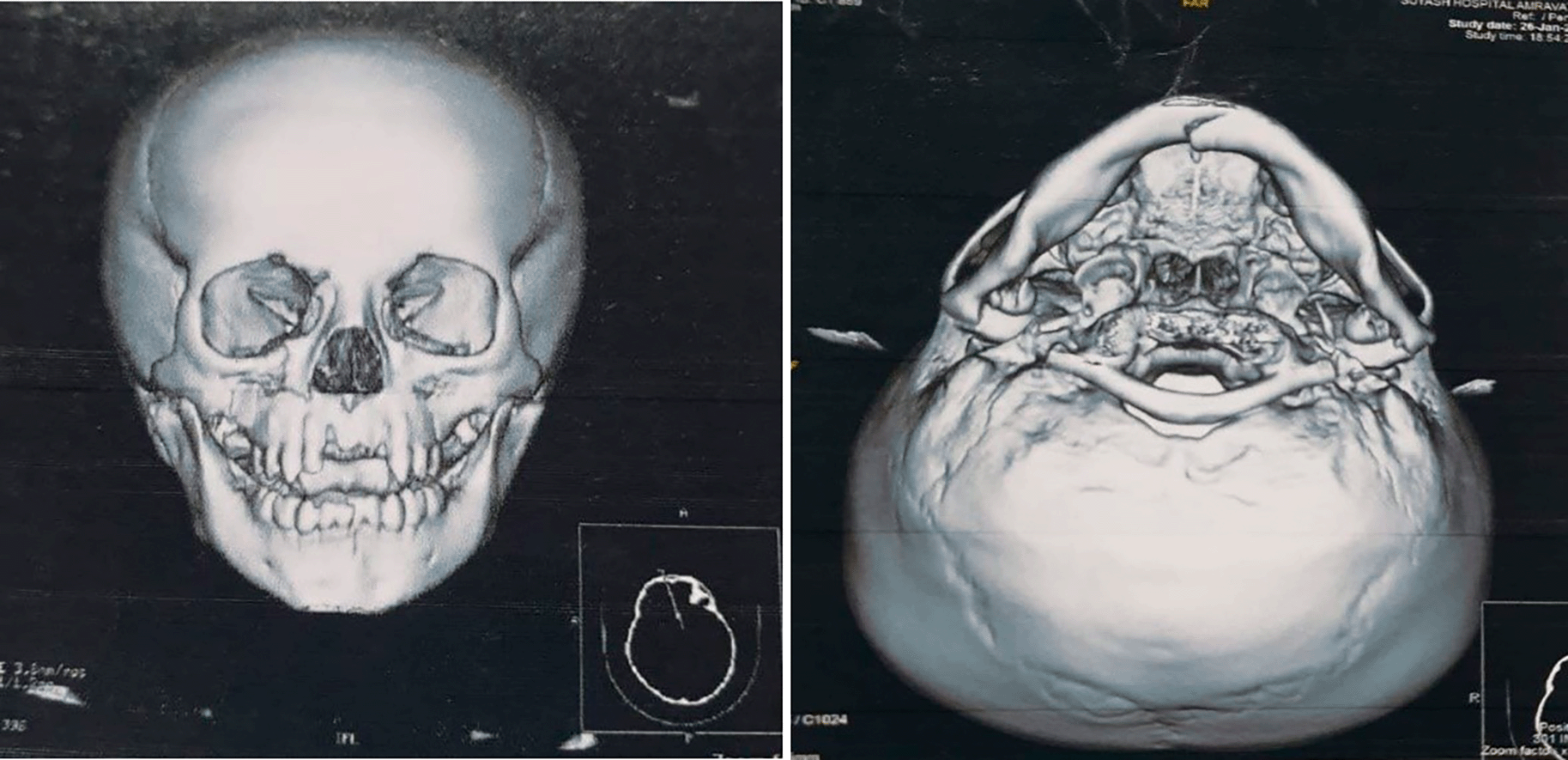
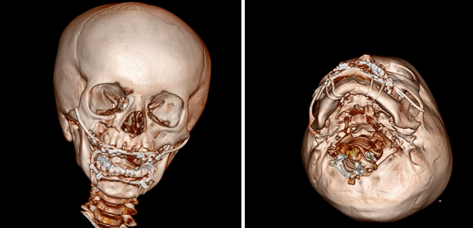
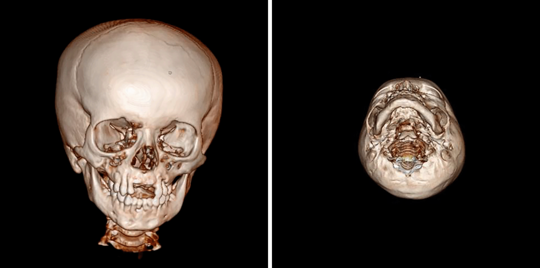
Computed tomography of the face revealed undisplaced bilateral Le-Fort I fracture, displaced fracture of mandibular symphysis and bilateral sagittal condylar fracture (Figure 4).
Considering the young age of the patient closed reduction of the symphysis fracture was planned using open cap splints secured with circum-mandibular wiring along with closed reduction of bilateral Le-Fort I fracture using open cap splints secured with circum-zygomatic wiring.
For the management of bilateral condylar fracture, intermaxillary fixation with elastics for 2 weeks was planned (Kang-Young Choi et al., 2012).7
Impressions of both the jaws was taken with alginate impression material and the cast was poured. Mock surgery was performed on the cast and anatomic reduction was achieved. An open acrylic cap splint for both the jaws was then fabricated. On the maxillary and mandibular splint cleats of arch bar were incorporated for intermaxillary fixation in the post-operative period. On the maxillary splint buccal tubes were also incorporated posteriorly to pass the wires for circumzygomatic wiring bilaterally (Figure 5 A–E).

After taking an informed, written consent from the patient’s father and obtaining the final fitness for the surgical procedure the patient was taken for surgery. The displaced mandibular segments were reduced by bidigital pressure with the guidance of the surgical splint. Stab incisions were placed at the inferior border of the mandible bilaterally and a William Velsey Fry Awl was introduced. Circum-mandibular wiring was done to stabilize the open acrylic mandibular cap splint.
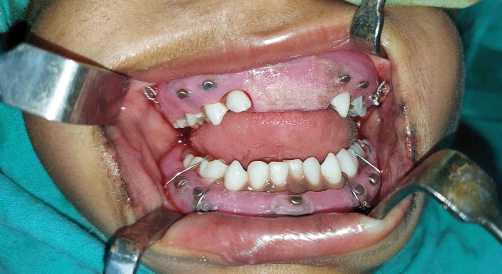
Then the maxillary splint was placed and stabilized with circumzygomatic wiring bilaterally with the wires engaging the buccal tubes on the splint (Figure 6).
Post-operative OPG was taken and computed tomography was done to check the reduction and proper positioning of the wires (Figures 7 and 8).
On the second post-operative day intermaxillary fixation with elastics was done for 2 weeks.
Postoperative follow-up was done on a weekly basis up to 2 weeks. No signs of any complications were observed during the healing period.
After 2 weeks the elastics were removed and intermaxillary fixation released.
The open acrylic cap splints were left in-situ and were removed on the sixth postoperative week under local anesthesia. There was no mobility of the fractured segments and the occlusion was bilaterally stable (Figure 9). The mouth opening was noted to be 22 mm (Figure 10). CT scan showed anatomic reduction of the fracture segments (Figure 11).
Management of a pediatric patient with Le Fort and mandibular fractures is very challenging and requires multiple considerations. Closed reduction of pediatric facial fractures is preferred in order to protect the growth centers.
Cap splints stabilized with circum-mandibular wiring is the standard treatment modality for the management of pediatric mandibular symphysis fracture. The use of open cap splints is preferred because it helps in occlusion guided reduction, promotes tooth eruption, patient is able to take soft diet and maintenance of oral hygiene is also possible. In our case we used the modified open cap splints stabilized with circum-mandibular wiring for the management of symphysis fracture.
The recommended treatment of fractures of pediatric mandibular condyle is either conservative, or intermaxillary fixation with splints for 7–10 days. It is followed by physical therapy to prevent TMJ ankylosis. In our case the patient was put in intermaxillary fixation with elastics for two weeks and then started on mouth opening exercises (Kang- Young Choi et. al, 2012).7
Currently, there is a paucity of studies on Le Fort-type fractures in children. More recent reports find that Le Fort-type fractures more commonly occur in older children with permanent dentition.
Thorough literature search revealed just 1 case report where a complex Le Fort II and III fracture was managed by a retention modified occlusal splint stabilized with circumzygomatic wiring for a 3-week period.8 In our case we used the modified open cap splints stabilized by circumzygomatic wiring for the management of bilateral Le Fort I fracture. This treatment modality provides a conservative operative approach, achieving excellent functional and cosmetic results without the potential morbidity of a more invasive surgery. It provides maximum stability during the healing period, is easy to apply and remove, reduces operating time, causes no trauma to the adjacent vital structures and provides added comfort to the young patients. The patient’s mouth opening and chewing capacity was restored and he was also able to occlude properly as narrated by the patient’s father during further follow-ups. The only disadvantage of the present technique was reduced patient compliance and its limited role in severely displaced fractures.
The clinical outcome in the present case indicates that the modified open cap splint is an effective and reliable treatment method in the management of pediatric maxillofacial fractures. It is an easy, quick, comfortable and cost-effective mode of stabilization. Extensive literature search reveals no splints that allow postoperative intermaxillary fixation in pediatric facial fractures. This case report offers a unique mode of postoperative stabilization and it should be considered as an alternative in the management of complex midface and mandibular fractures in the pediatric population.
| Views | Downloads | |
|---|---|---|
| F1000Research | - | - |
|
PubMed Central
Data from PMC are received and updated monthly.
|
- | - |
Provide sufficient details of any financial or non-financial competing interests to enable users to assess whether your comments might lead a reasonable person to question your impartiality. Consider the following examples, but note that this is not an exhaustive list:
Sign up for content alerts and receive a weekly or monthly email with all newly published articles
Already registered? Sign in
The email address should be the one you originally registered with F1000.
You registered with F1000 via Google, so we cannot reset your password.
To sign in, please click here.
If you still need help with your Google account password, please click here.
You registered with F1000 via Facebook, so we cannot reset your password.
To sign in, please click here.
If you still need help with your Facebook account password, please click here.
If your email address is registered with us, we will email you instructions to reset your password.
If you think you should have received this email but it has not arrived, please check your spam filters and/or contact for further assistance.
Comments on this article Comments (0)