Keywords
Petrous face meningioma, meningioma, retrosigmoid craniotomy, Ataxia, Gross total resection
Petrous face meningioma, meningioma, retrosigmoid craniotomy, Ataxia, Gross total resection
Meningiomas are second in prevalence only to gliomas.1 They make about 12% to 18% of cerebral tumours. They have a female-to-male ratio of 2:1 and are mostly harmless.2 Although most meningiomas appear above the tentorium, only around 10% are found in the posterior fossa.3 The cerebellopontine angle (CPA) accounts for 50% of these, the tentorial or cerebellar convexity for 40%, the clival for 9%, and the foramen magnum for 6%.4 Because of their closeness to important neurovascular systems and the brainstem, tumours in these areas are particularly difficult to surgically remove.5
The most effective methods for treating meningiomas are surgical excision or radiation therapy, both of which have a high success rate.6 The precise position along the petrous face determines the surgical method that is most appropriate for tumours of the petrous face, including meningiomas. The transpetrosal, far-lateral, and retrosigmoid methods are common.7–9 Careful consideration of the tumor's exact anatomical location informs the technique selection process.
This study presents the surgical outcomes and associated complications stemming from the application of the retrosigmoid craniotomy for the complete removal of petrous face tumors.
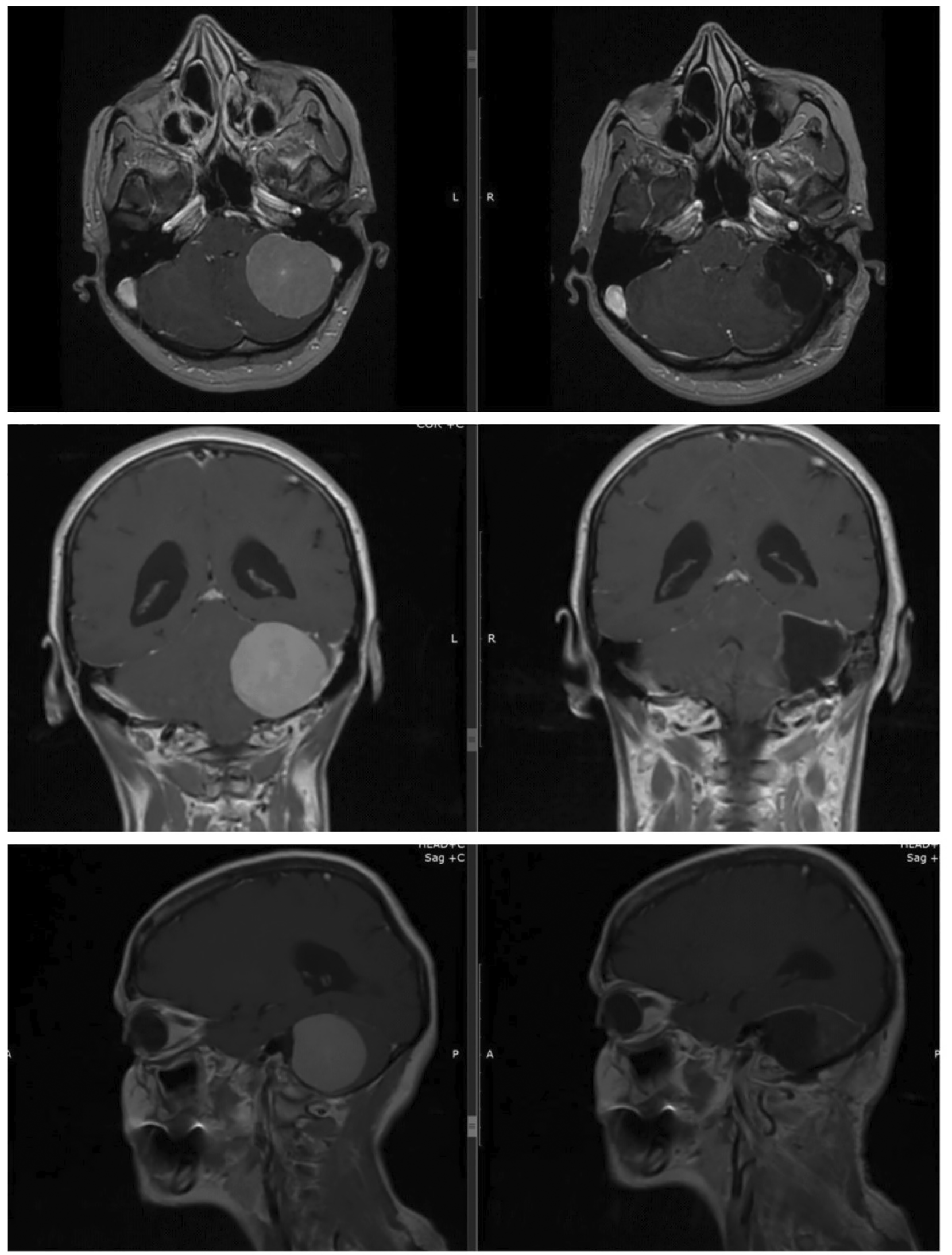
A 50-year-old female, employed as a housewife, presented at the regional clinical center of neurosurgery and neurology with a two-fold complaint of severe headache and vomiting persisting for two weeks, concomitant with a two-month history of ataxia. Notably, the patient did not report any symptoms of tinnitus or facial numbness. Preoperative magnetic resonance imaging (MRI) delineated the presence of tumors situated in the cerebellopontine angle (CPA) within the petrous face. These lesion exhibited iso- to hypointensity on T1-weighted imaging and iso- to hyperintensity on T2-weighted imaging. Upon contrast administration, the tumor manifested homogeneous enhancement, accompanied by a discernible dural tail. Importantly, these mass were localized on the left side and measured 4.2 cm as the greatest diameter (Figure 1).

Intraoperatively, a consistent pattern emerged with fibrous, exophytic lesions originating from the posterior petrous face of the temporal bone, resulting in varying degrees of cerebellar compression. Importantly, a discernible arachnoid plane was consistently identified between the tumor and the neurovascular structures of the cerebellopontine angle, facilitating the dissection of these structures without compromising neurovascular integrity. The surgical procedure culminated in the complete gross resection of the lesion, and subsequent postoperative MRI affirmed the absence of any residual tumor (Figure 2).
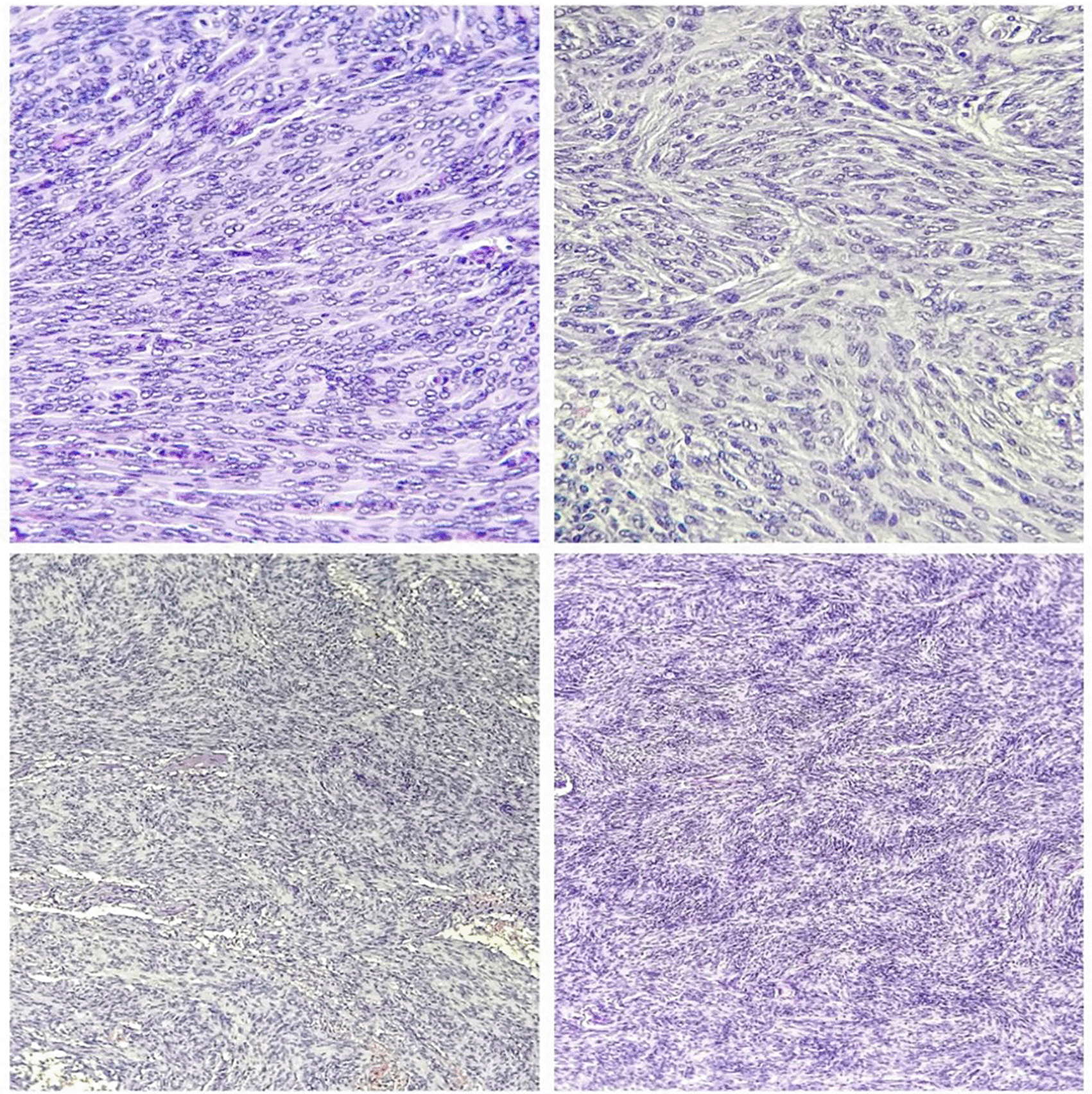
Tumour pathology showed transitional and syncytial patterns without angioblastic or malignant characteristics. Near the internal auditory meatus, jugular foramen, or dural venous sinus borders, these lesions formed. Meningiomas have whorls, spherical aggregates of meningothelial cells that mineralize to produce psammoma bodies (Figure 3).
The patient received perioperative steroid coverage, and the surgical approach involved operating in the prone position with a retrosigmoid craniotomy (Figure 4).

Following cerebellar retraction and the identification of cranial nerves, a piecemeal removal technique, incorporating bipolar coagulation, suction, and ultrasonic aspiration, was employed to meticulously excise the tumor. Noteworthy was the thorough coagulation of the dural origin upon completion of tumor removal, ensuring a comprehensive and successful surgical intervention. This case underscores the efficacy of the chosen approach in achieving complete gross resection and highlights the importance of neurovascular preservation in the management of petrous face meningioma.
Posterior cranial fossa meningiomas, predominantly located in the petrous bone (45% with dural attachment), tentorium (30%), occipital squamosa (10%), clivus (10%), and foramen magnum (5%), present unique challenges to skull base surgeons.10 Petroclival tumors, in particular, are intricate due to their depth within the skull and their ventral relationship to posterior fossa cranial nerves. Early surgical endeavors, before 1975, were associated with mortality rates exceeding 50%, with successful total resection a rarity. Advances in lateral skull base approaches and microsurgical techniques have significantly reduced mortality, with recent studies reporting rates below 10%.11–13
A meta-analysis encompassing six studies and 298 patients highlighted that complete resection of petroclival and clival meningiomas was achieved in only 68% of cases.11,12,14–18 Notably, meningiomas originating from the posterior half of the medial aspect of the temporal bone, known as retromeatal meningiomas, exhibit a more favorable disposition. These tumors, characterized by ventral displacement of cranial nerves and encirclement by cerebellum with an intact layer of arachnoid, are amenable to the classical retrosigmoid approach.19,20
This study emphasizes that posterior half skull base meningiomas can be safely resected with low morbidity, even when reaching considerable size. The retrosigmoid craniectomy, tailored to the site of origin, serves as an effective approach, with tumor size and location serving as pivotal prognostic indicators.5,21 Various approaches to the cerebellopontine angle (CPA) exist, with the lateral suboccipital or retromastoid technique being commonly employed. Operative mortality for CPA meningiomas ranges from 0 to 40%, with complete resections achieved in less than 50% of cases.5 Notably, our case study demonstrated no complications after the complete removal of the tumor. Modern surgical techniques have transformed the landscape of petrous meningioma treatment, allowing for curative complete resection with minimal mortality and morbidity. Despite these advancements, challenges persist, including postoperative cranial nerve function worsening and tumor recurrence due to incomplete resection. Contemporary electrophysiologic monitoring may mitigate cranial nerve injuries. An aggressive initial approach, aiming for complete tumor resection, is recommended, with dural coagulation at strategic sites aiding in the prevention of tumor recurrence.
The retrosigmoid approach emerges as a preferred strategy for treating petrous face meningiomas, yielding favorable outcomes in terms of neurological function and overall quality of life, particularly when aiming for gross total resection. This approach is characterized by its commitment to providing a safe, uncomplicated, and less invasive avenue for accessing the petroclival region during the resection of petrous face meningiomas.
We are happy to report that the patient's formal agreement to use their clinical information and photographs in our publication has been obtained. An essential part of our proposal, this consent statement demonstrates our firm resolve to conduct all medical research in accordance with the highest ethical standards. The patient's signature on the consent form confirms that she has read and understood the terms and conditions, and give her assent for the release of their medical records and photos for research and educational purposes. At every stage of the publishing process, we will remain committed to protecting patient privacy and following all applicable ethical guidelines to the letter.
| Views | Downloads | |
|---|---|---|
| F1000Research | - | - |
|
PubMed Central
Data from PMC are received and updated monthly.
|
- | - |
Provide sufficient details of any financial or non-financial competing interests to enable users to assess whether your comments might lead a reasonable person to question your impartiality. Consider the following examples, but note that this is not an exhaustive list:
Sign up for content alerts and receive a weekly or monthly email with all newly published articles
Already registered? Sign in
The email address should be the one you originally registered with F1000.
You registered with F1000 via Google, so we cannot reset your password.
To sign in, please click here.
If you still need help with your Google account password, please click here.
You registered with F1000 via Facebook, so we cannot reset your password.
To sign in, please click here.
If you still need help with your Facebook account password, please click here.
If your email address is registered with us, we will email you instructions to reset your password.
If you think you should have received this email but it has not arrived, please check your spam filters and/or contact for further assistance.
Comments on this article Comments (0)