Keywords
Pancreas, injury, blunt, stabbing trauma, management, surgery, endoscopy, non-operative treatment, outcome
Pancreatic trauma is notably less frequent than injuries affecting other solid organs, such as the liver or spleen. Despite their infrequency, pancreatic injuries can pose significant risks, with mortality rates ranging from 5 to 30% and morbidity rates reaching 50%. Managing such injuries remains contentious because of the anatomical complexity, lesion extent, and close proximity to neighboring organs.
Our study aimed to delineate the clinical and biological characteristics of pancreatic trauma and specify diagnostic and therapeutic modalities. This study was conducted retrospectively from 2010 to 2021 at Mahmoud El Matri Hospital’s General Surgery Department in Ariana, TUNISIA, and included five cases of blunt and open pancreatic trauma.
Although rare, pancreatic injuries can lead to serious consequences, prompting the development of a diverse array of therapeutic options. Pancreatic trauma presents a complex clinical challenge, necessitating a comprehensive approach that considers various factors, such as injury severity, anatomical location, and patient condition. Advanced imaging techniques, including computed tomography (CT) scanning, Wirsung-MRI, and ERCP, aid in the accurate diagnosis and assessment of canal involvement. Conservative management was considered for cases with limited canal rupture, whereas endoscopic treatments showed promise for hemodynamically stable cases. Surgical interventions, such as duodenopancreatectomy, are reserved for exceptional cases with severe injuries.
Pancreatic trauma requires a comprehensive approach that considers factors such as injury severity, anatomical location, and patient condition. Ongoing research and collaboration are vital for refining pancreatic trauma management and proposing guidelines for facilitating decision-making.
Pancreas, injury, blunt, stabbing trauma, management, surgery, endoscopy, non-operative treatment, outcome
Pancreatic trauma is far less common than trauma to other solid organs such as the liver or spleen.1,2 Despite their rarity, pancreatic injuries can be potentially serious, with mortality rates ranging from 5 to 30% and morbidity rates reaching 50%.1 The management of pancreatic injuries remains controversial. Therapeutic options are numerous, and the indications are often challenging owing to anatomical complexity, the extent of the lesions, and their intimate contact with neighboring organs.
This work has been reported in line with the PROCESS criteria.1
Our study aimed to describe the clinical and biological characteristics of pancreatic trauma and identify diagnostic and therapeutic modalities. This retrospective study was conducted from 2010 to 2021 in the General Surgery Department of Mahmoud El Matri Hospital, Ariana, TUNISIA. Five patients with blunt pancreatic and open trauma were included in the study. Analysis of these five observations yielded the following results (Table 1):
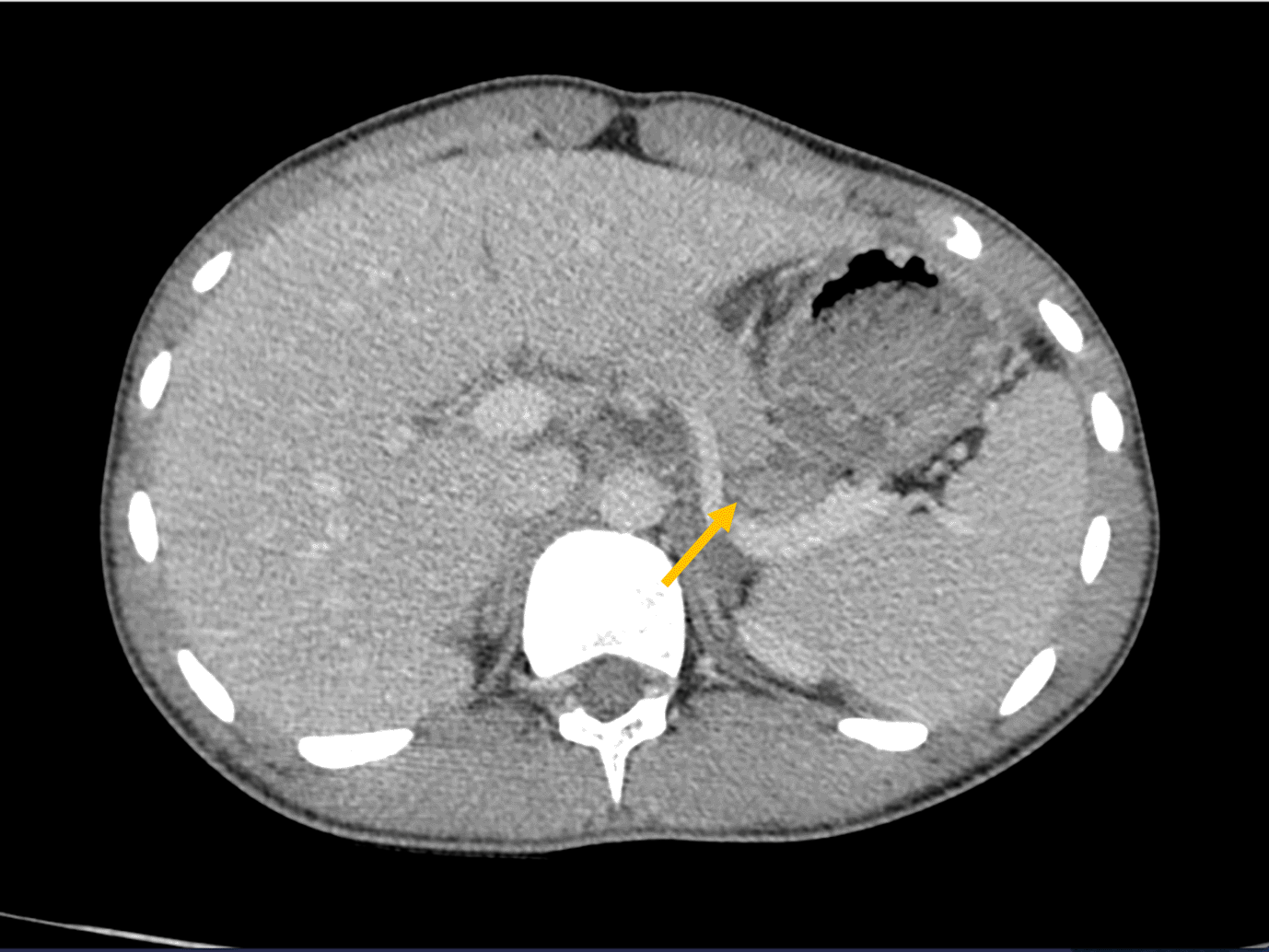
A 16-year-old boy, who was a victim of a physical assault (involving multiple punches and kicks to the abdomen), was admitted for blunt abdominal trauma. Upon examination, the patient was conscious and showed stable vital signs. Tenderness was evident in the upper abdominal region. Abdominal computed tomography (CT) revealed a transverse fracture at the junction of the pancreatic body and tail with moderate hemoperitoneum (Figure 1). The injury was classified as grade II according to the Lucas classification.3

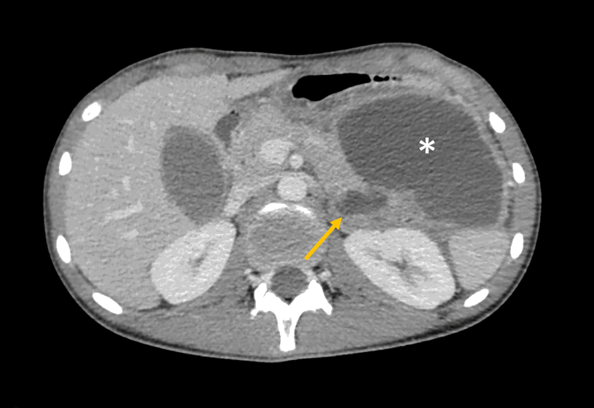
Based on the patient’s stable hemodynamics and relatively favorable physical examination findings, a non-operative treatment approach was considered. The clinical course was uneventful, and the patient was discharged on the 4th day. A follow-up abdominal CT scan conducted 14 days after the incident revealed a pancreatic fracture accompanied by two interconnected collections, one located caudally and the other occupying the lesser sac, of 9.7 × 7 × 9.3 cm (Figure 2). The patient is asymptomatic.

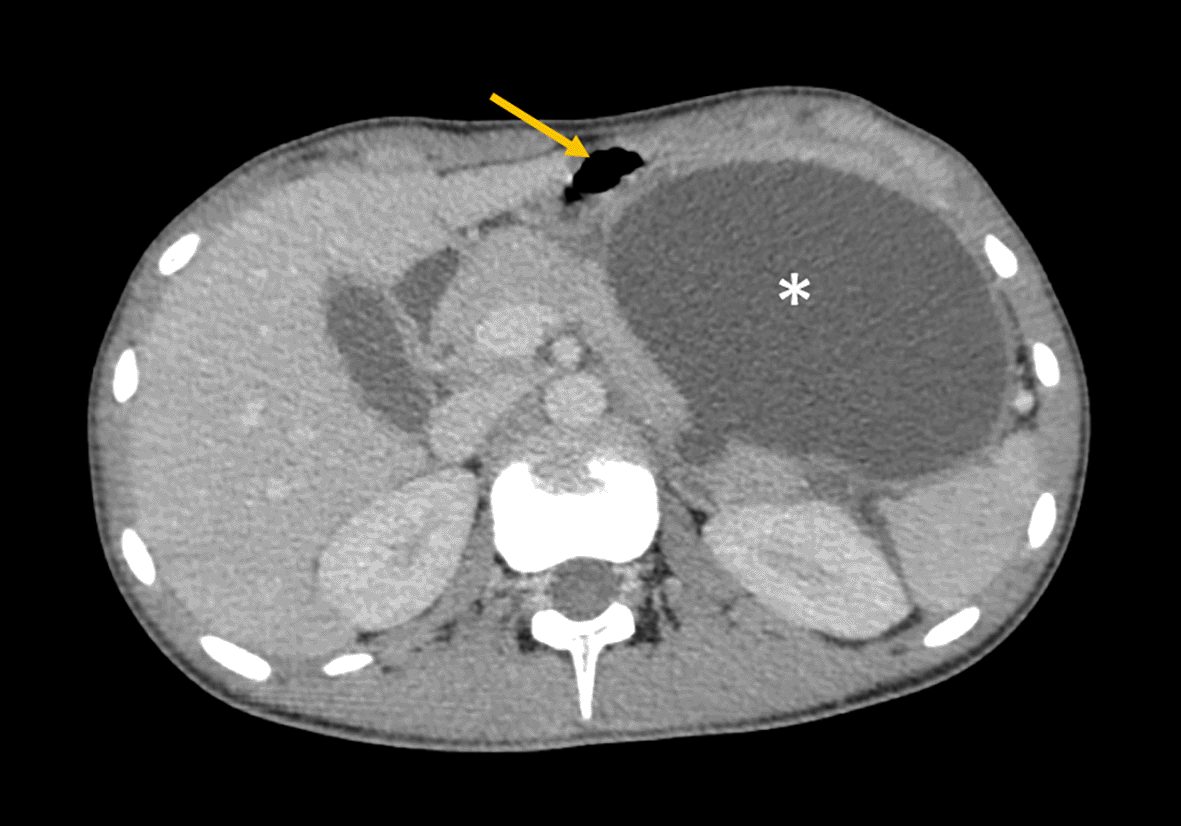
On the 27th day following the accident, the patient presented with epigastric pain and postprandial vomiting. A subsequent CT scan demonstrated a pancreatic fracture, with communication to a 24 mm collection in the tail (Figure 3). This collection was also found to be interconnected with a bulky pseudocyst of the pancreas, which measured 100 × 70 × 90 mm, resulting in a mass effect on the stomach and provoking anterior displacement (Figure 3).

Magnetic Resonance Cholangiopancreatography (MRCP) was performed to confirm the communicative nature of the pancreatic pseudocyst (Figure 4).
Esophagogastroduodenoscopy revealed no gastric varices, and the patient underwent endoscopic cystogastric drainage with double-ended pigtail placement. The clinical outcomes were favorable. Four years after the accident, the patient reported no complaints, and exhibited no signs of diabetes.
A 35-year-old woman with no prior medical history was admitted following a stabbing assault that resulted in open abdominal trauma. Initial physical examination in the emergency department revealed multiple penetrating abdominal wounds and hemorrhagic shock with a pulse of 125 bpm, blood pressure (BP) of 100/60 mmHg, and hemoglobin level of 3 g/dl. The patient received rapid general supportive treatment, started a blood transfusion, and underwent urgent laparotomy. Intraoperative exploration revealed abundant hemoperitoneum of approximately 3 liters and multiple visceral injuries: a severely damaged wound in the proximal small intestine, a wound in the distal ileum, a wound in the gastrocolic ligament accompanied by a nearby hematoma, and a transfixing wound at the junction of the isthmus and body of the pancreas, with the two pancreatoduodenal veins sectioned. The entire pancreatic parenchyma and the retroportal lamina were also divided. The stabbing weapons reach the vertebral body. Neither the superior mesenteric artery nor the superior mesenteric vein, portal vein, inferior vena cava, or aorta were affected. The injury was classified as grade II according to the Lucas’ classification.3
Peritoneal lavage, a pancreaticopancreatic suture, repair of the proximal ileal lesion, and rod loop stoma of the distal ileal loop were performed. A large drainage was maintained in proximity to the pancreas. There were no complications in the postoperative period.
An MRCP conducted one week postoperatively identified a 10 mm pseudocyst in the pancreas, and a subsequent MRI at 3 months displayed resolution of the pseudocyst, accompanied by complete restoration of the pancreas.
In the 6th month after the incident, the patient was admitted with acute intestinal occlusion secondary to postoperative adhesions. She underwent an explorative laparotomy with a section of adhesions and reverse-loop anastomosis. She later underwent large ventral incisional hernia repair using a polypropylene mesh.
The patient was doing well seven years after the assault. She reported no complaints, exhibited no signs of diabetes or pancreatic insufficiency, and had a solid midline scar.
A 40-year-old patient with no prior medical history was involved in a traffic accident (pedestrian hit by a motorcycle), resulting in both thoracic and abdominal injuries. Upon examination, the patient was conscious, displayed polypnea, and maintained stable hemodynamic parameters. Abdominal evaluation revealed tenderness in the epigastric and left hypochondrial areas without any peritoneal signs. A body CT scan revealed fractures in the 6th and 7th left ribs without a flail chest, pleural effusion, or pneumothorax. Additionally, a fracture was observed at the junction between the body and tail of the pancreas. No other abdominal lesions were found on CT scan; in particular, no splenic lesions were observed. The injury was classified as grade II according to the Lucas’ classification.3 A nonoperative approach was considered, and the patient was hospitalized for 16 days.
Subsequent follow-up CT showed the presence of an 8 cm pseudocyst in the lesser sac. As it was uncomplicated with an asymptomatic patient, the pseudocyst was respected. Subsequent follow-up demonstrated favorable clinical progress, with the pseudocyst nearly completely disappearing at 8 months. The patient denied any complaints during the final visit five years after the accident.
A 33-year-old patient was admitted to the emergency ward following a physical assault, resulting in blunt abdominal trauma. Physical examination revealed a conscious patient with low blood pressure and tachycardia, indicating a severe hemodynamic condition. Abdominal examination revealed tenderness of the epigastric region. Abdominal ultrasound revealed abundant intra-abdominal fluid, suggestive of abundant hemoperitoneum.
The patient underwent urgent laparotomy. Intraoperative exploration revealed abundant hemoperitoneum secondary to pancreatic isthmus injury. The duodenum is unaffected. No other injuries were noted. Peritoneal lavage was performed and a large drain was positioned in close proximity to the pancreas. The postoperative course was uneventful and the surgical drain was removed after 18 days.
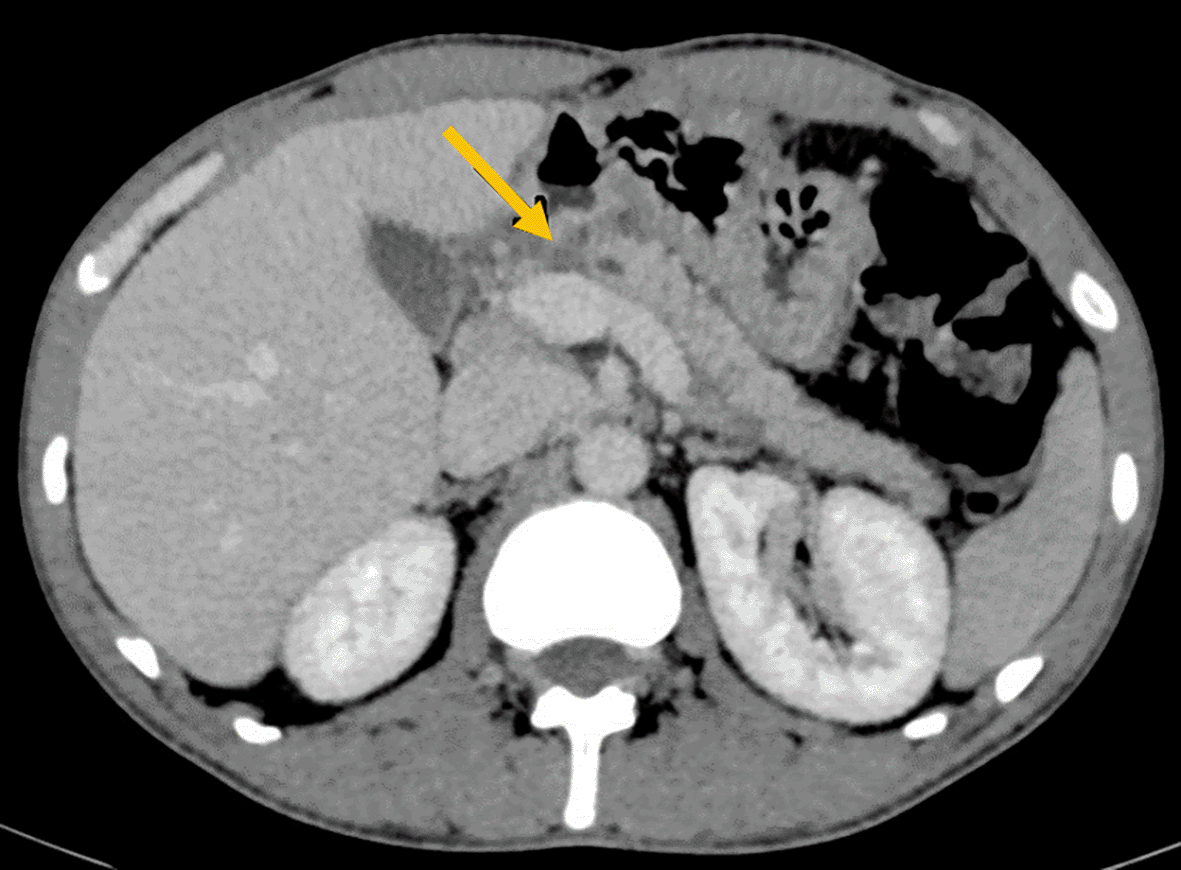
A follow-up CT scan before discharge (Figure 5) revealed a transverse fracture at the isthmus-body junction of the pancreas. Injury was categorized as grade II according to the Lucas classification.3

Another CT scan, conducted two months after the accident, revealed a 17 mm pseudocyst. The decision was made to respect the pseudocyst.
After 12 months of follow-up, the pseudocyst remained asymptomatic and did not increase in size, as evidenced by subsequent pancreatic MRI (Figure 6).
A CT scan performed 2 years after the initial trauma showed complete disappearance of the pseudocyst.
A 30-year-old man was admitted to our hospital following a road traffic accident. He was the driver of a car involved in a head-on collision with the steering wheel impacting his abdomen, resulting in blunt abdominal trauma. Physical examination revealed tenderness in the epigastric region. His hemodynamic parameters were within the normal ranges. His hemoglobin level were 14.1 g/dL, and his lipase level was recorded as 528 U/L, respectively.
An abdominal CT scan (Figure 7) revealed a laceration in segment III of the liver and a transverse fracture of the body of the pancreas, accompanied by a disconnection syndrome, and peri-pancreatic and pouch Douglas effusions. No other visceral injury was observed. Injury was categorized as grade I according to the Lucas classification.3
Non-operative treatment was initiated, and a follow-up CT scan at 72 hours showed regression of the hepatic laceration and stability of the pancreatic lesion.
After clinical and biological improvements, the patient was discharged 14 days after hospitalization.
An abdominal CT scan after 3 weeks revealed 20 mm fluid collection in the lesser sac, suggesting a pancreatic pseudocyst.
Eleven months post-accident, the pseudocyst had disappeared on the CT scan, with complete restitution of the pancreas, and the patient remained completely asymptomatic.
Pancreatic trauma is much rarer than trauma involving other solid organs, such as the liver or spleen.1,2 In the United Kingdom Trauma and Research Network (TARN), among 356,000 trauma patients, PD injuries accounted for 0.32%.4 Although liver, spleen, and kidney injuries are more common, pancreatic injuries occur in less than 10% of all abdominal traumas.1
In an acute setting, the diagnosis of a pancreatic injury should adhere to the general principles applicable to all trauma patients. Trauma patients with hemorrhagic shock should be promptly taken to the operating room for vigorous resuscitation and exploratory laparotomy, with foregoing radiological exploration to prevent treatment delays.
It is essential to recognize that the initial clinical indicators of pancreatic injury are nonspecific and laboratory assessments often lack specificity. Elevations in lipase and amylase levels are generally modest and nonspecific during the first 6 h following an accident,5 and they do not necessarily correlate with more severe pancreatic injuries.6
An abdominal CT scan with a pancreatic protocol exhibits a sensitivity of approximately 80% and reduced sensitivity for injuries to the Wirsung duct.7 CT scans may reveal peri-pancreatic effusion, pancreatic laceration, contusion with varying degrees of parenchymal engagement, or even pancreatic fractures with or without contrast extravasation. CT scans also evaluate canal involvement and duodenal injury, establishing the foundation for the Lucas classification.3
Pancreatic MRI or Wirsung-MRI, along with endoscopic retrograde cholangiopancreatography (ERCP), exhibits a sensitivity of nearly 100%.7 MRI has the advantage of being non-invasive and can be conducted on trauma patients in stable condition to assess pancreatic or canal injuries.8,9
The presence of an intra-parenchymal hematoma could compress the Wirsung duct, resulting in a loss of the duct signal at this level. This signal loss should be distinguished from an actual discontinuity or ductal section. Endoscopic retrograde cholangiopancreatography (ERCP) was performed both diagnostically and therapeutically. Despite being an invasive procedure, it is the gold standard for Wirsung exploration. It allows differentiation between compression and (complete or incomplete) ductal sectioning while also initiating the management of these lesions by placing a stent within the Wirsung duct.10
Given the infrequency of pancreatic traumas, management recommendations and strategies rely on consensus guidelines with limited levels of evidence.10–15 The severity of pancreatic trauma can range from mild symptoms resembling pancreatitis to severe damage involving the pancreatic parenchyma, occasionally leading to complete gland section. Therapeutic options include nonoperative conservative management, surgical treatment, and endoscopic interventions. Decisions hinge on the patient’s hemodynamic stability, imaging quality, type and severity of the pancreatic lesion, canal involvement, associated injuries, and available technical capabilities.
Conservative treatment is recommended for patients with equivalent of “traumatic pancreatitis” with limited pancreatic involvement without canal or duodenal injuries. It is also appropriate for Lucas type I pancreatic trauma (pancreatic contusion or laceration with limited parenchymal involvement, without canal or duodenal injury).
Management in a surgical intensive care unit includes pain control with analgesics, gastrointestinal rest until symptoms regress, and digestive intolerance if present. Proton pump inhibitors were administered to prevent stress ulcers, and the volume status was restored. The administration of somatostatin or its synthetic analogs was also carried out to inhibit pancreatic exocrine secretion and reduce postoperative morbidity in patients with pancreatic trauma,13,14 although some authors and the consensus of the Eastern Association for the Surgery of Trauma in 2017 questioned the beneficial effects of somatostatin.12–16
After blunt pancreatic trauma, when canal involvement is confirmed, endoscopic treatment may be considered for hemodynamically stable patients without other associated injuries. Endoprosthesis placement can play a central role in the management and healing of canal injuries.17 In a systematic review, Björnsson et al. suggested that early ERCP and canal stenting can lead to symptom resolution and canal healing in 30–100% of cases.18 The success rate of endoscopic treatment for duct lesions is 98%, both in the acute phase and in cases of complications such as pseudocysts or pancreatic fistulas.17 Encouraging results have been reported in published series, but cases of secondary canal stenosis have also been reported.19–21
For patients with limited canal rupture, simple superficial debridement of the pancreatic abrasion with pancreatic drainage in the lesser sac is recommended. Medium-depth parenchymal injuries without canal involvement can be sutured with epiploplasty, which may reduce the duration of drainage.22,23 However, the nature (aspiration vs. passive) and duration of drainage remain controversial. Pancreatic resection may be necessary in cases of a duct rupture.
Left pancreatectomy is recommended for distal pancreatic lesions, especially corporocaudal lesions. For cephalic pancreatic lesions, cephalic duodenopancreatectomy, a major and morbid procedure, is still a viable option, aiming to prevent peritoneal contamination by digestive fluid leaking through a duodenal perforation. Some authors support delaying immediate restoration of digestive continuity when the patient’s hemodynamic status is unstable, following the concept of “shortened laparotomy”.24 In our study, pancreatic resection was avoided even in cases of duct rupture (cases 2 and 5).
Cephalic duodenopancreatectomy (DPC) should remain an exceptional indication for dilation of the head of the pancreas and duodenum, with severe vascular injuries.
Pancreatic trauma presents a complex clinical challenge, requiring a comprehensive approach that takes into account various factors, such as the severity of the injury, anatomical location, and patient condition. Diagnostic modalities, including advanced imaging techniques such as CT, Wirsung-MRI, and ERCP, play a pivotal role in accurate assessment. Although conservative management remains a viable option for select cases, endoscopic interventions have emerged as effective tools for treating canal injuries, offering a high success rate and contributing to optimal patient outcomes. Surgical interventions, such as duodenopancreatectomy, should be reserved for specific and severe cases, ensuring a balanced treatment approach. Continued research and collaboration are essential to further refine our understanding of pancreatic trauma management and to enhance patient care in these challenging scenarios, with the aim of proposing guidelines to facilitate decision-making for management.
Ethical approval: Patient approval was provided. This study was exempt from ethical approval by our institution (Medical University of Tunis).
Written informed consent for the publication of this case and the associated images was obtained from the patient.
Anis BELHADJ: Supervision, Validation Visualization
Med Dheker TOUATI: Writing – Original Draft Preparation
Mohamed Raouf Ben Othmane: Conceptualization, Data curation
Fahd KHEFACHA: Conceptualization, Writing – Original Draft Preparation
Ahmed Bouzid: Formal Analysis
Ahmed SAIDANI: Supervision, Validation Visualization
Faouzi CHEBBI: Supervision, Writing – Review & Editing
| Views | Downloads | |
|---|---|---|
| F1000Research | - | - |
|
PubMed Central
Data from PMC are received and updated monthly.
|
- | - |
Provide sufficient details of any financial or non-financial competing interests to enable users to assess whether your comments might lead a reasonable person to question your impartiality. Consider the following examples, but note that this is not an exhaustive list:
Sign up for content alerts and receive a weekly or monthly email with all newly published articles
Already registered? Sign in
The email address should be the one you originally registered with F1000.
You registered with F1000 via Google, so we cannot reset your password.
To sign in, please click here.
If you still need help with your Google account password, please click here.
You registered with F1000 via Facebook, so we cannot reset your password.
To sign in, please click here.
If you still need help with your Facebook account password, please click here.
If your email address is registered with us, we will email you instructions to reset your password.
If you think you should have received this email but it has not arrived, please check your spam filters and/or contact for further assistance.
Comments on this article Comments (0)