Keywords
Branchial cleft cyst, ultrasonography, thyroid gland, papillary carcinoma, metastases
This article is included in the Datta Meghe Institute of Higher Education and Research collection.
Branchial cleft cyst, ultrasonography, thyroid gland, papillary carcinoma, metastases
Incomplete obliteration of the first four pharyngeal arches gives rise to branchial cleft anomalies in the form of cysts, sinus or fistulae formation depending on the degree of obliteration during embryonic development.1,2 The most common branchial cleft anomaly is the branchial cleft cyst arising from the second pharyngeal cleft.1,3 Their most common location is below the mandible just anterior to sternocleidomastoid but can occur at any location along the path of second branchial apparatus.4 Since most of them are benign, their presentation to a clinician is usually when it increases in size or post-infection when it becomes tender with or without surrounding inflammatory skin changes.5,6 Investigating a symptomatic branchial cleft cyst becomes important to rule out neoplastic etiology and for early management.
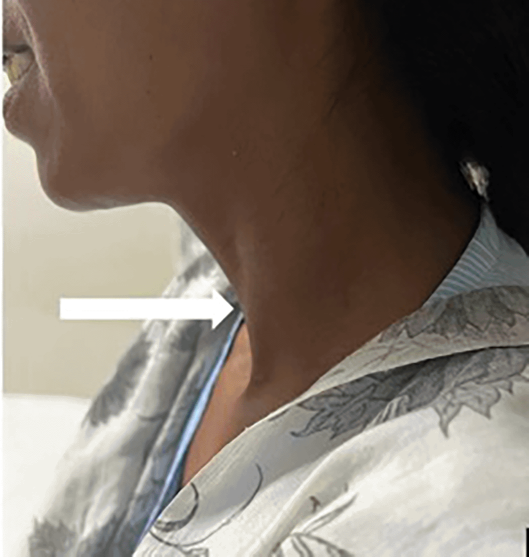
A 35-year-old female presented to the surgery OPD with a 2-3 cm swelling on the left side of the neck along the middle third of sternocleidomastoid muscle (Figure 1). The patient said that the swelling was present since many years. However, in the past three months there was gradual increase in the size of the swelling. It was round to oval in shape, soft in consistency, non-tender and freely movable over the underlying muscle. There was no evidence of adjacent skin changes or any other neck swelling on physical examination.
The patient had no complains of difficulty in breathing or deglutition and no restriction in movement of the neck. She had no history of trauma, fever or any event of tuberculosis. Her routine blood investigations as well as thyroid profile was within normal ranges.
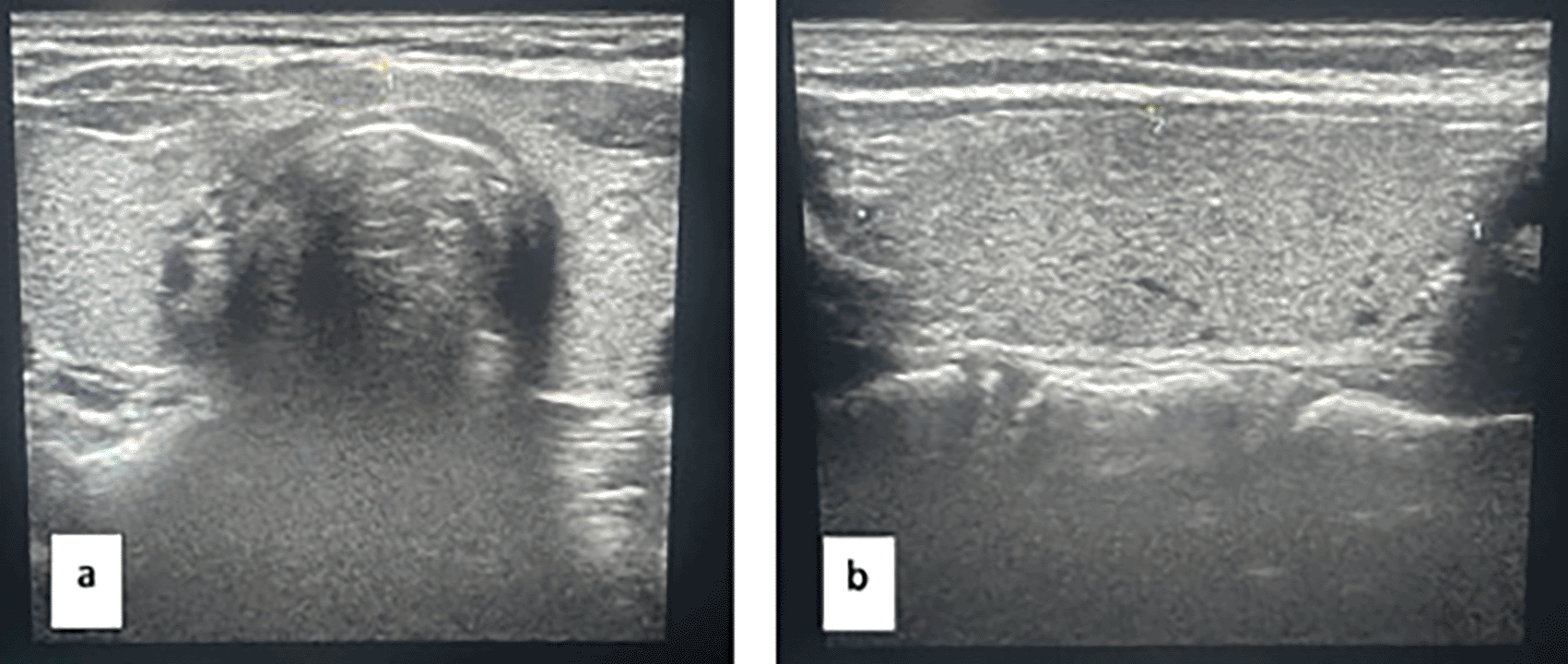
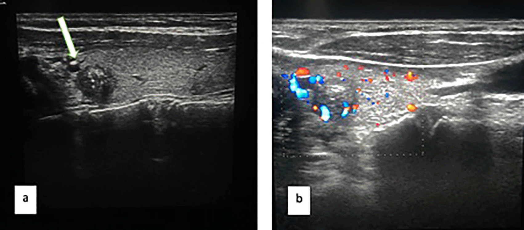
On ultrasound of the neck, the lesion measured 2 x 1.5 cm, was thin walled solid-cystic (cystic component> solid) with echogenic debris and abutting sternocleidomastoid muscle (Figure 2a). The solid component showed internal vascularity on color doppler (Figure 2b).

Thyroid gland was further examined on ultrasonography and appeared normal in maximum dimensions. The isthmus and right lobe were normal in echotexture (Figure 3).
However, the left lobe of thyroid revealed an ill-defined round (1 x 1 cm) hypoechoic solid lesion with punctate microcalcifications and increased vascularity within (Figure 4a). Rest of the left lobe appeared normal in echotexture as well as vascularity (Figure 4b). The lesion was graded BIRADS-4 on ultrasound indicating moderate suspicion for malignancy.
The patient was then further evaluated on computed tomography before subjecting the patient to FNAC (fine needle aspiration cytology) of the neck lesion. On contrast enhanced CT of the neck, there is a well-defined round solid-cystic lesion lying posterior to middle one-third of the left sternocleidomastoid muscle and focally abutting the left jugular vein. The lesion has thin enhancing wall and enhancing internal solid component (Figure 5).

After these radiological investigations, the patient was then taken for FNAC of the swelling. The first sample that was fluid aspirate came out to be indeterminate in nature. However, in the next sampling, the solid component was targeted and it came out to be positive for malignant cytology. Since the patient had concurrent BIRADS-4 thyroid lesion, there was suspicion of thyroid malignancy with metastasis to the branchial cleft cyst. The thyroid lesion was then taken up for aspiration and was proven to be papillary thyroid carcinoma.
Branchial cleft cysts are one of the most commonly encountered lateral neck masses.6,7 These are embryological remnants that occur due of failure of closure of the pharyngeal arches before birth.8 Metastases to the branchial cleft cyst is rare and identifying, differentiating it from cystic metastatic lymph nodes is important to plan treatment.9,10 Papillary thyroid carcinoma frequently metastasizes and therefore it is necessary to evaluate the thyroid gland in such instances to narrow down the diagnoses.11,12
| Views | Downloads | |
|---|---|---|
| F1000Research | - | - |
|
PubMed Central
Data from PMC are received and updated monthly.
|
- | - |
Provide sufficient details of any financial or non-financial competing interests to enable users to assess whether your comments might lead a reasonable person to question your impartiality. Consider the following examples, but note that this is not an exhaustive list:
Sign up for content alerts and receive a weekly or monthly email with all newly published articles
Already registered? Sign in
The email address should be the one you originally registered with F1000.
You registered with F1000 via Google, so we cannot reset your password.
To sign in, please click here.
If you still need help with your Google account password, please click here.
You registered with F1000 via Facebook, so we cannot reset your password.
To sign in, please click here.
If you still need help with your Facebook account password, please click here.
If your email address is registered with us, we will email you instructions to reset your password.
If you think you should have received this email but it has not arrived, please check your spam filters and/or contact for further assistance.
Comments on this article Comments (0)