Keywords
Colopleural fistula, empyema, diaphragmatic hernia, resection, anastomosis, thoracotomy
Colopleural fistulas are rare and generally correspond to thoracic empyema alongside an acute abdomen. Early diagnosis and prompt treatment are essential to prevent recurrence of thoracic empyema.
An 18-year-old male was admitted to our hospital with dyspnea and abdominal pain. The patient had a history of a left diaphragmatic hernia repair. Physical examination revealed signs of left-sided pleural effusion and abdominal pain. Chest computed tomography (CT) revealed a left lung empyema with a suspected connection to the intra-abdominal cavity through the diaphragm. Colopleural fistula and diverticulosis were confirmed by colonoscopy. Subsequently, primary resection, transverse-descending anastomosis, and fistula suturing were performed, accompanied by empyema evacuation through thoracotomy and diverting colostomy.
Colopleural fistula is an incredibly rare phenomenon that can result from diaphragmatic injury, malignancies, perforated diverticulosis, or colonic/pulmonary infections. The management of a colopleural fistula depends on anatomical, clinical, and other factors. Diverticulitis is usually treated using antibiotics and other conservative therapies. Diverticular disease usually requires surgery if there is perforation, progressive signs of sepsis or peritonitis, fistula, or failure of conservative treatment. A surgical procedure was performed in this case because of empyema arising from a colopleural fistula formation caused by diverticulosis.
Primary colon resection, colon anastomosis, fistula suturing, and decortication thoracotomy were shown to effectively treat colopleural fistula.
Colopleural fistula, empyema, diaphragmatic hernia, resection, anastomosis, thoracotomy
Fistulas are abnormal communications that occur between two epithelial surfaces. Gastrointestinal fistulas comprise all connections that involve the gastrointestinal tract. Which can manifest as either congenital of acquired in nature.1 Colopleural fistula is an incredibly rare phenomenon resulting from a wide variety of colon disorders such as injury, tumor infiltration, and infection. Despite being rare, empyema following acute abdomen or infection of the gastrointestinal tract could indicate a fistula connecting the pleura and abdominal cavity with infection involved. A proper workup is required to establish the diagnosis, and comprehensive management is essential to prevent empyema recurrence. Evaluation of pleural fluid and radiological examination of the chest and abdomen, along with contrast studies, can provide early visualization and diagnosis of the fistula. Management of colopleural fistula depends on the underlying pathology.2 Therefore, we report a rare case of a patient admitted with empyema and colopleural fistula secondary to transverse colon diverticulosis. This case report was prepared in accordance with the SCARE 2020 guidelines and submitted subsequent to the completion of the SCARE 2020 checklist.3
Case report
An 18-year-old Indonesian male presented to the emergency department with abdominal pain in all four quadrants over the previous 4 weeks. Abdominal pain was accompanied by dyspnea, cough, and fever. One week prior, he began experiencing dyspnea and cough with left-sided chest discomfort. The severity of dyspnea was alleviated when the patient sat in a tripod position. He had a history of left diaphragmatic repair and colostomy formation surgery due to an empyema complicated by a diaphragmatic hernia 10 months prior to presentation. The patient presented with abdominal pain, and a routine chest radiograph revealed a high contour of the diaphragmatic dome on the left diaphragm, without suspicion of diaphragmatic hernia. On the sixth day of hospitalization, the patient developed dyspnea and sharp pain, which worsened with deep inhalation. The patient underwent chest radiography, which revealed pleural effusion. The patient underwent needle decompression and Water Sealed Drainage (WSD) insertion, resulting in discharge of pus and fecal matter. The patient underwent a diaphragmatic hernia repair and colostomy. Intraoperative findings also revealed intestinal adhesions, raising the suspicion of intestinal tuberculosis. Colostomy was performed in the right abdominal quadrant on the same side as the adhesions. The patient was treated with rifampicin 450 mg, isoniazid 400 mg, and etambutol 500 mg for 9 months because of extrapulmonary tuberculosis. The patient had no complaints regarding defecation through colostomy, but some parts of the colostomy had pus.
Upon admission, a physical examination revealed that the patient was fully conscious and had moderate pain. The vital signs showed normal blood pressure, heart rate, tachypnea of 28x/min, and slight fever of 37.5°C. Physical examination of the chest showed delayed asymmetrical left hemithorax movement with decreased vesicular sound on the ipsilateral side and normal breath findings on the right side. During lung auscultation, no rales, ronchi, or wheeze was heard. Abdominal examination revealed normal bowel sounds and tenderness in the epigastric region. Colostomy examination showed positive production of fecal material on the proximal side and pus on the distal side.
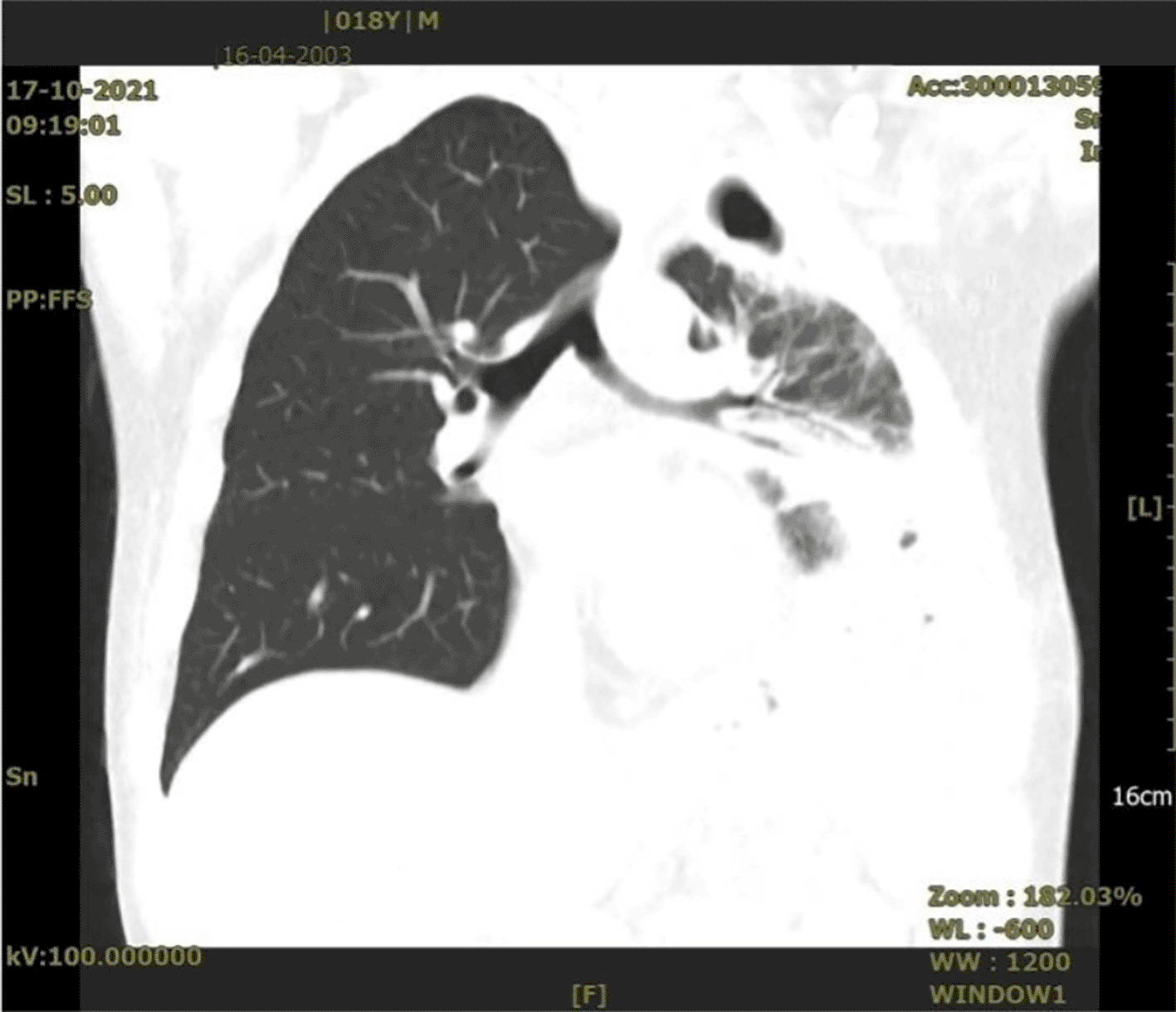
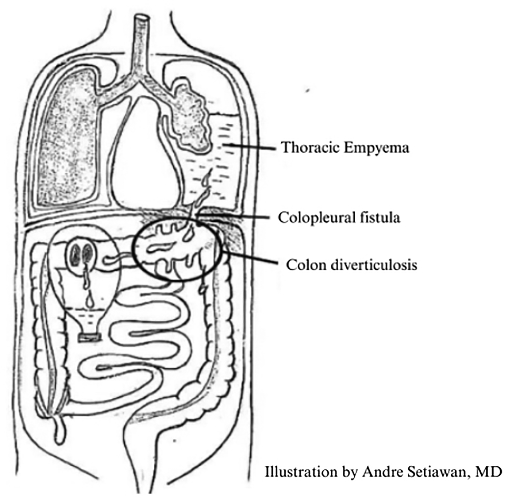
During admission, laboratory workup revealed a slight decrease in albumin (3,2 g/dL) and an increase in globulin (4,3 g/dL). Other laboratory parameters were within the normal range. The contrast-enhanced Chest CT scan revealed an atelectasic left lower lobe in the inferior central region; haziness of the left lung base with an air component raises suspicion of bowel gas and empyema on the left side of the thorax with a suspected connection to the intra-abdominal cavity through the diaphragm (Figure 1). Colonoscopy revealed pus production and two lumens in the transverse colon around the anastomosis area that appeared to be a fistula (Figure 2), and no abnormalities were found in the descending and sigmoid colons. The colopleural fistula was found to connect the pleural space to the intra-abdominal cavity near the perforated diverticulum in the left transverse colon (Figure 3).


Surgical intervention was decided after the preoperative diagnosis of an intestine-diaphragm fistula and left-sided thoracic empyema. The midline incision extended from three fingers infraumbilical to two fingers below the xyphoid process, and no evidence of exudate fluid/pus was observed after the peritoneum was incised. Full intraoperative inspection revealed adhesions in the intestines, particularly the ileum and colon, towards the left subdiaphragm, followed by sharp adheolysis. There were further adhesions in the transverse colon to the left diaphragm, and a fistula connecting the left pleural cavity to the intraluminal left transverse colon was discovered (Figure 4B and 4C). The fistula was then cannulated using a 5F nasogastric tube as a marker. Adhesion was then performed via adhesiolysis by releasing the fistula from the left diaphragm into the colon. The transverse colon was resected, and end-to-end anastomosis between the transverse colon and descending colon was made with a 3.0 vicryl suture and overhected with a 3.0 silk suture (Figures 4D and 4E). The fistula-caused defect in the left diaphragm was repaired and sutured using a simple interrupted technique with prolene 1.0. The colostomy in the previous surgery, which was proximal to the anastomosis, was retained as transient fecal diversion. After controlling the hemorrhage and washing the abdominal cavity, the operative region was closed layer by layer, leaving one drain in the subhepatic region, which was then connected to a water-seal drainage (WSD) system in the left thoracic region. Left decortication thoracotomy was performed after adhesiolysis and resection of the transverse colon, end-to-end anastomosis of the transverse-descending colon, and primary suturing of the defect in the diaphragm. The patient underwent thoracotomy and debridement of the left thoracic space under general anesthesia. During the thoracotomy, approximately 100 mL of exudate fluid was observed (Figure 4A).
The Histopathological findings revealed diverticula protruding from the colonic mucosa through the muscularis propria and multinucleated giant cells with acute and chronic inflammatory cells. No tubercle, Langhans large cells, or caseous necrosis was observed. This finding is in concordance with the pathology of the diverticulosis (Figure 5).
One month after surgery, the WSD tube was removed because of the absence of exudate production and improvement of dyspnea. After two months, the colostomy was closed, and the patient was able to return to his usual activities without any complaints and with a good surgical wound (Figure 4F).
Colopleural fistula is an incredibly rare phenomenon caused by a variety of colon disorders, and patients are typically hospitalized for dyspnea and chest pain. Colopleural fistulas can be formed secondary to diaphragmatic injury, which can result in connections between the pleural space and intra-abdominal cavity through hernia. This was described in a case report by El Hiday et al., who reported a colopleural fistula secondary to diaphragmatic injury caused by surgery.4
Hydrothorax rather than empyema occurred because of the absence of an infection during the formation of a fistula. Colopleural fistulae with empyema may result from tumor infiltration or infection in the surrounding area. The development of a colopleural fistula resulting from tumor infiltration was documented in a recent study by Lian et al. in 2017. The report stated that the chief complaints were chest pain, dyspnea, and fever, which were also experienced by the patient. The CT scan revealed an empyema caused by infiltration of colon cancer pT4bN0M0.5
Colopleural fistulas may also result from infection-related perforations. Infection may occur in the colon but may also occur in the lungs. Hayashi et al. reported a case of colopleural fistula secondary to a pulmonary Aspergillus infection. According to their report, aspergillus infection may produce protease, fibrinase, and various toxins, which can destroy the lung and adjacent structure, resulting in the formation of a fistula.6 In our case, a colopleural fistula was formed secondary to transverse colon infection and diverticulosis perforation of the colon near the left side of the diaphragm, which can spread the infection and lead to diaphragmatic injury along with perforation.
Diverticulosis is a pathological state characterized by the development of multiple sac-like protrusions (diverticula) throughout the gastrointestinal tract. Although diverticula may form in areas of vulnerability in the walls of both the small and large intestines, this condition is commonly reported in the sigmoid colon of the large intestine. Throughout in the intestines, these spaces are reinforced by the external longitudinal layer of the muscularis propria. In the colon, this muscle layer is discontinuous and divided into three bands known as taeniae coli. Excessive peristaltic contractions can cause an increase in the intraluminal pressure. This can lead to the formation of diverticles in the colon. Colonic diverticula are characterized by a thin wall that consists of flattened or atrophic mucosa, compressed submucosa, and typically, an attenuated muscularis propria is absent. This histopathological finding was in accordance with the present case, in which the muscularis propria was absent. Obstruction of the diverticula with stasis of contents results in inflammatory changes that cause diverticulitis and peridiverticulitis. Perforation can easily occur when there is inflammation, increased pressure, and mucosal ulceration within an obstructed diverticulum, because the wall of the diverticulum is only supported by the muscularis mucosa and a thin layer of subserosal adipose tissue. Recurrent diverticulitis, regardless of perforation, can lead to complications, such as abscess formation, fistula formation, or bowel obstruction. In our case, the presence of a colopleural fistula occurred due to a perforated diverticulum in the left transverse colon that connects the intra-abdominal cavity to the pleural space.7,8
The occurrence of colopleural fistula secondary to diverticulosis was documented in previous studies conducted by Olesen et al. in 1989 and Papagiannopoulos et al. in 2004.2,9 Olesen et al. reported a 63-year-old male patient admitted due to dyspnea and tachycardia. Two years prior, the patient had been admitted for irregular defection and faint left-sided upper abdominal pain, and barium enema examination revealed slight diverticulitis. Chest radiography revealed left-sided pleural effusion. Chest drainage revealed foul pus containing gram-negative and gram-positive cocci. Subsequent microbiological cultures confirmed the presence of anaerobic bacteria. A pleural drain was inserted, and saline irrigation was administered daily until the pleural fluid became sterile. During the course of antibiotic treatment, the patient developed abdominal symptoms including nausea and left-sided upper abdominal pain. A fistula was identified via barium enema, which originated from the descending colon below the left flexure and passing through the left diaphragm. The patient was managed with parenteral nutrition for two weeks, and the fistula was barely visible on fistulography.9 Colopleural fistula secondary to diverticulosis was also reported by Papagiannopoulos et al. in a 46-year-old male who was admitted with an acute abdomen. Emergency laparotomy and Hartmann procedure were performed, which revealed a perforated sigmoid diverticulum. Two months later, the patient was readmitted with fever and left-sided abdominal and thoracic pain. Chest radiography revealed left-sided pleural effusion, and CT revealed a basal empyema pocket with no intra-abdominal collection. Based on the patient's clinical condition, empyema evacuation with rib resection and drain placement was performed. Chest drainage revealed pus discharge with coliforms and Streptococcus group D growth. Computed tomography (CT) of the chest and abdomen followed by a colostomy enema revealed a colopleural fistula extending from the colostomy site to the pleural space with communication with the distal colon. After performing an urgent laparotomy with adhesiolysis, en bloc resection of the fistula and colon was performed, followed by primary anastomosis and loop ileostomy.2
A diaphragmatic hernia is a protrusion of abdominal organs into the chest cavity resulting from a defect in the diaphragm. This condition often arises as a congenital phenomenon, although it can also be acquired by trauma or surgery involving the diaphragm, thorax, or abdomen.10,11 There are two types of congenital diaphragmatic hernias: Bochdalek and Morgagni. Bochdalek hernia is the most common type of diaphragmatic hernia. It occurs through a posterior defect in the diaphragm and manifests at an early stage in life. While Morgagni’s hernia is due to a defect in the anterior part of the diaphragm, the foramina Morgagni, and presents later in life.11,12 An acquired diaphragmatic hernia may result in various complications, including diaphragmatic rupture, acute obstructive symptoms, respiratory arrest, strangulation, and cardiac tamponade. Several complications may arise as a consequence of acquired diaphragmatic hernia, including acute obstructive symptoms, diaphragmatic rupture, cardiac tamponade, strangulation, and respiratory arrest. A delayed diagnosis results in permanent complications associated with prolonged herniation. The etiology of acquired diaphragmatic hernia is dependent on the type of surgery (the technique used to close the diaphragm) and patient-related factors, such as penetrating trauma; however, the left hemidiaphragm is involved more frequently (>80%) than the right.10,13 The occurrence of contamination leading to infection increases the probability of developing empyema in the future. Additionally, it has been observed that the colon becomes trapped in the chest due to a diaphragmatic hernia.10 This condition can be caused by a range of disorders including colitis, diverticulitis, colon polyps, and colon cancer.14
The management of a colopleural fistula depends on several factors, including anatomical characteristics and clinical conditions. Graham et al. suggested that conservative treatment should be considered for all uncomplicated fistula. The majority of colonic fistula close spontaneously within two weeks if there is no mucosal growth within the fistula, obstruction, or malignancies of the bowel distal to the fistula.15 This approach was used by Olesen et al., where the patient only had conservative treatment using parenteral nutrition, pleural drain, and antibiotics.9 Diverticulitis usually treatable with antibiotics and other conservative therapies. Diverticulitis or diverticular disease usually requires surgery if there is a clear sign of perforation, a progressive sign of sepsis or peritonitis, failure of conservative treatment, or the formation of a fistula.16 In our case, surgical intervention was indicated because of the presence of a perforated colonic diverticulum, along with the spread of infection leading to fistula development in the diaphragm, and the patient showed clinical signs of peritonitis in the epigastric area. Furthermore, a surgical procedure was performed to evacuate the pus from the thoracic region via thoracotomy.
The most common technique for diverticulosis surgery is the resection of the diverticulum and anastomosis between the proximal and distal segments of the resected region. Nevertheless, the primary distinction between “standard” diverticulitis and diverticulitis that causes colopleural fistula is the diverticulum site. Diverticulitis commonly occurs in the sigmoid colon; hence, the standard surgical procedure is to perform a sigmoidectomy followed by colorectal anastomosis.17 In our case, a perforated diverticulum that resulted in a colopleural fistula occurred in the transverse colon in the region near the left diaphragm. Therefore, transverse colectomy and transverse descending colon anastomosis were performed. The same technique was also performed by Papagiannopoulos et al. also conducted a similar procedure with resection of the colon and fistula, and primary anastomosis was performed with the addition of a loop ileostomy.2 Diverting stoma can be performed to prevent anastomosis leak, which is a significant cause of morbidity and mortality following colorectal resection. Several randomized controlled trials and meta-analyses corroborate the evidence that stoma diversion can reduce the incidence of anastomotic leak and the need for emergent abdominal reoperation.18–21 Fortunately, the patient in our case had already undergone surgery to treat colon adhesion and colostomy formation prior to admission to our center. Therefore, we retained the colostomy proximal to the resected region for a temporary fecal diversion.
Written informed consent was obtained from the patient's parents/legal guardians for the publication of this case report and accompanying images. A copy of the written consent is available for review by the Editor-in-Chief of this journal upon request.
Data sharing is not applicable to this article, as no datasets were generated or analyzed during the current study.
Respository: Updating Consensus Surgical Case Report (SCARE) 2020 guidelines checklist https://www.sciencedirect.com/science/article/pii/S1743919120307718.
| Views | Downloads | |
|---|---|---|
| F1000Research | - | - |
|
PubMed Central
Data from PMC are received and updated monthly.
|
- | - |
Is the background of the case’s history and progression described in sufficient detail?
Yes
Are enough details provided of any physical examination and diagnostic tests, treatment given and outcomes?
Yes
Is sufficient discussion included of the importance of the findings and their relevance to future understanding of disease processes, diagnosis or treatment?
Partly
Is the case presented with sufficient detail to be useful for other practitioners?
Yes
Competing Interests: No competing interests were disclosed.
Reviewer Expertise: Gastroentestinal Surgeon, Lecturer
Alongside their report, reviewers assign a status to the article:
| Invited Reviewers | |
|---|---|
| 1 | |
|
Version 1 17 Jun 24 |
read |
Provide sufficient details of any financial or non-financial competing interests to enable users to assess whether your comments might lead a reasonable person to question your impartiality. Consider the following examples, but note that this is not an exhaustive list:
Sign up for content alerts and receive a weekly or monthly email with all newly published articles
Already registered? Sign in
The email address should be the one you originally registered with F1000.
You registered with F1000 via Google, so we cannot reset your password.
To sign in, please click here.
If you still need help with your Google account password, please click here.
You registered with F1000 via Facebook, so we cannot reset your password.
To sign in, please click here.
If you still need help with your Facebook account password, please click here.
If your email address is registered with us, we will email you instructions to reset your password.
If you think you should have received this email but it has not arrived, please check your spam filters and/or contact for further assistance.
Comments on this article Comments (0)