Keywords
Blast injury, blast-related craniofacial trauma, blast-induced neurotrauma, blast-related ocular trauma, developing countries, case report
Blast-related craniofacial and ocular injuries have traditionally been associated with battlefields. In civilian populations, these injuries were infrequent, primarily arising from accidental explosions or terrorism incidents. Explosions can inflict severe damage to the neurological, ocular, and auditory systems through blast waves, high-velocity foreign bodies, and thermal radiation. The pathophysiology of blast-related craniofacial injuries is specific and complex, with severe cases often involving both penetrating and blunt trauma, leading to high mortality and morbidity rates. These injuries necessitate multidisciplinary management and can be defined as intracranial polytrauma. Recently, Niger has seen a surge in blast injuries, predominantly due to the increase in clandestine artisanal gold mining. Managing these injuries in resource-limited settings poses significant challenge. Comprehensive national data on these injuries are sparse due to high pre-hospital mortality rates and their infrequent occurrence, resulting in limited experience among our local physicians in their management. We present a rare case involving an artisanal gold miner with a history of smoking and previous concussions from explosion exposures. The patient was transferred to our hospital following severe craniofacial and ocular injuries caused by an accidental dynamite explosion at a mining site. On admission, the patient presented altered consciousness, agitation, unstable vital signs, multiple craniofacial wounds, including a large frontal wound with brain substance extrusion, diffuse facial burns, left globe rupture, and rhinorrhagia. After resuscitation and stabilization, brain imaging revealed multiple complex craniofacial fractures with foreign bodies. The patient underwent multidisciplinary surgical management. However, the postoperative course was complicated by post-concussive syndrome, and infection of the surgical wounds, necessitating surgical revision. Following the second surgery, the postoperative course was uneventful, although the patient experienced reduced visual acuity. This case highlights the management challenges in Niger and underscores the urgent need for clinical studies and training for gold miners to enhance safety practices in their activities.
Blast injury, blast-related craniofacial trauma, blast-induced neurotrauma, blast-related ocular trauma, developing countries, case report
In recent years, blast-related craniofacial and ocular injuries have increasingly occurred outside of battlefields, becoming a significant concern in civilian settings.1–3 The pathophysiology of these injuries is distinct due to the interaction of blast waves and thermal radiation with biological tissues, which exacerbates both penetrating and blunt injuries.2,4,5 Blast waves cause rapid changes in the pressure within the brain, ocular globe, and middle ear, leading to tension and shearing forces that can result in contusions, intracranial hemorrhage, vitreous hemorrhage, globe rupture, retinal detachment, tympanic membrane perforation, etc.2,6 Consequently, severe blast-related craniofacial injuries can be defined as intracranial polytrauma, necessitating immediate multidisciplinary management.
The medical community’s interest in civilian blast-related craniofacial injuries has grown due to the rise in terrorism (improvised explosive devices), accidental explosions, and civilian casualties from military conflicts (heavy munitions firing).4,5,7 In our country, the primary causes of these injuries are explosion accidents and terrorism, particularly accidental dynamite explosions at artisanal gold mining sites. However, comprehensive national data on these injuries are sparse due to high pre-hospital mortality rates and their rarity, resulting in limited experience among our physicians in managing such cases.
Our objective is to report a rare and well-documented case of blast-related severe craniofacial and ocular injuries at an artisanal gold mining sites. This report aims to highlight the management challenges in developing countries like Niger, emphasize the urgent need for clinical studies on blast-related craniofacial trauma in resource-limited settings, and advocate for safety training for gold miners.
This case report has been reported in line with the SCARE Criteria.8
A 25-year-old married male, an illegal artisanal gold miner, with a history of smoking and two prior concussions from explosions exposure (without any apparent serious injury), was referred to our hospital by a peripheral health center sustaining severe craniofacial injuries from a dynamite explosion. The accident occurred three hours prior, while he was installing dynamite accidentally it a gold shaft, resulting in multiple deaths. The patient, found comatose with multiple sharp missile craniofacial and ocular injuries and diffuse facial burns, was the sole survivor and was transported by a personal vehicle to a health center 120 km away. There, he was resuscitated and stabilized with intravenous crystalloid fluids (0.9% sodium chloride saline solution), analgesia (paracetamol 1 g), and antibiotic prophylaxis (ceftriaxone 2 g). He was then transferred by ambulance to our hospital’s emergency department without intubation.
On admission, the patient was agitated with an altered state of consciousness (Glasgow Coma Scale [GCS] 9/15: Y1 V3 M5) and hemodynamically and respiratory unstable, with a blood pressure of 79/44 mmHg, pulse rate of 156 beats/min, respiratory rate of 24 breaths/min, and oxygen saturation 86% on 10 L/min oxygen via a non-rebreathing mask. He was sedated with midazolam at 4 mg/h via electric syringe pumps. Oro-tracheal intubation and administration of crystalloid fluids and emergency drug (Adrenaline 0.75 mg) stabilized his vital parameters to a blood pressure of 144/64 mmHg, pulse rate of 105 beats/min, respiratory rate of 14 breaths/min, and oxygen saturation 96%. His core temperature was 37-37.5°C, and his body mass index (BMI) was 20.75 kg/m2. Craniofacial examination revealed multiple wounds, including a large penetrating left frontal wound with brain substance extrusion, a penetrating left maxillary sinus injury, diffuse facial burns, and moderate rhinorrhagia (Figure 1). Ophthalmological examination showed laceration of the left eyelids, globe rupture with disorganization of the ocular structures, and a complex fracture of the left orbital frame. The right eye had corneal edema and intracorneal foreign bodies. Examinations of the cervical spine, thorax, abdomen, limbs, and motor function were normal.
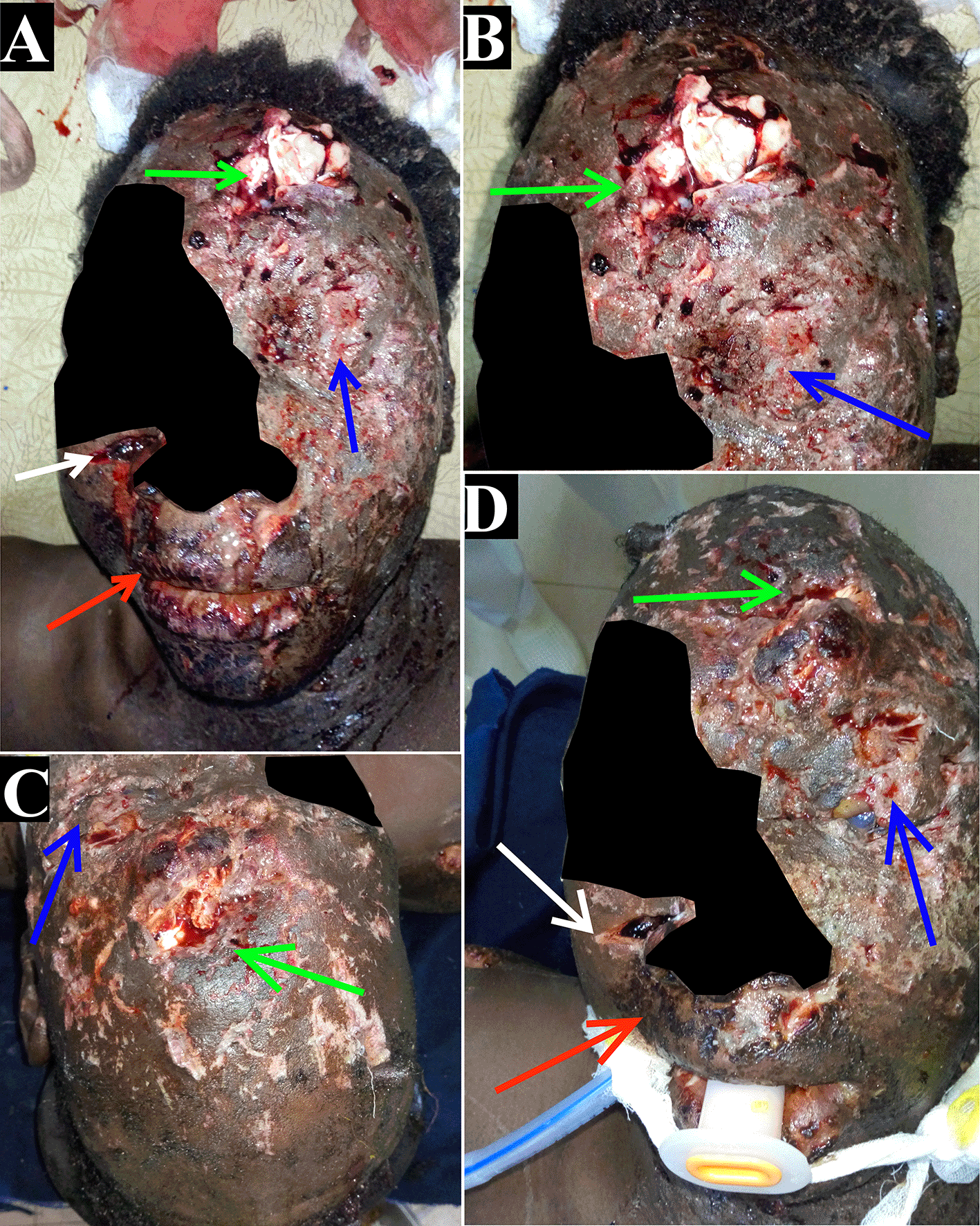
(A-D) Large penetrating left frontal wound with brain substance extrusion (green arrows), laceration of the left eyelids, globe rupture with disorganization of the ocular structures (blue arrows), penetrating the left maxillary sinus injury (white arrow), rhinorrhagia (red arrows), and diffuse facial burns from the explosion.
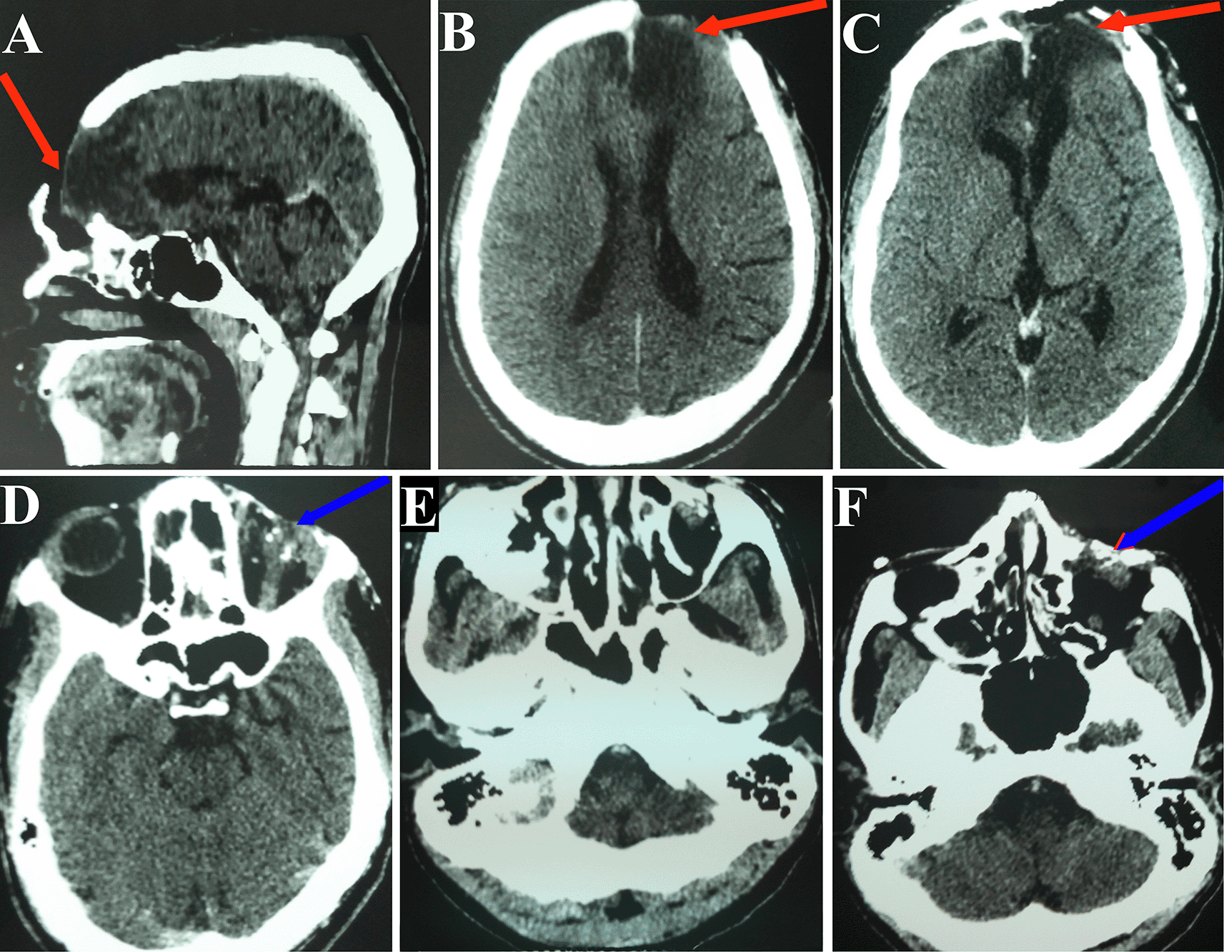
Stabilization was continued following the ATLS (Advanced Trauma Life Support) guidelines, including intravenous crystalloid fluids, multimodal analgesia (paracetamol 6 g/24 h, tramadol hydrochloride 400 mg/24 h), 96-hour prophylactic antibiotic (ceftriaxone 4 g/24 h), and nasal packing. Blast-related severe craniofacial and ocular injuries were diagnosed. Non-contrast brain computed tomography (CT) revealed a large left frontal fracture with intraparenchymal bone fragments and foreign bodies, a left orbital frame fracture with intraorbital foreign bodies, maxillary sinus fractures with foreign bodies, and a skull base fracture associated with a fracture of the left orbital inner wall (Figure 2). Laboratory tests showed an inflammatory process with hyperleukocytosis (17,950 cells/μL, reference: 4,000 – 10,000 cells/μL), granulocytosis (80.04%, reference: 50-70%), elevated C-reactive protein (CRP) (24 mg/L, reference: < 6 mg/L), and anemia (10.2 g/dL, reference range: 11.0-16.0 g/dL). Renal and hepatic function tests were normal, with no ionic or coagulation disorders. A whole blood transfusion was administered, and prophylactic antiepileptic (phenobarbital 100 mg/24 h) was systematically instituted.

(A-F) Large left frontal fracture with intraparenchymal bone fragments and foreign bodies (red arrows), fracture of the left orbital frame with intraorbital foreign bodies (blue arrows), fracture of the left maxillary sinus with foreign bodies (white arrows), and skull base fracture associated with a fracture of the left orbital inner wall (green arrows).
After informing the patient’s family of the poor prognosis, multidisciplinary surgery involving neurosurgeons, ophthalmologists, and a maxillofacial surgeon was performed under general anaesthesia with the patient in the spine position. Initially, the right eye was lavaged with saline, and superficial intracorneal foreign bodies were removed (Figure 3A). Subsequently, a large penetrating frontal injury was approached via a bicoronal incision; limited surgical debridement was performed, followed by a watertight duroplasty, cauterization and closure of the frontal sinus (Figure 3B, 3C). Enucleation of the left eye, and surgical care of maxillary sinus injury were performed. Postoperative care in the general intensive care department included intubation, crystalloid fluids, multimodal analgesia, intravenous antibiotic prophylaxis, sedation, local antibiotic prophylaxis for right eye with ciprofloxacin 0.3% (eye drops and ointment) four times daily, alternating with artificial tears. On the third day, the patient was transferred to neurosurgery department.
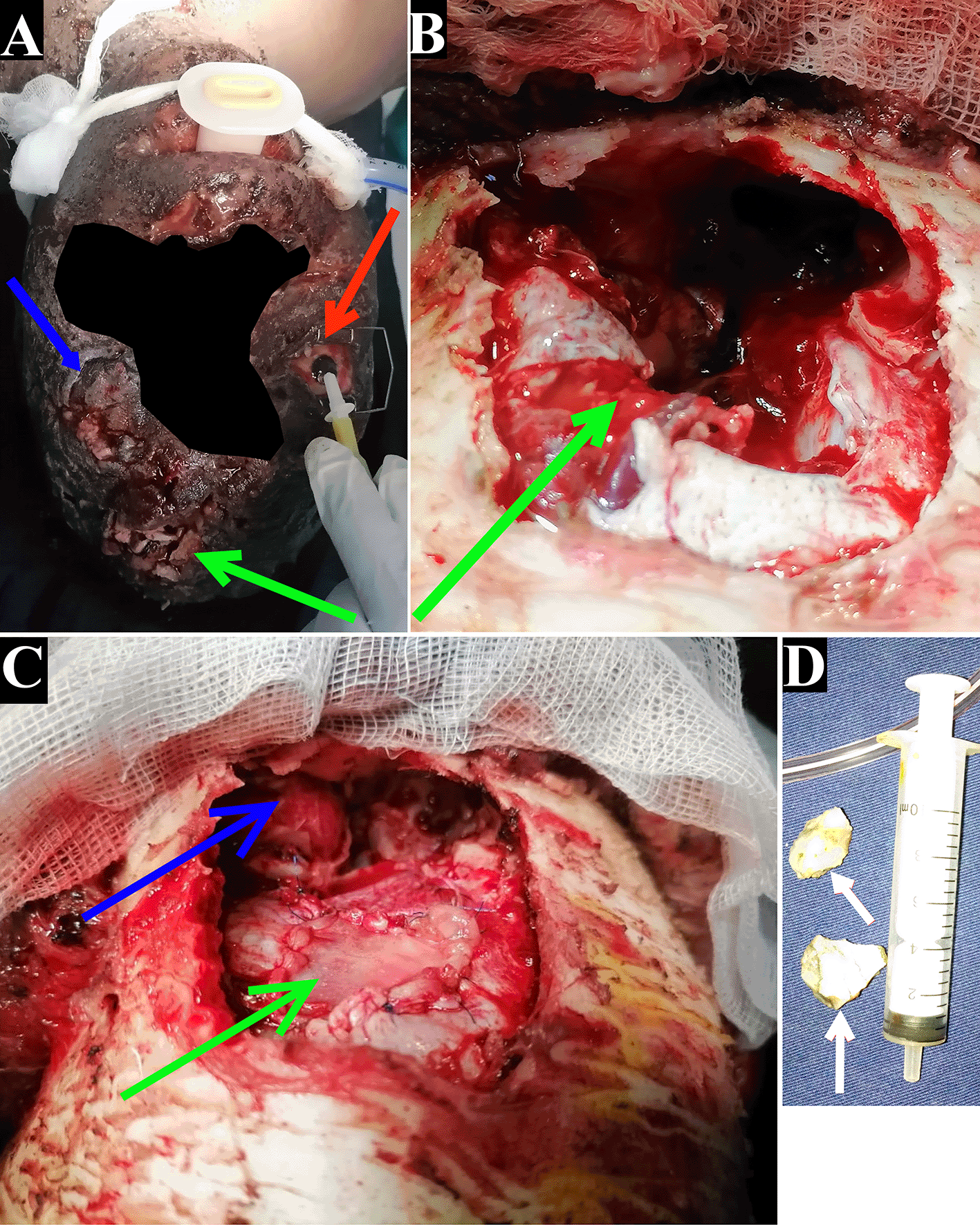
(A) Large penetrating left frontal wound with brain substance extrusion (green arrow), laceration of the left eyelids and globe rupture with disorganization of the ocular structures (blue arrow), right eye lavage with saline, and removal of superficial intracorneal foreign bodies (red arrow);
(B) Lost-bone craniotomy showing a large dural breach with loss of left frontal lobe substance;
(C) Watertight duroplasty after limited surgical debridement (green arrow), complex fracture of the left orbital roof communicating between the orbit and skull base (blue arrow);
(D) Removal of granite fragments from craniofacial injuries (white arrows).
During the first postoperative week, sedation continued due to agitation, and recovery of clear consciousness was slow. Phenobarbital was replaced with sodium valproate 1500 mg/24 h for two weeks. On the third and fifth days, examination of the surgical sites revealed necrosis then dehiscence and infection of surgical wounds (skin around the frontal surgical wound, and the left eyelids) (Figure 4). Biological control tests showed an accelerated erythrocyte sedimentation rate (ESR) of 43 mm/hour and elevated CRP (58 mg/L, reference: < 6 mg/L), prompting intravenous probabilistic antibiotic therapy (amoxicillin/clavulanic acid 3g/187.5 mg/24 h, metronidazole 1500 mg/24 h). A surgical revision was performed on the seventh day, and greasy dressing was applied postoperatively until the surgical wounds healed. By the second week, the patient became less agitated and regained clear consciousness but experienced post-concussive syndrome with headaches, fatigue, insomnia, and concentration difficulties. Psychiatric evaluation recommended conservative treatment. The first ophthalmological examination after regaining clear consciousness revealed light perception. The third-week check-up was good, except for the electroencephalogram (EEG) which showed epileptic activity, necessitating a six-month antiepileptic treatment. All antibiotics (intravenous and local) were discontinued on the 21st day of hospitalisation. Ophthalmological control examination showed corneal clearing and improved visual acuity (6/10). However, the frontal surgical wound showed significant delay in healing, despite good granulation during the first week after surgical revision (Figure 5A). Discharge was authorized on the 40th day of hospitalization, with the surgical wounds were almost healed (Figure 5B). A brain CT with contrast prior to discharge showed good evolution (Figure 6). The patient continued with fat dressings at a nearby peripheral center and regularly consulted our hospital. Post-concussive syndrome symptoms decreased significantly by the second month, with residual headaches and slight fatigue disappearing by the third month. He resumed normal activities in the fourth month, as the symptoms had disappeared and the wounds had completely healed. A follow-up EEG at six months showed spike waves, leading to another six-month renewal of antiepileptic treatment. An eighth-month brain CT with contrast showed good progress but indicated a forming porencephalic cavity (Figure 7). Cranioplasty was planned for the ninth postoperative month. Unfortunately, the patient later died in a gold mine collapse.
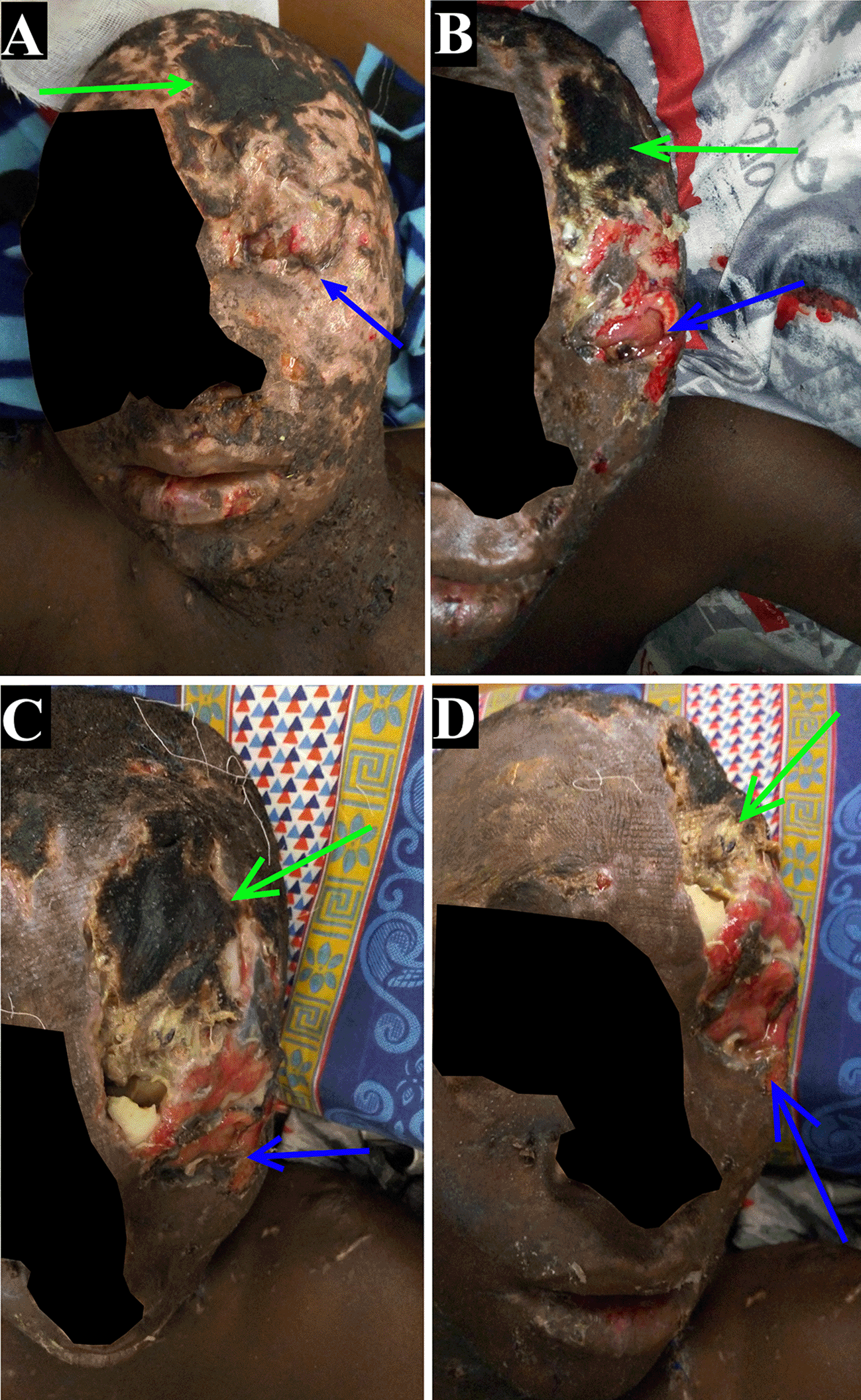
(A) On third postoperative day, desiccation of the frontal surgical wound (green arrow), and early dehiscence of the left eyelid (blue arrow);
(B) On fifth postoperative day postoperative, early necrosis and dehiscence of the frontal surgical wound (green arrow), and necrosis of the left eyelid (blue arrow);
(C and D) On seventh day postoperative, advanced necrosis and infection of the frontal surgical wound (green arrows), and the left eyelid (blue arrows).
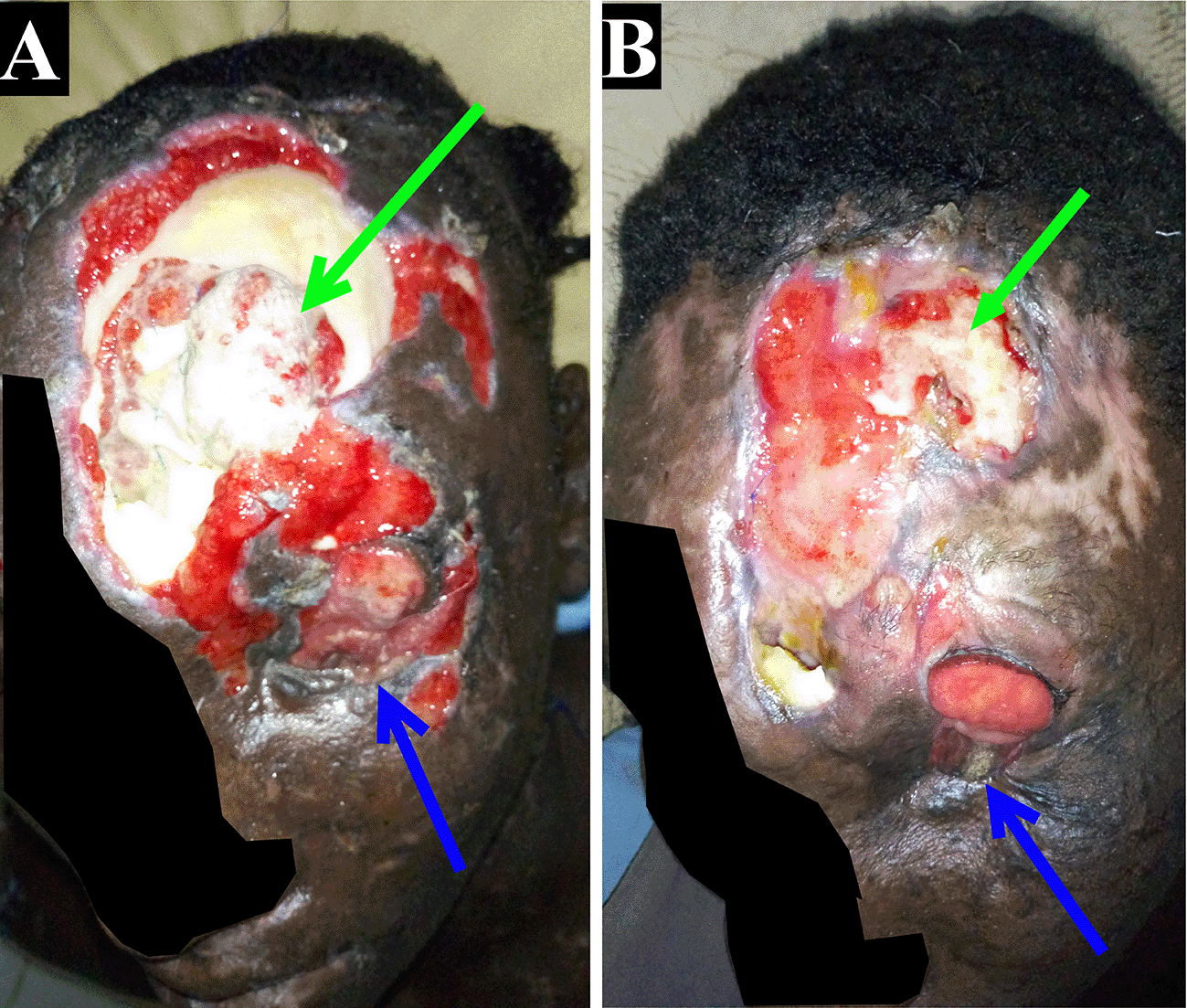
(A) On ninth day post-surgical revision, good granulation of the frontal surgical wound (green arrow), and the left eyelid (blue arrow);
(B) On thirty-second day post-surgical revision, advanced healing of the frontal surgical wound (green arrow) and the left eyelid (blue arrow).
(A-F) Evolution of contusion and loss left frontal lobe substance forming fronto-polar hypodensity (red arrows), and remodeling of the orbital cavity after enucleation (blue arrows).
Blast-related civilian craniofacial and ocular injuries in Niger predominantly arise from terrorism and accidental explosions at artisanal gold mining sites. Globally, such injuries in general civilian population is more commonly linked to fireworks, firecrackers, mine gas, detonators, and containers.9,6 In Niger, the high pre-hospital mortality rate and lack of consultation for mild cases result in under-diagnosis and incomplete national data, resulting in limited experience among our physicians in managing such cases. Explosions are sudden and devastating, often causing numerous casualties and significant morbidity among survivors.6 According to Zhong et al.,6 limb injuries are the most common complication of systemic explosion damage in the general civilian population, followed by craniocerebral injuries. Our patient sustained only craniofacial injuries, with the rest of his body spared, and could not recall his position during the explosion. Dynamite explosions can cause a variety of traumatic injuries due to the blast wave, foreign bodies propelled by the explosions, and released heat, with injuries generally categorized as primary to quaternary.5,7,10 Our patient was the sole survivor of the accident, presented all possible categories of craniofacial lesions (penetrating, blunt injuries, blast injuries), resulting a high morbidity state. Unfortunately, the other gold miners sustained fatal injuries, and we do not know the proportion of craniofacial injuries compared to other injuries (limbs, thorax, abdomen, etc.).
Blast-related craniocerebral trauma involves multiple pathophysiological mechanisms, including blast wave transmission through the cranium, skull flexure, thoracic mechanism, translation and rotation head acceleration, and cerebrospinal fluid (CSF) cavitation.11 During blast wave transmission through the cranium, shock waves travel almost unimpeded through nerve tissue, its envelopes, globe, and ear, resulting in a rapid pressure peak followed by negative pressurization. This leads to the compression and expansion of the brain, globe, and tympanic membrane in rapid succession.4,12 Changes in pressurization caused by shock waves can result in tension and shearing of brain tissue, blood vessels, and globe, as well as cavitation of CSF. These changes can cause hemorrhagic contusions, intracranial hemorrhages due to alterations in blood-brain barrier (BBB) permeability, diffuse axonal damage, retinal detachment, tympanic membrane perforation, etc.1,2,5,11,13 Additionally, neuroinflammation, vasospasm, neural loss, oxidative stress, and metabolic changes are observed in moderate and severe trauma, with the most significant changes noted in the frontal cortex and basal ganglia.11 Patient preoperative assessment of the lesions revealed mainly fractures and hemorrhagic contusions, primarily caused by the penetration of high-velocity fragments rather than blast waves. Cerebral edema, intracranial hemorrhages, and diffuse axonal damage were minimal.
The ocular globe is particularly vulnerable to blast trauma due to its exposure, incompressibility, fluid content, rich vascular network, fragile tissues, and fine structures. Blast-related ocular injuries can lead to impaired visual acuity and even blindness.6,9,13 Ocular damage is almost inevitable in cases of blast-related civilian craniofacial trauma, especially since illegal gold miners do not wear personal protective equipment, hence our patient’s bilateral ophthalmic damage. Over the past decade, ocular injuries have been the leading cause of vision loss globally, affecting developed, developing, and high-income countries alike.9 Unfortunately, the incidence of civilian blast injuries is increasing in our region, as the low socio-economic status of the rural population has led to gold rushes.14 Gold mining activities are predominantly carried out by young adults, and life at clandestine gold mining sites is precarious, exposing miners to numerous diseases (infectious, environmental, etc.) and blast injuries. This observation aligns with scientific literature, indicating that blast injuries mainly affect young adults.1,5,7,13,15 The remoteness and inaccessibility of certain illegal gold mining sites makes pre-hospital treatment of injured individuals extremely challenging. Patients are transferred in personal vehicles or poorly equipped ambulances, resulting in unstable conditions on admission.
Patients with severe craniofacial and ocular injuries from explosions requires comprehensive management, involving a multimodal approach that incorporates data from neuroimaging (magnetic resonance imaging [MRI], functional MRI [fMRI], diffusion tensor imaging [DTI], positron emission tomography [TEP], magnetoencephalography, and electroencephalography), biomarkers (p-tau, glial fibrillary acidic protein, etc.), and neurobehavioral assessments.2,5,16 Certain abnormalities linked to neuroinflammation and oedema caused by BBB damage and oxidative stress have been identified as the causes of neuropsychiatric symptoms, but are very difficult to detect with basic imaging (brain CT, MRI). Detection of these abnormalities in the acute phase is crucial for diagnosis, prognosis and patient follow-up, requiring advanced neuroimaging techniques such as fMRI, TEP, and DTI.2,16 Nevertheless, sequelae lesions are more easily detected in the late phase, even on basic imaging. However, access to brain imaging is limited in Niger due to the absence of many advanced neuroimaging techniques, restricted accessibility for basic imaging, and the financial difficulties patients face in obtaining examinations. Our patient was only able to undergo a preoperative brain CT and faced difficulties obtaining two postoperative brain CT contrast injections.
Blast-related severe craniofacial and ocular injuries necessitate immediate multidisciplinary management and can be defined as intracranial polytrauma. A psychiatric evaluation should be included in their management regardless of the trauma’s severity, as patients with blast-related head trauma are likely to develop various neuropsychiatric disorders (behavioral deficits, post-concussion syndrome, post-traumatic stress disorder, chronic traumatic encephalopathy, etc.) within 2-4 weeks.17,18 Our patient developed post-concussion syndrome during the second week of postoperative follow-up, which was managed conservatively. However, with his history of multiple blast exposures, he is also at risk of developing chronic traumatic encephalopathy.16,19 During follow-up, some patients with neuropsychiatric disorders also show certain neuroimaging signs, notably the loss of white matter integrity and alterations in cortical volume and thickness.11 Unfortunately, we were unable to follow the patient long enough to see the sequellar lesions on the brain CT, due to his accidental death on the gold mining site after resuming his activities. He had also presented with globe rupture and the presence of intraocular foreign bodies only in the left eye, while the right eye was relatively spared by the explosion. Fortunately, the right eye showed no signs of poor prognosis, such as globe rupture or perforation, retinal detachment, or the presence of intraocular foreign bodies.9 Persistent poor postoperative visual acuity in the right eye would have led to severe visual handicap or even total work incapacity for our patient. Despite our limited resources, our patient has a relatively good postoperative outcome, as he had fewer severity factors, including an initial GCS > 9/15 and the absence of tympanic membrane perforation.7
Artisanal gold panning in our country causes numerous health problems, particularly among young adult miners. These problems may require frequent medical attention, entail high financial costs, and result in loss of working days.14,15 Health issues observed on artisanal gold mining sites include disabling after-effects (visual impairment, motor impairment, etc.) resulting from severe head trauma (accidental dynamite explosions, falls from great heights, etc.) and chronic disabling illnesses (chronic pneumocardiopathy, mental disorders, etc.) due to prolonged exposure to chemicals, dust, alcohol and tobacco.15 Moreover, clandestine mining is one of the most dangerous working environments. According to the International Labour Organization, the accident rate is six to seven times higher than in industrial mining, particularly during the excavation stage.15,20 Many adult young artisanal gold miners, like our patient, have suffered severe, lifelong visual impairments as a result of accidents. Some injuries were severe enough to impair their ability to work, while others rapidly progressed to total work incapacity, becoming a burden to their families. Unfortunately, in Niger, the working population (aged 15-64) represented 47.8% of the population in 2022, making it difficult to manage the consequences of these associated health problems, and potentially placing a burden on the healthcare system and the economy.21 However, artisanal gold panning has developed over the years since the first gold rush in Koma Bangou, and it is now a source of income for hundreds of thousands of unemployed people.14 For our patient, clandestine artisanal gold panning is the only activity in his locality that enables him to provide a decent living for his family and relatives. Niger does not have the necessary means to implement coercive measures to put an end to clandestine gold panning due to the vastness of the territory to be controlled and the difficult access to certain areas. It is, therefore necessary to implement a participative management approach involving public decision-makers, the local population, and the gold miners to better regulate and control gold panning activities. Additionally, it is crucial to make gold miners aware of the dangers of some of their mining tools and methods (explosives, mercury, cyanide, etc.) through health and safety training. This training should encourage them to purchase personal protective equipment, such as helmets and goggles, as well as quality machinery. It is also essential to provide isolated gold mining sites with health support and pre-hospital care facilities.
Our study has several limitations. Firstly, the small number of cases and the absence of multimodal neuroimaging did not allow us to obtain comprehensive data on craniofacial trauma caused by accidental explosions on gold mining sites, and to identify aggravating factors. Secondly, the lack of prolonged follow-up (several years) did not allow us to identify late sequelae, particularly disabling ones. It is essential to advocate for funding to conduct multi-center observational pilot studies to better analyse these lesions for improved management.
The incidence of blast-related severe craniofacial and ocular trauma at artisanal gold mining sites is increasing in Niger due to the proliferation of these sites. However, data on such injuries remain limited due to high pre-hospital mortality rates and their rarity. The absence of advanced neuroimaging techniques and limited healthcare resources further complicate the comprehensive care of these patients. The severity of the injuries and postoperative complications observed in our patient underscores the urgent need for therapeutic protocols tailored to resource-limited settings. Future clinical studies are essential to develop these protocols and improve patient management in such contexts.
Written informed consent for publication of their clinical details and/or clinical images was obtained from the patient/parent/guardian/relative of the patient.
Conceptualization: OIH, GI, AII, IDB, ABK, MSC, RS; Data Curation: OIH, IMD, FLA, KNA; Investigation: OIH, YBT, HAM, TMH, IMD, FLA, KNA, GI, AII, IDB, ABK, MSC, RS; Methodology: OIH, YBT, HAM, TMH, IMD, FLA, KNA, GI, AII, IDB, ABK, MSC, RS; Project Administration: GI, AII, IDB, ABK, MSC, RS; Resources: OIH, YBT, HAM, TMH, IMD, FLA, KNA, GI, AII, IDB, ABK, MSC, RS; Supervision: OIH, GI, AII, IDB, ABK, MSC, RS; Validation: OIH, YBT, HAM, TMH, IMD, FLA, KNA, GI, AII, IDB, ABK, MSC, RS; Visualization: OIH, YBT, HAM, TMH, IMD, FLA, KNA, GI, AII, IDB, ABK, MSC, RS; Writing – Original Draft Preparation: OIH, YBT, HAM, TMH, IMD, FLA, KNA, GI, AII, IDB, ABK, MSC, RS; Writing – Review & Editing: OIH, YBT, HAM, TMH, IMD, FLA, KNA, GI, AII, IDB, ABK, MSC, RS.
Figshare: CARE checklist for ‘Check list severe blast-related craniofacial and ocular injuries’, https://doi.org/10.6084/m9.figshare.26323417.v1. 22
Data are available under the terms of the Creative Commons Attribution 4.0 International license (CC-BY 4.0).
The authors are grateful to the following teacher-researchers: Pr. DECHAMBENOIT Gilbert, and the late Pr. SANOUSSI Samuila, for their unconditional support.
| Views | Downloads | |
|---|---|---|
| F1000Research | - | - |
|
PubMed Central
Data from PMC are received and updated monthly.
|
- | - |
Is the background of the case’s history and progression described in sufficient detail?
Partly
Are enough details provided of any physical examination and diagnostic tests, treatment given and outcomes?
Partly
Is sufficient discussion included of the importance of the findings and their relevance to future understanding of disease processes, diagnosis or treatment?
Yes
Is the case presented with sufficient detail to be useful for other practitioners?
Yes
Competing Interests: No competing interests were disclosed.
Reviewer Expertise: Minimally invasive neurosurgery, neurotrauma, spine, neurooncology, hydrocephalus, neuroendoscopy
Alongside their report, reviewers assign a status to the article:
| Invited Reviewers | |
|---|---|
| 1 | |
|
Version 1 31 Jul 24 |
read |
Provide sufficient details of any financial or non-financial competing interests to enable users to assess whether your comments might lead a reasonable person to question your impartiality. Consider the following examples, but note that this is not an exhaustive list:
Sign up for content alerts and receive a weekly or monthly email with all newly published articles
Already registered? Sign in
The email address should be the one you originally registered with F1000.
You registered with F1000 via Google, so we cannot reset your password.
To sign in, please click here.
If you still need help with your Google account password, please click here.
You registered with F1000 via Facebook, so we cannot reset your password.
To sign in, please click here.
If you still need help with your Facebook account password, please click here.
If your email address is registered with us, we will email you instructions to reset your password.
If you think you should have received this email but it has not arrived, please check your spam filters and/or contact for further assistance.
Comments on this article Comments (0)