Keywords
Cholecystoduodenal fistula, Gastrointestinal bleeding, Cystic artery, Gallstone disease, Peptic ulcer
Cholecystoduodenal fistula, Gastrointestinal bleeding, Cystic artery, Gallstone disease, Peptic ulcer
The cholecystoenteric fistula, an abnormal communication between the gallbladder and the gastrointestinal tract, represents a rare complication of cholelithiasis. The symptoms of cholecystoenteric fistula vary, and an accurate diagnosis before surgery is rarely established. It also rarely presents as upper gastrointestinal bleeding.
In this context, we present a rare case of cholecystoduodenal fistula that manifested as hypovolemic shock due to massive upper gastrointestinal bleeding, and the origin was identified as the cystic artery. Furthermore, this case reflects the necessary multidisciplinary approach to address the complexity of this condition. This work has been reported in line with the SCARE 2023 criteria.1
This is a case report of a 63-year-old diabetic patient on metformin (850 mg daily) with a history of a left forearm fracture that required surgical intervention, including postoperative intramedullary nailing. The patient received postoperative pain management with non-steroidal anti-inflammatory drugs (550 mg once daily) and a proton pump inhibitor (20mg once daily). He presented to our emergency department with hematemesis persisting for two days prior to admission, along with melena, and no fever. On examination, the patient was hemodynamically unstable with a blood pressure of 70/50 mmHg, a heart rate of 95 bpm, and was afebrile. Rectal examination revealed melena. Laboratory findings showed blood type A+, hemoglobin of 6.5 g/dL, and a prothrombin time of 80%. Renal function was within normal limits.
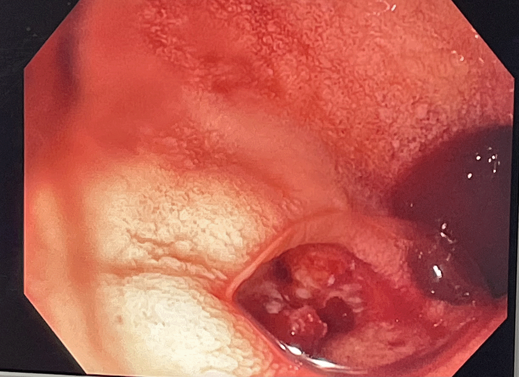
The patient was admitted to the intensive care unit with two peripheral intravenous lines, a urinary catheter, normal saline infusion, and a bolus of omeprazole (80 mg), followed by continuous infusion at a rate of 2 mg/hour. Additionally, he received a transfusion of 2 units of packed red blood cells. After stabilization of the hemodynamic status without the need for catecholamines, a diagnostic esophagogastroduodenoscopy was performed, revealing a large ulcer at the anterior wall of the first part of duodenum with active bleeding, classified as Forrest Ib (Figure 1). Adrenaline (1/1000 dilution) was injected for chemical hemostasis, and two clips were applied to achieve hemostasis.
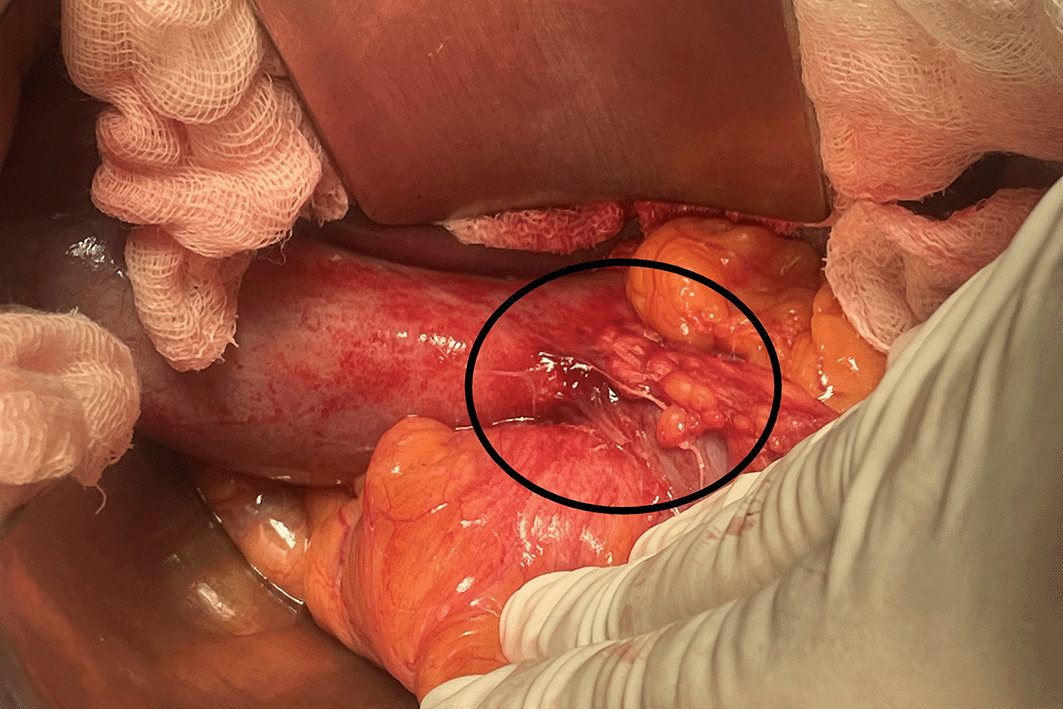
The patient remained stable for 17 hours but subsequently experienced a recurrence of hematemesis and developed tachycardia and hypotension (blood pressure 80/50 mmHg) with a hemoglobin level of 6.1 g/dL.Catecholamines were initiated, and the patient was taken to the operating room for an exploratory laparotomy. A midline incision revealed features of cholecystitis, with the gallbladder neck adherent to the anterior aspect of the duodenal bulb (Figure 2).
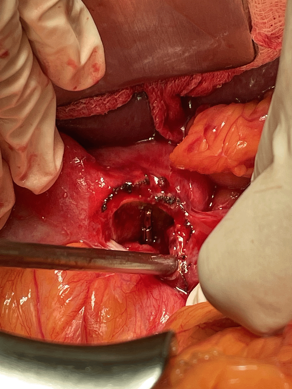
The release of the gallbladder from the duodenal bulb revealed the presence of an approximately 1cm ulcer with a visible vessel (Figures 3, 4). Cholecystectomy was performed along with en bloc resection of the ulcer and gallbladder. The source of bleeding was identified as the cystic artery (Figure 5). The procedure concluded with pyloroplasty and truncal vagotomy.
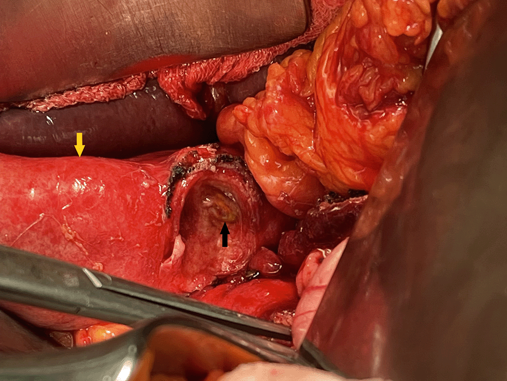

Catheterization of the cystic artery (yellow arrow) through a venous catheter across the ulcer (white arrow).
The evolution was favorable with no recurrence of hemorrhage, along with stabilization of hemodynamic status and hemoglobin levels. The patient was discharged on postoperative day 10. Postoperative follow-up was favorable with no recurrence of hemorrhage after 1 year of follow-up.
We present an exceptionally rare case of gastrointestinal bleeding manifested as hematemesis, related to the erosion of the cystic artery caused by a cholecysto-duodenal fistula and an ulcer on the anterior aspect of the bulb. The study is limited by the patient’s postoperative demise, preventing subsequent follow-up.
Cholecystoenteric fistula is a rare complication of biliary lithiasis, with autopsy-reported incidences ranging from 0.1% to 0.5%, and an occurrence of 1.2% to 5% among cases undergoing cholecystectomy.2 This complication is more prevalent in women, typically around the age of 60,2 and the cholecystoduodenal type is the most frequently observed, followed by cholecystocolonic and cholecystogastric fistulas.3
Cholecystoenteric fistulas form when the obstructed cystic duct undergoes repetitive inflammation, leading to the adherence of the gallbladder to an adjacent organ.4,5 Approximately 91% to 94% of internally formed biliary fistulas occur spontaneously due to the presence of stones in the biliary tract, with gastric ulcers ranking as the second most common cause. Other potential causes include tumors, biliary abscesses, and echinococcus cysts.6 Most patients present with general features of gallstone disease, but less commonly, patients may also exhibit cholangitis, severe upper gastrointestinal bleeding, or gallstone ileus.
The cholecystoduodenal fistula is rarely associated with gastrointestinal bleeding. Invasion of the cystic artery by a duodenal ulcer can lead to massive bleeding, while a gallstone can cause erosion of the same artery.2 Severe gastrointestinal bleeding from a cholecystoduodenal fistula is uncommon, as reported by Park et al., who conducted a literature review and identified only 11 case reports.7 Gallstones were the primary cause, with gastric ulcers as the etiology in only two cases. Endoscopic findings varied, ranging from ulcers to fistulous openings and bleeding of unknown origin. Although endoscopic hemostasis was attempted in four cases, surgical intervention was necessary in all cases, consistent with our case report.
The rarity of this condition, despite the high incidence of cholecystitis, could be explained by the more frequent occurrence of cystic artery thrombosis due to nearby inflammation.8
Bleeding from the biliary tract typically occurs into the duodenum, leading to hematemesis and/or melena in patients.9 In cases of upper gastrointestinal bleeding with inconclusive endoscopy results, consideration should be given to a cholecystoduodenal fistula with a bleeding marginal ulcer.9 In a hemodynamically stable patient, a CT scan is required to establish the diagnosis.
Endoscopic ultrasound can detect clots in the cystic duct and the gallbladder lumen, indicating potential bleeding from the gallbladder. A definitive diagnosis is often achieved through selective hepatic angiography, which can reveal a pseudoaneurysm or active bleeding from a nondilated cystic artery. This diagnostic method is particularly valuable because it allows for subsequent selective embolization.10
Angioembolization serves as an alternative treatment in some centers for identifying and potentially stopping active bleeding from a fistula that cannot be managed with endoscopic hemostasis. If angioembolization is performed, it should be followed promptly by surgical intervention.11 This is essential as the clinical presentation of bleeding caused by gallstones might indicate a more complex gallbladder condition than initially suspected.12
Cholecystoduodenal fistula leading to severe upper gastrointestinal bleeding is exceedingly rare.9 Most case reports indicate that the bleeding source is typically the erosion of the cystic artery, either by a duodenal ulcer or gallstones. Managing bleeding cholecystoduodenal fistulas usually requires surgical resection of the fistula and repair of the duodenal perforation due to significant bleeding from the cystic artery, which is unlikely to be resolved by conservative methods. In our case, despite the use of diluted adrenaline injections and the placement of hemostatic clips, the patient continued to experience active bleeding.6
While laparoscopic methods are often employed due to advanced surgical techniques, open operations are preferred in cases involving anatomical distortion and severe inflammation.
We present a unique case of massive bleeding from the cystic artery eroded by a duodenal ulcer through a concurrent cholecystoduodenal fistula.
Given the rarity of cholecystoduodenal fistula as a cause of massive gastrointestinal bleeding, it is imperative for clinicians to be attentive to certain signs that can facilitate the differential diagnosis. A thorough consideration of medical history remains essential, as a history of gallstone disease can guide the diagnosis.
Written informed consent to publish this case and associated images was obtained from the patient.
Wassim dziri: Conceptualization, Writing – Original Draft Preparation
Med Dheker TOUATI: Writing – Original Draft Preparation
Nadhem khlifi: Writing – Original Draft Preparation
Rahma yousfi: Conceptualization, Writing – Original Draft Preparation
Sana Landolsi: Conceptualization, Writing – Review & Editing
Faouzi CHEBBI: Supervision, Writing – Review & Editing
| Views | Downloads | |
|---|---|---|
| F1000Research | - | - |
|
PubMed Central
Data from PMC are received and updated monthly.
|
- | - |
Provide sufficient details of any financial or non-financial competing interests to enable users to assess whether your comments might lead a reasonable person to question your impartiality. Consider the following examples, but note that this is not an exhaustive list:
Sign up for content alerts and receive a weekly or monthly email with all newly published articles
Already registered? Sign in
The email address should be the one you originally registered with F1000.
You registered with F1000 via Google, so we cannot reset your password.
To sign in, please click here.
If you still need help with your Google account password, please click here.
You registered with F1000 via Facebook, so we cannot reset your password.
To sign in, please click here.
If you still need help with your Facebook account password, please click here.
If your email address is registered with us, we will email you instructions to reset your password.
If you think you should have received this email but it has not arrived, please check your spam filters and/or contact for further assistance.
Comments on this article Comments (0)