Keywords
Metastasis, Follicular Carcinoma, Thyroid, Cutaneous, Dermoscopy
Metastasis, Follicular Carcinoma, Thyroid, Cutaneous, Dermoscopy
Follicular thyroid cancer (FTC) is the second most common differentiated thyroid cancer, accounting for approximately 10–15% of all thyroid cancers.1 In 1997, a review of the English literature was conducted, and only 30 cases of Metastasis of Follicular Carcinoma of the Thyroid to the skin were documented.2 The most common location in the head and neck area.3
Poor prognosis and mortality rates are common for distant metastasis, such as to the lungs and bone, and disease-specific mortality is around 40% at a median follow-up of 5 years;1 however, no data are available for prognosis and mortality of metastasis to the skin. Here, we present a rare case of Metastatic Follicular Carcinoma of the Thyroid using dermoscopic, radiographic, and histopathological images.
A 49-year-old Southeast Asian woman with a past medical history of diabetes, hypertension, hyperlipidemia, and follicular thyroid cancer, which was fully removed surgically in 2019 in a complete thyroidectomy, shortly followed by one session of radioactive iodine 131, demonstrated no evidence of local or distant metastatic lesions, and presented with a suspicious solitary lesion that was noted on the anterior side of the neck. The lesion was originally a small macule that was black to brown in color and grew to become a red papule over the previous 4 months, coinciding with almost 18 months after her radioactive iodine session.
She is not currently on any immunosuppressants.
Family history was notable for a skin lesion on the father’s back, and surgery was performed. The patient was unaware of the histopathology results, but was told that it was benign in nature.
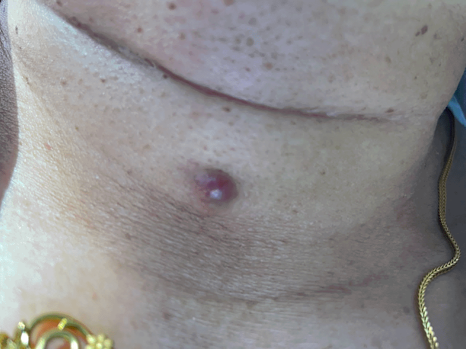
On further examination the lesion was an erythematous papule, 0.9 × 0.8 cm and blanchable in the center, it was non-tender to touch, and the lymph nodes were non-palpable during the examination (Figure 1).
Examination using a dermatoscope revealed arborizing vessels on the periphery, with a pink structureless area in the center (Figure 2).
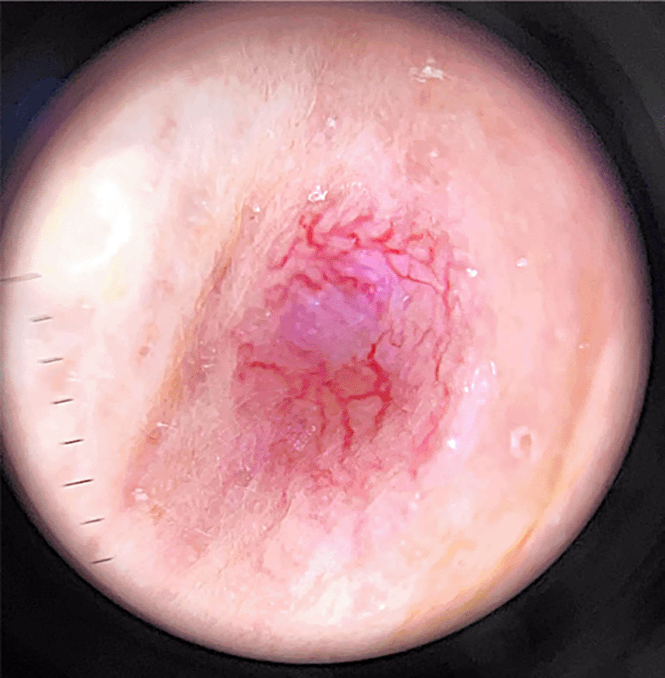
Absence of features in the center.
A 4 mm punch biopsy from the center of the lesion was performed with a clinical differential diagnosis of a basal cell Carcinoma and a Hidradenoma.
Laboratory tests revealed a steady rise in thyroglobulin levels from 1.5 ng/ml to 15 ng/ml, 6 months and 1 month prior to the visit, respectively, with a low TSH level at 0.01 mlU/L on the same visits.
The biopsy results revealed a normal epidermis with an underlying dermal tumor that was rich in follicles with colloids (a T3, T4 precursor) and was strongly positive for Thyroglobulin and Thyroid transcription factor-1 (TTF-1) on immunohistochemistry. Highly suggestive of follicular carcinoma of the thyroid metastasis (Figure 3).
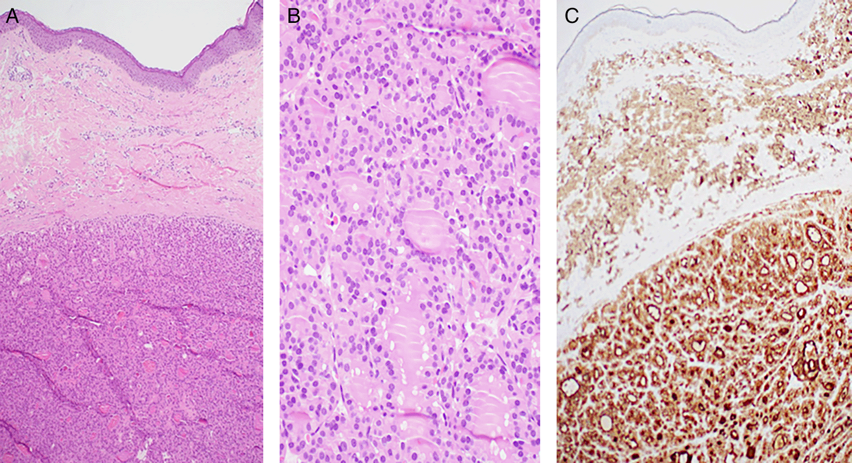
A, B, hematoxylin-eosin stain; magnification; C, Thyroglobulin and Thyroid transcription factor-1 immunohistochemistry.
After informing the patient of the diagnosis, she was referred to the thyroid multidisciplinary team (MDT).
The MDT decided to initiate therapeutic and diagnostic radioactive Iodine-131 therapy for thyroid cancer.
48 hours after administration of Iodine-131, 1093 megabecquerels (MBq), the scan demonstrated a small remnant of thyroid tissue in the thyroid bed. Single-photon emission computed tomography (SPECT/CT) imaging revealed focal uptake corresponding to a 5 mm diameter subcutaneous nodule in the midline of the neck, 1 cm below the lower margin level of the thyroid cartilage (Figure 4). The patient will continue to receive radioactive Iodine-131 therapy as decided by the thyroid multidisciplinary team.

Follicular thyroid cancer (FTC) is the second most common differentiated thyroid cancer, accounting for approximately 10–15% of all thyroid cancers.1 In 1997, a review of the English literature was conducted, and only 30 cases of metastasis of follicular carcinoma of the thyroid to the skin were documented.2
The rarity of this disease and its cutaneous presentation may present as a difficulty when encountered in the clinical setting, history, and clinical examination may not be sufficient to confidently verify the diagnosis and hence a low threshold of secondary diagnostics may be needed, metastatic lesions are even more difficult to diagnose as they can have a variable presentation; however, when the clinical and even dermoscopic appearance do not fit with diseases typically seen in the dermatology realm, then this should evoke further investigations. Some dermoscopic structures are shown (Figure 2) may have been suggestive of a BCC However, the features seen in arborizing vessels are nonspecific and arise in many other tumors,4 and with the increasing use of dermoscopy in various practices, more patterns will eventually be recognized, both for rare and common disorders. However, we could not identify any articles describing the dermoscopic findings of metastasis of follicular carcinoma of the thyroid to the skin. We hope that the images we shared and the findings described could facilitate the identification of such tumors in the future.
Patient consent statement: The authors obtained written consent from patients for their photographs and medical information to be published in print and online and with the understanding that this information may be publicly available. Patient consent forms were not provided to the journal but were retained by the authors.
| Views | Downloads | |
|---|---|---|
| F1000Research | - | - |
|
PubMed Central
Data from PMC are received and updated monthly.
|
- | - |
Provide sufficient details of any financial or non-financial competing interests to enable users to assess whether your comments might lead a reasonable person to question your impartiality. Consider the following examples, but note that this is not an exhaustive list:
Sign up for content alerts and receive a weekly or monthly email with all newly published articles
Already registered? Sign in
The email address should be the one you originally registered with F1000.
You registered with F1000 via Google, so we cannot reset your password.
To sign in, please click here.
If you still need help with your Google account password, please click here.
You registered with F1000 via Facebook, so we cannot reset your password.
To sign in, please click here.
If you still need help with your Facebook account password, please click here.
If your email address is registered with us, we will email you instructions to reset your password.
If you think you should have received this email but it has not arrived, please check your spam filters and/or contact for further assistance.
Comments on this article Comments (0)