Keywords
GBR, The sausage technique, Anorganic bovine bone mineral, Horizontal bone augmentation.
GBR, The sausage technique, Anorganic bovine bone mineral, Horizontal bone augmentation.
Alveolar Bone resorption is an inevitable consequence after tooth extraction1 as a result of the lack of mechanical load transmitted by the teeth to the bone.2 A noticeable reduction of the alveolar bone width has been observed within the first six months.3 However; the bone continues to resorb over time.3 The bony defects could be categorized as horizontal, vertical, or both. This may help clinicians to choose the proper treatment modality to manage the deficient ridge.4 The class IV alveolar bone atrophy according to the Cawood-Howell classification commonly referred to as the knife edge ridge represents a unique situation with an adequate height but a deficient width. In such cases, lateral bone augmentation is essential before dental implant placement.5 Guided bone regeneration (GBR) has been regarded as an authoritative approach for managing horizontal defects.6 The concept of GBR uses graft materials as a scaffold to preserve the created space and a non-resorbable or resorbable membrane to exclude the growth of epithelium and connective tissue cells inward the defect. The use of resorbable membranes in GBR is well documented, and its application has increased widely to overcome the risks and complications associated with non-resorbable membranes.7 Despite that, the conventional GBR using resorbable membranes did not obtain the desired bone growth at the alveolar crest due to the collapse of the resorbable membrane that forces the graft material to migrate apically.5 Therefore, Urban and colleagues proposed an improved technique to stabilize the graft material to the crest by modifying the collagen membrane fixation method.5 The original sausage technique combines autograft chips with anorganic bovine bone mineral (ABBM) particles to provide the graft mixture with osteogenic properties.6 Although autogenous graft is considered the gold standard of graft materials, harvesting the bone may increase the morbidity of the donor site and postoperative complications. Furthermore, autograft has a high resorption rate, which can reduce its ability to sustain space for an extended period.4,8 The ABBM is derived from xenogenous sources and treated with heat and chemical products to produce a human bone-like structure. The ABBM has a low resorption rate, serving only as a scaffold with a prolonged space-maintaining feature.9 Several authors deemed that GBR using ABBM is a reliable method to augment horizontal alveolar bone atrophy.4,10,11 Thus, the purpose of this case report was to scrutinize the effectiveness of the sausage technique using ABBM without autograft chips radiologically in increasing the bone width at the crestal part of a posterior mandibular knife edge ridge, report implants primary stability in the healed ridge when using such graft materials, and present a detailed description of the surgical technique.
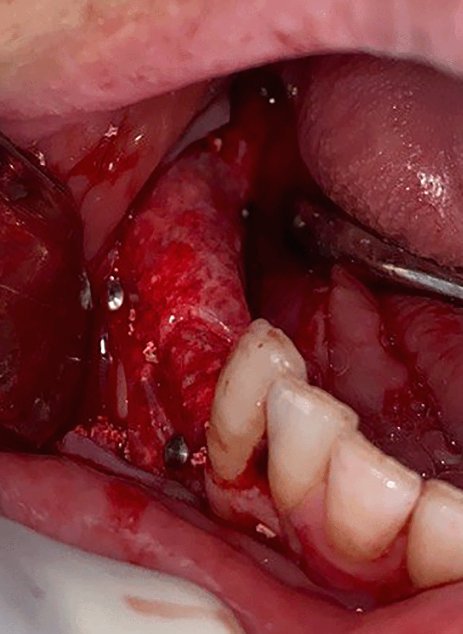
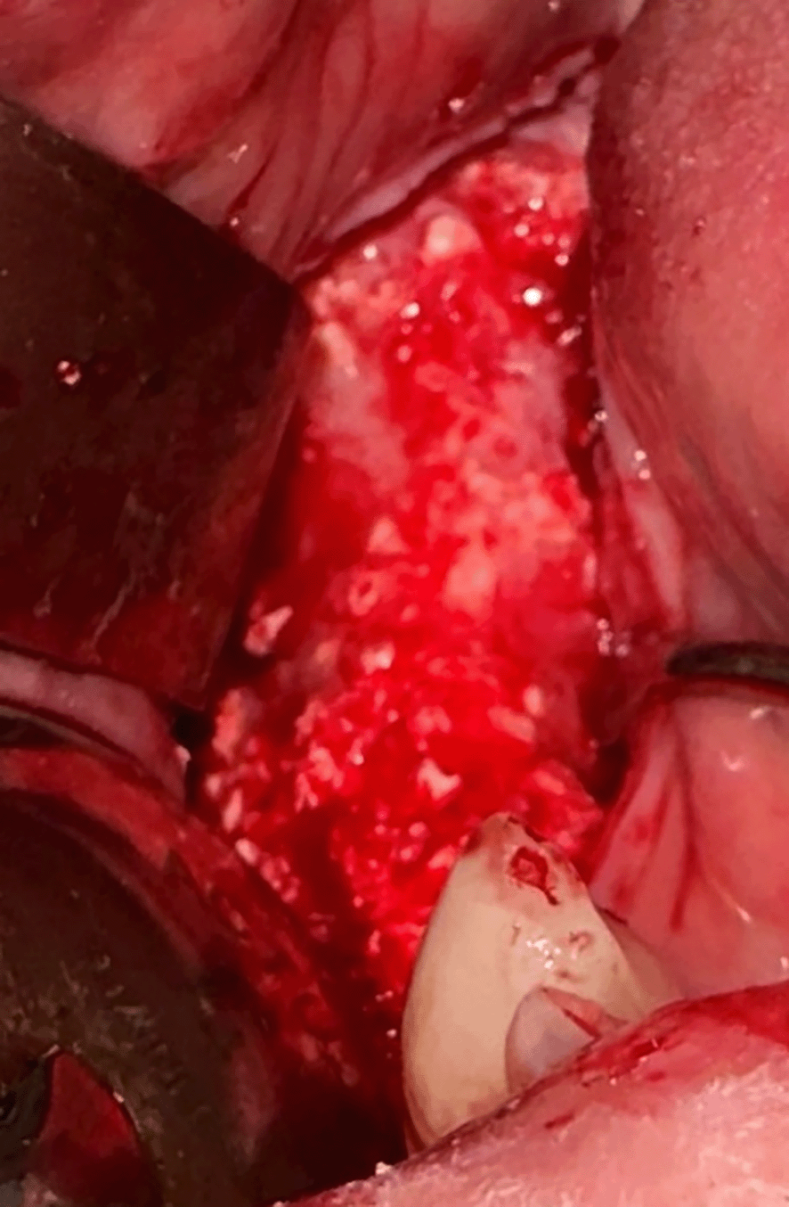
A healthy, non-smoker, thirty-year-old, unemployed, female patient attended the oral and maxillofacial surgery department of the faculty of dentistry at Damascus University to address the issue of missing teeth in the right lower quadrant. A comprehensive clinical examination and dental history evaluation determined that the premolars and molars had been absent for 5 years due to extensive caries. The edentulous area exhibited a keratinized tissue of 3 mm (Figure 1). The CBCT (Pax-i3D Green; VATECH, Gyeonggi Province, South Korea) revealed a knife edge ridge in the edentulous area, which was insufficient for dental implant placement without prior horizontal bone augmentation. The buccal-lingual measure was performed on multiple CBCT sectional views 2 mm below the crest using Ez3D Plus 3D Viewer Ver. 1.2.6.33 (VATECH Co., South Korea) (Figure 2), and the average of the bone width was calculated (Table 1). A staged GBR using the sausage technique was proposed as a treatment plan to rebuild the ridge to receive dental implants after nine months of healing. Before starting the surgical procedure, the patient was instructed to rinse for 2 minutes with 0.12% chlorhexidine solution. Inferior alveolar nerve and buccal nerve block were administrated with 2% lidocaine and 1:100000 epinephrine (Lidocaine 2% E-100, New Stetic SA, Guarne, Colombia). The safety flap5 was reflected, including a mid-crestal, two vertical, and intrasulcular incisions. The mesial vertical incision was made two teeth away from the intended site at the mesiobuccal angle of the right lateral incisor, and the distal one was made by extending the crestal incision toward the anterior border of the ascending ramus. An additional 3-4 mm hokey stick-like vertical incision was made at the mesiolingual aspect of the right canine to aid in advancing the lingual flap. A periosteal elevator was used to reflect a full-thickness flap buccally and lingually to expose the deficient ridge. The lingual flap was advanced by separating the mylohyoid muscle attachments from the inner part of the lingual flap without separating them from the bone. A curette was used to clean the soft tissue debridement (Figure 3). Bone decortication was performed using a round surgical bur to procure blood supply to the graft particles (Figure 4). A porcine pericardium membrane (Jason® membrane; 20×30 mm; Botiss Biomaterials GmbH, Zossen, Germany) was secured to the lingual side using two titanium pins (titan pin set, botiss biomaterials GmbH, Zossen, Germany). The xenograft particles (Cerabone®; particles sized 1-2 mm; Botiss Biomaterials GmbH, Zossen, Germany) were hydrated with saline and packed into the defect at an appropriate volume (Figure 5). The membrane was then molded over the graft, affixed to the buccal bone with one pin distobuccally, stretched mesially to push the graft particles towards the ridge, and stabilized using another pin. The membrane margin was lifted at the area between the former buccal pins, filled with an additional quantity of graft particles that were pushed coronally toward the crest. lastly, the membrane was again stretched apically, and a final pin was placed between the two buccal pins (Figure 6). The periosteum on the buccal flap was scored, ensuring proper flap mobility for tension-free closure. The flaps were closed with a (4.0) Nylon suture, using horizontal and simple interrupted mattress sutures (Figure 7). The patient received a prescription for amoxicillin 500 mg, four times daily for seven days, ibuprofen 400 mg, three times daily for five days, and chlorhexidine 0.12% for rinsing, starting 24 hours post-operatively and lasting for ten days. The augmented site was evaluated after 3, 5, 7, and 10 days. The incisions healed normally without any complications. There was no wound dehiscence or membrane exposure and the sutures were removed on the tenth day after the procedure. Nine months later, another CBCT image was acquired, and the ridge was re-measured as described earlier (Figure 8). The bone gain outcome is shown in Table 1. In the second surgical procedure, a full-thickness flap was reflected, as previously described (Figure 9), the clinical appearance shows a thick periosteal lining the grafted ridge with more difficulty in raising the flap and good incorporation of the graft particles into the newly formed bone. The unincorporated graft particles were removed using a curette. Two implants sized 3.5×10 and 4×8.5 (AnyOne®; MegaGen implant, Daegu, Korea) were placed at the site of #44 and #46 respectively (Figure 10). Although the drilling protocol for implant placement was similar to that in pristine bone, the grafted ridge showed less resistance confronting the drills. The implant osteotomies were bleeding, revealing a living bone formation in the grafted site. There were no separation parts from the augmented ridge during drilling and the implants were placed with the handpiece and completed with the torque wrench. The insertion torque (using the torque wrench) and ISQ value (using MegaGen® ISQ device) were recorded (Table 2). The flap was closed with (4.0) silk sutures, utilizing interrupted sutures (Figure 11). The patient was prescribed the same medication regimen as after the first surgical procedure, and the sutures were removed after a week. Despite the high ISQ value for both implants, the author prefers to place cover screws and follow the delayed loading protocol.


| Before the augmentation | After nine months | |
|---|---|---|
| The average ridge width 2 mm below the crest | 3.58 mm | 7.9 mm |
| The bone gain | 4.32 mm | |


The bone growth can be noted.

| The implant at the site of #44 | The implant at the site of #46 | |
|---|---|---|
| The insertion torque value | 35 N/cm | 35 N/cm |
| The ISQ value | 76 | 78 |

The graft material, Jason membrane, and titanium pins are kindly provided by Botiss Biomaterials GmbH (Zossen, Germany).
The sausage technique enhances graft stability and prevents the collapse of the resorbable membrane. Urban and colleagues used a composite graft material consisting of autograft chips and ABBM particles in a ratio of 1:1 and they suggested examining the effectiveness of different bone substitutes with this technique.5 Bovine bone promotes bone formation by osteoconductive characteristics, providing a mechanical scaffold that maintains the created space and facilitates the migration and occupation of bone regeneration cells to form new bone.9 Long-term stability, decreased morbidity, and surgical complications are the main advantages of using xenograft bone.4 Jason membrane is a native collagen membrane derived from porcine pericardium with a slow degradation due to its natural cross-linked collagen fibers. It has a superior tear resistance property.12 The measurements of this report were conducted on the cone beam sectional view 2mm below the top of the crest and the average width was 3.58 mm and 7.9 mm before the treatment and after nine months of healing respectively with a horizontal bone gain of approximately 4.32 mm which was similar to the outcome found in a systematic review in which the bone gain was 4.4 mm in the horizontal direction when using xenograft particles with or without autogenous graft.4 Urban13 proposed that the sausage technique combining autograft and ABBM is more predictable in securing the graft on the crestal part of the alveolar ridge than the classic pouch technique. Nevertheless, a bone gain of 5.68 mm was obtained utilizing both techniques mentioned earlier in cases with horizontal defects.13 In a preclinical study in dogs, song and colleagues10 highlighted that there was no additional advantage in bone formation from adding autogenous chips to xenograft particles in treating horizontal defects. Even though all the ridges restored their original form, the bone formation was restricted beyond the ridge contour.10 Therefore, in the author’s perspective, Urban obtained greater bone gain due to using a larger membrane thus further graft materials were packed into the defect. It may be suggested that a large-sized membrane should be utilized with long defects. Steigmann,14 Amoian and colleagues15 as well as Hammerle and colleagues16 recorded 3.0364 mm, 3.37 mm, and 3.7 mm of bone gain respectively when using the classic GBR with particulate xenograft without autograft to augment deficient ridges in the horizontal direction. The superior result of this report could be elucidated by using the sausage technique rather than the classic GBR. In a study conducted by Mendoza-Azpurthe and colleagues,11 the average bone gain was 5.6 ± 1.35 mm when using xenograft particles and collagen membranes, it is worth mentioning that Mendoza-Azpurthe and colleagues11 measured the buccal-palatal width 5,7, and 11 mm below the crest using the CBCT,11 whereas the current report measurement was conducted 2mm apical to the top of the ridge. It was established that the greatest bone growth was gained apical to the crest as a result of the inability of the resorbable membrane to withstand at the crest and the apical relocation of the graft particles.5 Primary closure of the incisions, angiogenesis, stability of the graft particles, and blood clot, in addition to sustaining the created space in which bone will form, are the fundamental principles for accomplishing successful GBR.5 Thus without a proper membrane fixation, the stability and space maintained will be compromised resulting in poor bone growth outcomes at the crest.5 The sausage technique is a cumulative learning procedure with several critical steps to be aware of.5,13 To the best of the author’s understanding, the final push of the graft particles toward the crest, stretching the membrane apically, and fixing with a pin or two between the distal-buccal and mesial-buccal pins are the most critical steps in this technique to achieve a stable and firmly fixed graft-membrane complex. This report’s main limitation was the absence of a histological study to define the bone structure at the augmented ridge. Nonetheless, earlier studies found a formation of new and organized bone between the graft particles similar to that in pristine bone.10,8,15 It is worth noting that the present report focused on the surgical phase and the horizontal bone growth outcome without considering the prosthetic phase after implant placement which represents another limitation. Further studies are encouraged with a controlled randomized clinical trial design to confirm the data of the bone gain with the sausage technique using ABBM and a collagen membrane in treating horizontal alveolar bone defects. In addition, comparative researches are highly required to assess the insertion torque and primary stability of the implants in the augmented ridges and their influence on the osseointegration of the implants and loading protocols.
Within the limitations of this case report, it could be concluded that the sausage technique using ABBM particles without autograft chips showed a great capability to maintain the created space, prevent the collapse of the collagen membrane, and acquire an adequate width at the crestal part of the alveolar bone.
| Views | Downloads | |
|---|---|---|
| F1000Research | - | - |
|
PubMed Central
Data from PMC are received and updated monthly.
|
- | - |
Provide sufficient details of any financial or non-financial competing interests to enable users to assess whether your comments might lead a reasonable person to question your impartiality. Consider the following examples, but note that this is not an exhaustive list:
Sign up for content alerts and receive a weekly or monthly email with all newly published articles
Already registered? Sign in
The email address should be the one you originally registered with F1000.
You registered with F1000 via Google, so we cannot reset your password.
To sign in, please click here.
If you still need help with your Google account password, please click here.
You registered with F1000 via Facebook, so we cannot reset your password.
To sign in, please click here.
If you still need help with your Facebook account password, please click here.
If your email address is registered with us, we will email you instructions to reset your password.
If you think you should have received this email but it has not arrived, please check your spam filters and/or contact for further assistance.
Comments on this article Comments (0)