Keywords
Antiphospholipid syndrome, retinal ischemia, anticoagulation
Antiphospholipid syndrome, retinal ischemia, anticoagulation
Antiphospholipid syndrome (APS) is a rare autoimmune disease that can be primary or secondary if associated with connective tissue disease (CTD), most commonly systemic lupus erythematosus (SLE). The clinical manifestations of APS vary widely. However, the most frequent spectrum of manifestations seems to be vaso-occlusive and obstetric manifestations. Vaso-occlusive manifestations vary according to the localization of the vascular occlusion. The vessels of all the organs can be involved at different frequencies. Ocular involvement is rare.
We report a case of retinal arterial thrombosis in the context of APS.
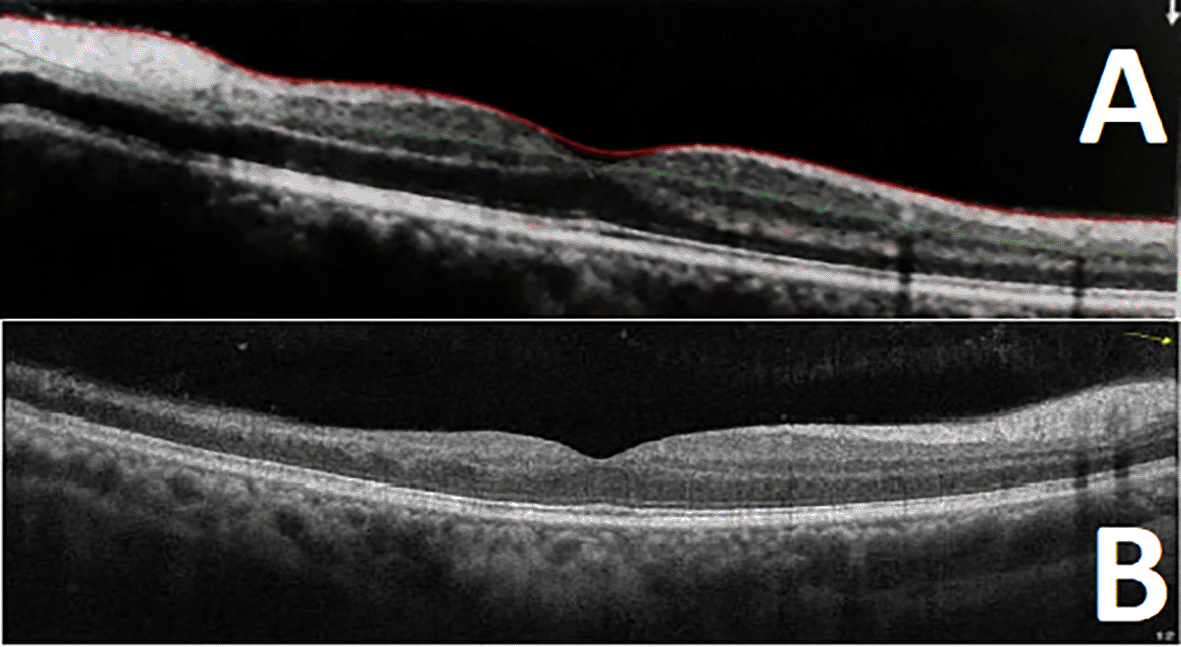
A 46-year-old patient with no previous medical history was admitted to the ophthalmology department for visual field amputation of the right eye, without other associated signs. On the fundus, the left side showed no abnormality, and the right side was the site of mixed arterial and superior temporal venous occlusion. Retinal angiography revealed mixed occlusion of the arterial and venous branches with ischemia of the superior temporal quadrant on the right and a retinal focus in the superficial inferior temporal region without occlusion on the left (Figure 1). On Optical Coherence Tomography, the left eye was normal, and the right eye was the site of ischemia of the superficial layers with alteration of macular microvascularization in the superior temporal quadrant (Figure 2). The rest of the physical and biological examinations were without particularity.
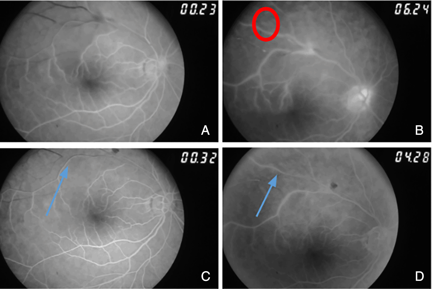
The initial etiological investigation revealed a cat scratch one month ago and a positive bartonella serology. The patient was treated with doxycycline and received 20 sessions of hyperbaric oxygen therapy. The evolution was marked by partial improvement in the right ischemic focus. Immunological work-up showed antinuclear antibodies to be positive at 160 and anti-B2GP11 positive with an IgG level of 118 UA/mL, IgM higher than 118 UA/mL and IgA at 101 UA/mL. Tests for anti-native DNAn, lupus anticoagulant and anticardiolipin antibodies were negative. The patient was administered effective anticoagulation therapy with good clinical outcome (Figures 1 and 2).
Antiphospholipid syndrome is a rare condition characterized mainly by vascular thrombotic and pregnancy accidents. Thrombotic events can occur in both arterial and venous vessels of all organs. Ocular involvement is considered among the rarest organs to be involved, affecting approximately 1% of patients.1 However, targeted studies revealed a higher prevalence of asymptomatic APS-associated ocular changes, ranging from 14 to 18%.2 Ocular involvement can be not only the inaugural manifestation but also the only clinical manifestation of APLS. In fact, vascular changes have been reported in patients with positive APS antibodies without fulfilling other clinical criteria to retain the diagnosis. This has been proven through a study of 13 patients with anti-cardiolipin antibodies and found abnormalities, such as retinal vasculitis in 60% of cases, vitritis, and retinal detachment.3 Ocular lesions can also occur in the context of catastrophic APS (CAPS), with poorer organ and worse overall prognosis.4
Posterior segment damage is the most frequent and severe4 in ocular ALPS. However, damage to other segments, as well as to visual pathways in the central nervous system, has been reported. Anterior segment involvement is rare and generally described as mild, with conjunctivitis sicca, conjunctival telangiectasias, microaneurysms, episcleritis, and limbal keratitis.3 These manifestations seem to occur more frequently in secondary APS due to the other inflammatory mechanisms related to the associated connective tissue disease.2 Visual pathways involvement consists mostly of ischemic events such as non-arteritic or arteritic ischemic optic neuropathy and central nervous system (CNS) infarctions along the visual pathway.5 All these manifestations can occur simultaneously especially with posterior lesions or isolated.
Posterior segment’s clinical signs lack specificity. Patients usually present with blurry vision, vision loss, red eye and eye pain, and headaches.2 It must be noted that even for arterial ischemia, asymptomatic cases have been described, which were found through systematic OCT screening.6 Presentation can be either acute, especially in arterial events, or progressive. Examination usually reveals vaso-occlusive diseases involving the retinal and choroidal vessels. Both the venous and arterial vessels may be involved simultaneously or isolated.7 C Fundus assessment is the core part of examination with specific mandatory imaging tools, such as retinal angiography and optical coherence tomography (OCT).
Venous disease has been described as either central retinal vein occlusion (CRVO), central retinal artery occlusion (CRAO), or branched retinal vein occlusion (BRVO).1 It can be the first manifestation of APS, leading to testing of patients with APS antibodies.8 In fact, in the ACR/EULAR 2023 classification criteria for APS, retinal vein thrombosis/occlusion is one of the clinical criteria for classifying patients with APS.9 Untreated vaso-occlusive retinopathies may lead to several complications such as neovascularization,10 vitreous hemorrhage, neovascular glaucoma, and tractional retinal detachment.11
Interestingly, and similar to our patient, retinal arterial occlusions do not usually occur at bifurcations, as observed in embolic situations.1 In fact, retinal areas that require the most oxygen are the internal plexiform layers and the internal segment of the photoreceptors. As a result, the choroidal vascular system is more developed in these regions, but the retinal thickness increase in the parafoveal region limits the diffusion of oxygen toward the retina from the choroid.12 In addition, because the visual resolution in the parafoveal region must be high, the parafoveal capillary system density in this region is reduced.12 Examination usually reveals acute retinal ischemia, as was the case in our patient. However, other funduscopic presentations have been reported, such as serpiginous Choroidopathy vitritis and vasculitis.13 Lesions can be unilateral or bilateral.14 Regarding treatment, there are no established guidelines for APS associated with pure ophthalmic manifestations. The only available recommendations suggest preventing subsequent events. Systemic anticoagulation, particularly with warfarin, has been linked to an increased risk of recurrence of vascular occlusions even at therapeutic dosages.1 Direct oral anticoagulants (DOACs) might be a suitable therapeutic alternative to some specific subgroups of patients with APS, but further studies are required to establish such conclusions, as data regarding the overall safety of DOACs in treatment compared to VKAs is still scarce.15 Immunosuppressive therapies are currently being tested. Some authors reported good outcomes with glucocorticoids.16 Rituximab use is increasing with few trials showing positive impact on antibodies titers and future events.17,18 This agent has also shown positive outcomes in CAPS cases when.18 Finally, hydroxychloroquine, an antimalarial drug with proven immunomodulatory effects that has been used in other CTDs such as SLE, seems to also have a good impact in APLS when prescribed in adjunction to anticoagulant agents.19
APS is a systemic disease that requires exhaustive assessment, especially for organ damage that might threaten functional and life prognoses. Ocular involvement appears to be one of the regular assessment targets. Although it is not frequent, it is difficult to diagnose and manage. Larger studies are required to establish guidelines on how and when to screen asymptomatic patients with APS for ocular damage, as well as on how to prevent and treat it.
| Views | Downloads | |
|---|---|---|
| F1000Research | - | - |
|
PubMed Central
Data from PMC are received and updated monthly.
|
- | - |
Provide sufficient details of any financial or non-financial competing interests to enable users to assess whether your comments might lead a reasonable person to question your impartiality. Consider the following examples, but note that this is not an exhaustive list:
Sign up for content alerts and receive a weekly or monthly email with all newly published articles
Already registered? Sign in
The email address should be the one you originally registered with F1000.
You registered with F1000 via Google, so we cannot reset your password.
To sign in, please click here.
If you still need help with your Google account password, please click here.
You registered with F1000 via Facebook, so we cannot reset your password.
To sign in, please click here.
If you still need help with your Facebook account password, please click here.
If your email address is registered with us, we will email you instructions to reset your password.
If you think you should have received this email but it has not arrived, please check your spam filters and/or contact for further assistance.
Comments on this article Comments (0)