Keywords
testicular artery, variation, renal artery, origin, duplication
This article is included in the HEAL1000 gateway.
testicular artery, variation, renal artery, origin, duplication
Typical and variant anatomy of the testicular arteries (TAs) or internal spermatic arteries should be carefully studied respectively to their laterality (side of appearance), because of their clinical role in testicular physiology and kidney and testicle surgery. Typically, the long and thin TA originates from the abdominal aorta (AA) anterolateral surface, at a distance of 2.5 to 5 cm distally to the renal artery (RA) origin, and above the inferior mesenteric artery origin.1 The TA supplies the perirenal fat, ureter, iliac lymph nodes, and the cremaster muscle.1–3 It is directed with the testicular vein (TV), retroperitoneal, on the psoas major muscle, enters the inguinal canal through the deep ring, and supplies the testicle.4 The right TA (RTA) originates anterior to the inferior vena cava (IVC) and lies posterior to the duodenum horizontal part, right colic and ileocolic arteries, root of the mesentery, and terminal ileum.1,2 The left TA (LTA) courses posterior to the inferior mesenteric vein, left colic artery, and descending colon.1,2 The right and left testicular veins (RTV and LTV) typically drain into the IVC, and the left renal vein (LRV), respectively.5
TA variants are not rare, as cadaveric studies have shown a prevalence ranging from 4.7% to 8.8%.5,6 The TAs may exhibit an arched course ranging from 1.7% to 20.3%.1 TA variants may be accompanied by TV alterations and the awareness of these venous anomalies may help surgeons accurately ligate abnormal venous anastomosis and avoid iatrogenic injury. TA variants may also coexist with RA alterations. Concerning the RAs, typically, a single RA emanates from the AA, on both sides.7 Frequent variants include the RA variant number7 among different ethnicities (4% in Malaysians, and 61.5% in Indians).8 The presence of multiple RAs is a well-documented phenomenon with a reported frequency of around 20%.7,8
The present cadaveric report explicitly describes the atypical origin and course of TAs along with an RA duplication and further discusses their clinical implications.
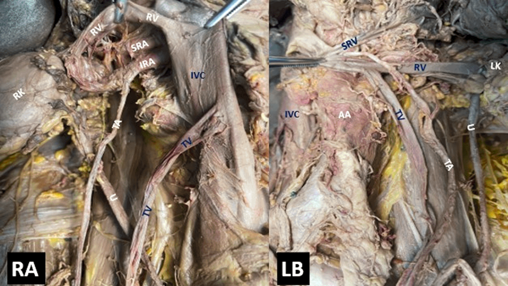
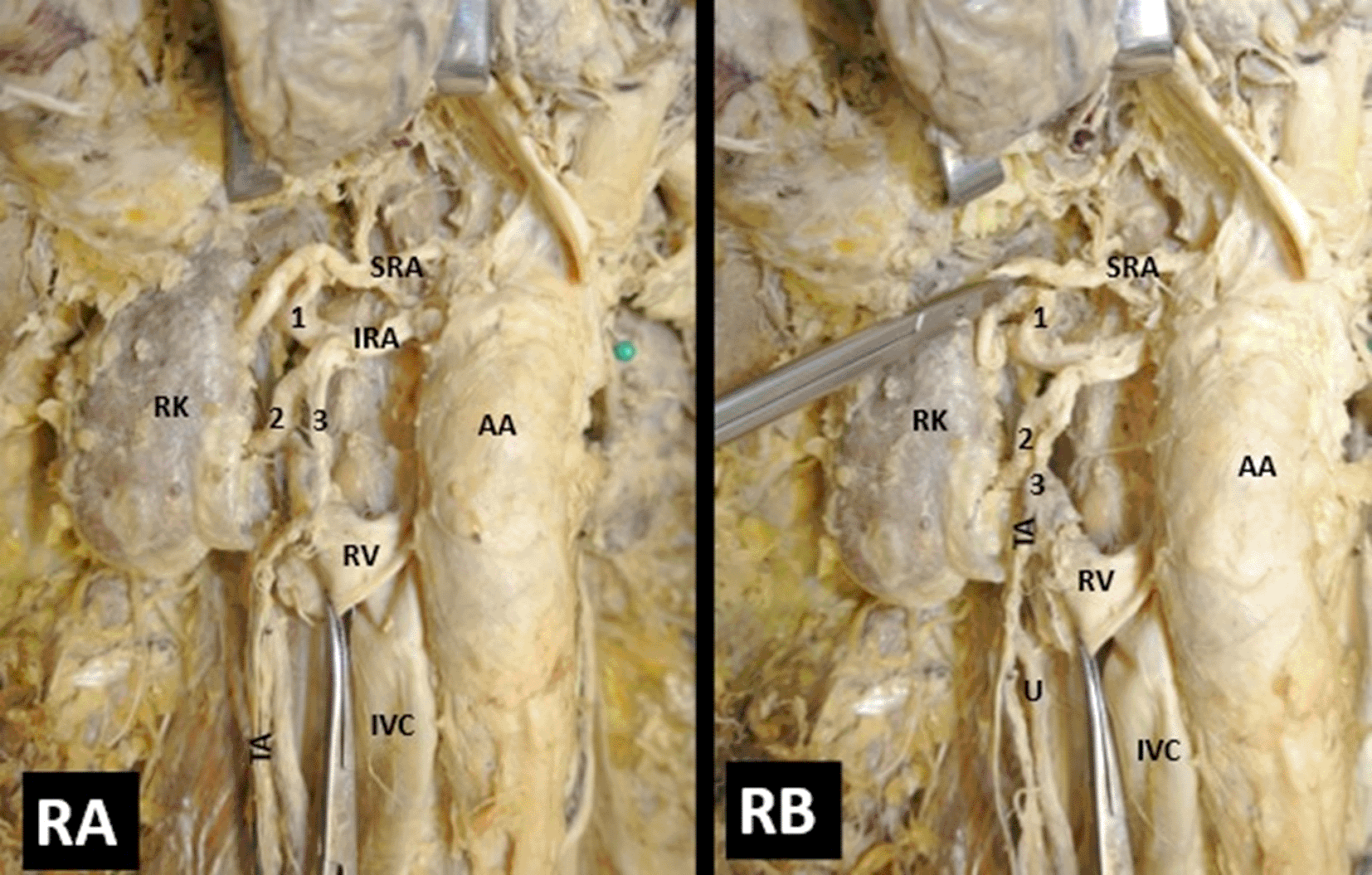
During a routine dissection of the IVC and AA branching patterns in two formalin-embalmed donated male cadavers 90-year- and 76-year-old, several variants of the TAs origin and course were identified, as coexisting with RA variants. The cadavers were donated to the Anatomy and Surgical Anatomy Department of the Medical School of the Faculty of Health Sciences of the Aristotle University of Thessaloniki, and the Anatomy Department of the Medical School of the National and Kapodistrian University of Athens, after signed informed consent. The Ethics Committees of the two Universities gave full approval for the study. Case 1: A right-sided RA duplication (RRA) (recorded as superior and inferior renal artery-SRA and IRA) with origin from the AA, coexisted with an RTA that originated from the IRA, and descended anteriorly to the ureter. The LTA typically originated from the AA proximal to the left RA (LRA) origin. The LTA coursed in front of the common drainage of the LTV and LRV to the IVC (Figure 1). Case 2: A bilateral duplication of the RA was identified and recorded as SRA and IRA (Figures 2,3). The right-sided SRA originated from the AA upper level and played the role of the accessory RA (ARA). The main perfusion of the right kidney was performed by the IRA which was further trifurcated into a superior polar artery (SPA), an inferior polar artery (IPA), and a TA. The RTA had an origin from the IPA and descended anterior to the RRV (Figure 2).

The current study describes the TA’s abnormal origin and course in coexistence with an RA duplication.
Understanding the developmental stages of arteries provides the basis for understanding morphological variants.5 Regarding the embryological progression of the gonads, which are drained by the mesonephric arteries originating from the AA lateral surface 2,9,10 there are nine pairs1–4,6,11,12 which can be categorized into a cranial group which consists of the 1st and 2nd mesonephric arteries that mature into the celiac trunk, the middle group which consists of the 3rd, 4th, and 5th arteries that develop into the RAs, and a caudal group which consists of the 6th, 7th, 8th, and 9th mesonephric arteries which become the TAs.2 An extra artery in the middle group will typically evolve into the ARA.2,4 If a cranial mesonephric artery does not regress, it will lead to a high-origin TA.1,2,6,12
Regarding the 1st cadaveric report, we hypothesize an additional mesonephric artery in the middle group on the right side led to the RRA duplication. On the left side, there was a persistent cranial lateral mesonephric artery which would explain the LTA’s abnormal origin. The LTA arched course anteriorly to the LRV and the LTV common drainage could be due to the kidney’s position and the higher vertebral level of the LRV compared to the LTA’s origin.1,2,12 This anterior course mimics an unusual variety of nutcracker phenomena.13 The RTAs’ atypical origin of both cadavers can be explained by a possible bifurcation of an additional artery from the middle group of the mesonephric arteries.1,4 One branch would drain the genital ridge and the other would supply the kidney. The TA would come from the former of the two branches.
In the 2nd cadaver, an extra pair of mesonephric arteries in the middle group would explain the RA bilateral duplication.2,4 On the right side, there was not only one more mesonephric artery that evolved into the ARA but also the other mesonephric artery bifurcation would explain the IRA duplication. TA variants are more common in males than females,2,6,12 and the LTA variants (20.7%) are more common than the RTA variants (12.7%).9
The TAs typically originate from the AA anterolateral surface, just below the RA origin, at the 2nd or 3rd lumbar vertebrae level (L2 or L3).2,9 The most common variants include alterations in the level, source of origin, number, and course of arteries. TA’s origin can be more proximal than the RA and RV.14 TA’s abnormal origins include the right subcostal artery, the suprarenal artery, the celiac trunk, the right inferior epigastric artery, the superior mesenteric artery (SMA), the inferior mesenteric artery (IMA), the right common iliac, and right external iliac artery.1–3,9,15 TA may originate at the level of the SMA, level of the RA or above,9 in a common trunk with the IMA, and an ARA,15 or in a common trunk with the LTA and RTA from the AA.16 Atypical origins proximal to the L2/L3 level are classified as high-origins, and distal to the L2/L3 or the IMA level are considered low-origins.2,9 The highest origin for the TA was identified at the left side, from the thoracic aorta, proximal to the inferior phrenic artery,10 or above the aortic diaphragmatic hiatus (level of the 12th thoracic vertebra).4 An LTA was identified originating from the LRA in 5.26% and the RTA from the RRA in 2.63%.9 The TA may originate from the main RA, an ARA, or a polar RA.1–4,6,9,15,16 The most common TA variant origins were found in association with renal vessels. Regarding their number, double TA was the most common.1 Concerning the combined TA variants with RA alterations, in the current report, Naito et al.17 recorded the coexistence of multiple RAs (8 in total, arising from the AA) with a bilateral renal origin for the TAs, and Notokovich18 reported gonadal arteries of renal origin in 15%. The number of RAs can be up to six, the most common being 2RAs, similar to the current cadaveric cases. Multiple RAs are reported as the most common renal vascular variant by Satyapal et,19 with an average incidence of 28.2% (range 9-76%). Two or double RAs are reported to make up 89.5% of all multiple RAs.8 Merklin and Michels20 reported incidences for 2RAs, 3RAs, and 4RAs unilaterally of 20%, 2%, and 0%, respectively. Ozkan et al.21 and Sampaio and Passos22 reported an incidence of 0.2% and 0.4% for 4RAs, respectively.
Concerning the TA variant course, the most common variation was an arched course over the ipsilateral RV. Occurrence of the arched TA has been noted on both sides but with a predilection for the left side. An arched RTA had an occasional retrocaval course.1 Kayalvizhi et al.1 concluded an incidence ranging from 1.7% to 20.3% for the arched TA. The arched LTA course over the LRV, similar to the current two unilateral (left and right side) cases is particularly important as it can compress the LRV and impede the venous return from the left kidney and testis. Consequently, varicocele, orthostatic proteinuria, and hematuria can occur. All the observed variants in these two cases could be due to aberrations in the development of visceral vessels controlled by a common molecular regulator.23 Most of these variants are part of the age of the cadaver. Our findings reiterate the old dictum that if an anomaly is detected in one vessel, there are chances of encountering other related anomalies as well. Clinicians need to check for the presence of an arched TA when performing renal surgery, as any injury to this artery might cause testicular atrophy.24
TAs variants although occur frequently, are accidentally discovered intraoperatively.9 The knowledge of the origin and the course of the TAs is clinically important for kidney and testicle surgery and can prevent intraoperative and postoperative complications.1–4,6,7,9,16 Surgeons should consider the RAs’ origin and path when they operate proximal to the renal pelvis or retroperitoneal and particularly target their usage in case of cryptorchism.1,4 The TAs’ abnormal origin and course around the kidney may lead to unexpected serious intraoperative hemorrhage.1,6,9 Radiologists should consider the possibility of variants when performing an angiography with indications, such as recognition of the arterial branches and planning the surgical procedure.1–6,9,15 If any abnormal origin or path of an artery is perceived via angiography, further examinations like magnetic resonance imaging (MRI) and phlebography should be performed to reduce the possibilities of intraoperative and postoperative complications.1 Furthermore, knowledge of the numerous variants of the renal vessels is surgically important in cases of repair renal stenosis, renal infarctions, and renal hypertension, as well as other urological surgeries.1,9,12 Renal arterial hypertension may occur by RA compression and could progress to renal ischemia.1,4,15 The variations could also affect the perfusion and function of testicles.3,4,5 Knowledge of these variations can aid in the prevention of testicular atrophy.1,3,5 These variations could also have serious implications regarding the thermoregulation of the male reproductive tract and consequently have an impact on spermatogenesis.3–5 The lower frequency of the RTA variants compared to the LTA provides an advantage in the usage of the right kidney in transplantations.4 Kidneys with multiple vessels are in high demand for transplantation due to their ability to help more patients who require a transplant.4 Furthermore, a TA originating from the RA may disturb the testicles’ drainage when embolizing renal tumors.2,9,14,25 A case report recorded that during the embolization of a vessel in a renal tumor, an infarction occurred because of an abnormal TA origin from the RA, despite attempts to prevent it.25 Another similar case of testicular infraction has been reported which proceeded from an embolization of a renal angiolipoma.14 Because of the TA arched and anterior course with the RV, there is a potential for RV compression, as seen in the current case description, causing a varicocele secondary to vascular compression.1,2,15 RV compression could also cause venous hypertension, proteinuria, and orthostatic hematuria secondary to the tortuous vascular course.2,15 The TA’s abnormal course could compress the ureter and potentially lead to hydronephrosis.1,4,6
In the current cadaveric report, upon performing simultaneous dissections in different locations, similar variants were discovered in the TAs origin and course with RAs duplications in two cadavers. Surgeons should consider the origin and path of the TAs and the possible coexistence of the RAs alterations to prevent intraoperative and postoperative complications.
The cadavers were donated to the Department of Anatomy and Surgical Anatomy, School of Medicine, Aristotle University of Thessaloneiki and the Anatomy Department of the Medical School of the National and Kapodistrian University of Athens through the Body Donation Program of the universities. The donor signed an informed consent before death giving consent to medical, education and research projects.
| Views | Downloads | |
|---|---|---|
| F1000Research | - | - |
|
PubMed Central
Data from PMC are received and updated monthly.
|
- | - |
Provide sufficient details of any financial or non-financial competing interests to enable users to assess whether your comments might lead a reasonable person to question your impartiality. Consider the following examples, but note that this is not an exhaustive list:
Sign up for content alerts and receive a weekly or monthly email with all newly published articles
Already registered? Sign in
The email address should be the one you originally registered with F1000.
You registered with F1000 via Google, so we cannot reset your password.
To sign in, please click here.
If you still need help with your Google account password, please click here.
You registered with F1000 via Facebook, so we cannot reset your password.
To sign in, please click here.
If you still need help with your Facebook account password, please click here.
If your email address is registered with us, we will email you instructions to reset your password.
If you think you should have received this email but it has not arrived, please check your spam filters and/or contact for further assistance.
Comments on this article Comments (0)