Keywords
Diagnosis, intestinal tuberculosis, internal validation, laboratory panel, scoring system
This article is included in the Global Public Health gateway.
Diagnosis, intestinal tuberculosis, internal validation, laboratory panel, scoring system
World Health Organization (WHO) reported through the Global Tuberculosis Report 2022 that Indonesia is in second place with the highest burden of tuberculosis in 2021.1 Tuberculosis (TB) is an infectious disease caused by Mycobacterium tuberculosis (MTB). These bacteria primarily infect the lung tissue but can also spread to other body parts. When TB affects areas outside the lungs, it is referred to as extrapulmonary tuberculosis (EPTB). The prevalence of EPTB worldwide is 16%, while in Indonesia is 10–19%.2 Intestinal tuberculosis (ITB) as part of EPTB has a prevalence of around 3–16%.3–5 The proportion of TB colitis in Dr. Cipto Mangunkusumo National Central General Hospital, reported by Suparmin, was 8/60 (13.3%).6
Although pulmonary TB (PTB) is more common, ITB can present with vague, non-specific gastrointestinal symptoms, such as abdominal pain, diarrhea, weight loss, and bloating. It remains a diagnostic challenge due to its overlap with other gastrointestinal diseases, such as Crohn’s disease (CD) and colon cancer. The management of ITB, CD, and colon cancer is known to be very different.7–10 While ITB can be cured with anti-TB therapy, CD typically persists and may relapse. Misdiagnosis of CD as ITB results in unnecessary anti-TB therapy, increased risk of toxicity, and late treatment of the primary disease. Otherwise, misdiagnosis of ITB as CD results in fatal ITB.7–14 Misdiagnosis of ITB as colon cancer can lead to unnecessary surgical interventions, such as hemicolectomy, which could have been avoided with accurate diagnosis. Misdiagnosing colon cancer as ITB can delay necessary cancer treatments, potentially worsening patient outcomes. Both conditions require different treatment approaches, and incorrect treatment due to misdiagnosis can lead to increased morbidity and even mortality.15,16
The routine diagnosis of ITB is based on clinical manifestations, Ziehl-Neelsen (ZN) acid-fast bacterial (AFB) staining examination of stool, stool culture, colonoscopy, and histopathology.4,14,17–19 Each of these ITB diagnostic examinations has advantages and limitations. Considering that some laboratory parameters often available in healthcare facilities and the impact of inflammation in ITB produces an abnormal immune response in leukocytes and differential count, erythrocyte sedimentation rate (ESR), neutrophil to lymphocyte ratio (NLR), monocyte to lymphocyte ratio (MLR), interferon-gamma (IFN-γ), adenosine deaminase (ADA), and the antimicrobial protein human beta defensin-2 (HBD-2)20–26 have proven to be valuable in differentiating ITB and other gastrointestinal diseases. Furthermore, using non-invasive laboratory samples, especially stool and blood, has become a pivotal area in modern diagnostics. These biological specimens offer significant advantages in patient comfort, non-invasive, ease of sample collection, and the potential for early detection of various diseases.27
In recent years, numerous studies have been attempted to create model panels that could improve the accuracy and success rate of ITB management,28–33 but they used different diagnostic models and scoring systems, some of which are not user-friendly, as they may need a calculator or a computer, and cannot be applied in healthcare facilities that lack access to colonoscopy and histopathology services. Hence, in this study, we aimed to develop and validate a non-invasive laboratory panel using a scoring system to differentiate ITB from other gastrointestinal diseases.
We recruited 191 adult subjects > 18 years who underwent colonoscopy at Dr. Cipto Mangunkusumo National Central General Hospital from November 2020 to December 2022. All suspected subjects fulfilled the 3 of 4 main clinical criteria: (1) weight loss, (2) non-specific abdominal pain, (3) fever, (4) diarrhea or chronic constipation, and 1 of 3 additional histories: (1) pulmonary tuberculosis history, (2) active pulmonary tuberculosis with ongoing anti-tuberculosis therapy (ATT) < 3 months, (3) contact with positive TB patient. All diagnosed subjects with ITB fulfilled the 2 of 4 criteria or positive stool TB PCR only: (1) summary of colonoscopy result showed ITB suspect, (2) histopathological result showed lesions in the submucosa or submuscular include epithelioid cells, Datia Langhans cells, lymphocytes at the edge of the granuloma, and caseous necrosis, (3) positive stool TB PCR, and (4) subjects showed a good response to ATT with clinical manifestation consistent with active TB. In addition, all subjects who were ongoing ATT > 3 months and post-treatment with ATT < 6 months are excluded from this study. Forty-eight subjects dropped out due to incomplete specimens. There were 143 subjects with complete specimens that could be analyzed.
The clinical data included age, gender, abdominal pain, chronic diarrhea, constipation, blood in stool, mucus in stool, weight loss, fever, night sweats, appetite loss, cough, TB contact, and TB history. Laboratory data included stool and blood examinations. For stool sample collection, participants provided a stool collection kit consisting of a red screw-capped collection tube, ice pack, plastic, plastic gloves, and a plastic spoon. Stool samples were collected using a plastic spoon in the tube. The tube was capped and the sample was sent to the laboratory with ice pack. Stool samples were then aliquoted and stored at -20 °C until processing. For blood sample colection, venous blood was taken from each subject and collected in 1 EDTA tube, 3 Heparin tubes, and 1 serum tube.
Stool examination included TB PCR, IFN-γ, and HBD-2. Stool specimens were subjected to direct extraction via lysis bead-based method using AllPrep® PowerFecal® DNA/RNA Kit (Qiagen, Hilden, Germany) according to the manufacturer’s instruction. The extracted DNA was then used as template DNA for the AccuPower® MTB&NTM Real-Time PCR Kit (Bioneer, Daejeon, South Korea). This assay relies on real-time PCR (RT-PCR) and distinguishes between MTB and non-tuberculosis mycobacteria. Stool IFN-γ assay relies on Enzyme-linked Immunosorbent Assay (ELISA) method using Human Interferon Gamma (IFNG) ELISA kit MBS017987 (MyBioSource, San Diego, United States) according to the manufacturer’s instruction. Stool HBD-2 assay relies on ELISA method using β-Defensin 2 ELISA kit (Immunodiagnostik AG, Bensheim, Germany) according to the manufacturer’s instruction.
Blood examination included hematology tests (leukocytes, differential count, NLR and MLR), ESR, IGRA, and blood ADA activity. Hematology tests using automated blood cell counter Sysmex XN-1000 (Sysmex, Kobe, Japan). ESR test relies on Westergren method using automated sedimentation rate analyzer Starrsed RS (RR Mechatronics, Zwaag, Netherland). IGRA was measured relies on sandwich ELISA method using QuantiFERON-TB Gold PLUS® (Qiagen, Hilden, Germany). Blood ADA activity assay relies on enzymatic colorimetry method using automated biochemistry analyzer Mindray BS-200 (Mindray, Shenzhen, China). These laboratory data were dichotomized based on their cut-off value.
Statistical analyses were performed using the Statistical Program for Social Science (SPSS) version 20 (IBM, New York, USA) and STATA 14 (StataCorp LLC, Texas, USA) (free alternative, R programming language). Subject characteristics and clinical manifestation were presented descriptively. Categorical variables were reported as frequency and percentages. Numerical variables were reported as mean (standard deviation) or median (interquartile range) depending on the distribution of data. Bivariate analysis of clinical and laboratory parameters between 2 groups i.e., ITB and Non-ITB subjects was conducted using the Chi-square test. Variables with p-values < 0.25 in the bivariate analysis were included in the initial model in multivariate analysis using the multiple regression analysis. Variables with p-values < 0.05 in the multivariate analysis were considered independent diagnostic parameters. These independent diagnostic parameters were subsequently incorporated into a scoring system. The score for each parameter was calculated based on its regression coefficient β value divided by the standard errors, then the results are divided by the smallest value, and the values rounded off to the nearest integer. The optimal cut-off value of the scoring system was determined by the receiver operating characteristic (ROC) curve, selecting the point on the curve with the largest area under the curve (AUC). The sensitivity and specificity of the scoring system were also determined by the ROC curve.
Calibration in the final model was determined using the Hosmer-Lemeshow test with p > 0.05. Discrimination performance was evaluated by the ROC curve corresponding to the point on the curve with the AUC. The internal validation of the final model was processed using a bootstrapping method to ensure that the scoring system can be used on a population with the same characteristics as subject characteristics in this study, with a larger number of subjects.
Subject characteristics and clinical manifestations are summarized in Table 1. Among 143 subjects, 22 were diagnosed with ITB and 121 Non-ITB (inflammatory bowel diseases, non-specific ileocolitis, malignancy, and hemorrhoid). This study was dominated by females (65.03%), with a female-to-male ratio of 1.86: 1. The median age of subjects with ITB and Non-ITB were 33.5 and 41 years, respectively. The prevalence of ITB in this study was 15.38%. Abdominal pain (86.01%) was the most common presenting symptom, followed by constipation (66.43%), chronic diarrhea (60.14%), alternating diarrhea and constipation (59.44%), and weight loss (57.34%). A history of pulmonary and extrapulmonary TB was only found in 12 subjects (8.39%). Weight loss, cough, appetite loss, chronic diarrhea, and alternating diarrhea-constipation showed statistically significant differences among ITB and Non-ITB subjects.
| Variables | ITB (n = 22, 15.38%) | Non-ITB (n = 121, 84.62%) | Total (n = 143) | p-value |
|---|---|---|---|---|
| Subject characteristics | ||||
| Age (years), Median (IQR) | 33.5 (24–55) | 41 (29–57) | 41 (28–57) | 0.278a |
| Gender, n (%) | ||||
| Males | 11 (50.00) | 39 (32.23) | 50 (34.97) | 0.108b |
| Females | 11 (50.00) | 82 (67.77) | 93 (65.03) | |
| Clinical manifestations | ||||
| Weight loss, n (%) | 18 (81.82) | 64 (52.89) | 82 (57.34) | 0.012b* |
| Night sweat, n (%) | 2 (9.09) | 4 (3.31) | 6 (4.30) | 0.213b |
| Cough, n (%) | 3 (13.64) | 4 (3.31) | 7 (4.90) | 0.039b* |
| Fever, n (%) | 6 (27.27) | 15 (12.40) | 21 (14.69) | 0.070b |
| Appetite loss, n (%) | 13 (59.09) | 37 (30.58) | 50 (34.97) | 0.010b* |
| Non-spesific abdominal pain, n (%) | 17 (77.27) | 106 (87.60) | 123 (86.01) | 0.199b |
| Chronic diarrhea, n (%) | 18 (81.82) | 68 (56.20) | 86 (60.14) | 0.024b* |
| Constipation, n (%) | 18 (81.82) | 77 (63.64) | 95 (66.43) | 0.097b |
| Alternating diarrhea and constipation, n (%) | 18 (81.82) | 67 (55.37) | 85 (59.44) | 0.020b* |
| Blood in stools, n (%) | 10 (45.45) | 67 (55.37) | 77 (53.85) | 0.391b |
| Mucus in stools, n (%) | 10 (45.45) | 68 (56.20) | 78 (54.55) | 0.352b |
| Blood and mucus in stools, n (%) | 10 (45.45) | 66 (54.55) | 76 (53.15) | 0.432b |
| TB history | ||||
| Contact history with TB subjects, n (%) | 1 (4.55) | 1 (0.83) | 2 (1.40) | 0.172b |
| Pulmonary and extrapulmonary TB history, n (%) | 3 (13.64) | 9 (7.44) | 12 (8.39) | 0.335b |
In this study, laboratory findings among ITB and Non-ITB subjects were presented in Table 2. Hematology test results (leukocytes, differential count, NLR, MLR) and IGRA showed no significant difference in ITB and Non-ITB subjects. However, ESR and blood ADA activity in ITB subjects were significantly higher than Non-ITB subjects (p < 0.0001 and p = 0.001, respectively). Stool HBD-2 level in this study was not significantly different in ITB and Non-ITB subjects (p = 0.170). Stool IFN-γ level in ITB subjects was significantly higher than Non-ITB subjects (p = 0.016). Stool TB PCR result was significantly different among ITB and Non-ITB subjects (p < 0.0001). The proportion of positive results for stool TB PCR in ITB subjects was 10/22 (45.5%), while none of the Non-ITB subjects had MTB detected. ROC curve analysis was generated for these laboratory findings that were tabulated and cut-off value were identified as shown in Table 2.
| Biomarkers | ITB | Non-ITB | p-value | Cut-off | AUC | Sensitivity | Specificity |
|---|---|---|---|---|---|---|---|
| Stool HBD-2 level (ng/mL), median (IQR) | 13.19 (6.69–88.37) | 35.83 (13.65–90,06) | 0.170b | 31.14 | 0.592 | 68.15% | 54.55% |
| Stool IFN-γ level (pg/mL), median (IQR) | 153.36 (63.19–647.88) | 75.53 (31–163.05) | 0.016b* | 75.76 | 0.661 | 72.73% | 50.41% |
| Blood ADA activity (IU/L), median (IQR) | 15.42 (12.76–24.98) | 11.96 (8.85–15.83) | 0.001b* | 12.56 | 0.772 | 81.82% | 60.33% |
| Stool TB PCR (n, %) | |||||||
| Positive | 10 (45.5) | 0 (0.0) | < 0.0001a* | 45.45% | 100.00% | ||
| Negative | 12 (54.5) | 121 (100.0) | |||||
| Leukocytes (103/μL), median (IQR) | 7.63 (5.35–11.34) | 6.98 (5.58–8.34) | 0.406b | 6.98 | 0.556 | 54.55% | 49.59% |
| Basophil (%), median (IQR) | 0.45 (0.3–0.62) | 0.60 (0.40–0.80) | 0.057b | 0.55 | 0.627 | 36.36% | 48.76% |
| Eosinophil (%), median (IQR) | 1.25 (0.57–4.05) | 1.9 (1–3.75) | 0.193b | 1.85 | 0.587 | 36.36% | 46.28% |
| Neutrophil (%), mean (SD) | 65.70 (SD 13.99) | 60.89 (SD 11.49) | 0.084c | 63.55 | 0.622 | 68.18% | 60.33% |
| Lymphocytes (%), mean (SD) | 24.47 (SD 12.44) | 28.68 (SD 9.79) | 0.078c | 26.85 | 0.623 | 63.64% | 60.33% |
| Monocytes (%), median (IQR) | 6.85 (4.5–8.17) | 6.6 (5.4–7.9) | 0.978b | 6.65 | 0.502 | 54.55% | 53.72% |
| NLR, median (IQR) | 3.22 (1.60–4.75) | 2.09 (1.56–2.97) | 0.065b | 2.32 | 0.624 | 68.18% | 61.98% |
| MLR, median (IQR) | 0.31 (0.19–0.45) | 0.22 (0.18–0.28) | 0.057b | 0.265 | 0.629 | 63.64% | 66.12% |
| ESR (mm), median (IQR) | 65.00 (34.75–97.50) | 25 (13–44) | < 0.0001b* | 33.5 | 0.775 | 81.82% | 66.12% |
| Positive IGRA (n, %) | |||||||
| Yes | 12 (54.5) | 39 (32.2) | 0.055a | 54.55% | 67.77% | ||
| No | 10 (45.5) | 82 (67.8) |
In the initial model, the results of bivariate analysis (p < 0.25) to determine the potential diagnostic laboratory parameters for differentiating ITB and Non-ITB subjects are demonstrated in Table 3. Stool HBD-2 levels, stool IFN-γ levels, blood ADA activity, stool TB PCR, basophil, eosinophil, neutrophil, lymphocytes, NLR, MLR, ESR, and IGRA have potential diagnostic laboratory parameters. These parameters were subjected to multiple regression analysis, and the results of multivariate analysis are demonstrated in Table 4. However, the results of multivariate analysis showed that stool HBD-2 level, ESR, blood ADA activity, lymphocytes, stool TB PCR, and NLR were more significant candidates as diagnostic parameters for ITB than Non-ITB (p < 0.05).
The scoring system based on the regression coefficient β value and standard errors of the independent diagnostic parameters in multiple regression analysis is displayed in Table 4. After rounding off to the nearest integer, the scores of each parameter in the scoring system are defined as follows: 1 point for stool HBD-2 levels, ESR, blood ADA activity, lymphocytes, NLR, and 2 points for stool TB PCR. This combination of 6 biomarkers is referred to as the HEALTH scoring system.
The total score of ITB subjects showed significantly higher than Non-ITB subjects (mean, 4.27 vs. 2.17, p < 0.0001). In this scoring system, we calculated the probability of ITB in each score, as shown in Table 5. It was observed that there is a synergistic relationship between an increasing total score and a higher percentage of subjects with ITB. To determine a cut-off score that indicates whether a subject is considered to have ITB, a statistical cut-off score between sensitivity and specificity was searched for, which can be seen in Table 6.
| Total scores | ITB | Total | Probability (%) | |
|---|---|---|---|---|
| No | Yes | |||
| 0 | 1 | 0 | 1 | 0.46 |
| 1 | 25 | 0 | 25 | 3.19 |
| 2 | 53 | 2 | 55 | 2.20 |
| 3 | 36 | 5 | 41 | 13.63 |
| 4 | 6 | 5 | 11 | 52.55 |
| 5 | 0 | 5 | 5 | 88.68 |
| 6 | 0 | 5 | 5 | 98.20 |
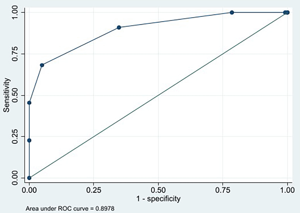
According to the ROC curve, the cut-off score of ≥ 4 yielded the largest AUC of 0.8978 (95% CI 0.823–0.971) with a sensitivity of 68.18% (95% CI 45.13%–86.14%) and specificity of 95.04% (95% CI 89.52%–98.16%) ( Figure 1). The HEALTH scoring system was then applied to our data and the subjects were split into two groups: Scores < 4 (Non-ITB) and scores ≥ 4 (ITB) as shown in Table 7. It was found that the HEALTH scoring system with a cut-off score of ≥ 4 successfully predicted 15 of 22 ITB cases and the scores < 4 successfully predicted 115 of 121 Non-ITB cases, yielding positive predictive value of 71.43% (95% CI 52.14%–85.16%) and negative predictive value of 94.26% (95% CI 89.90%–96.81%). The new proposed scoring system for differentiating ITB and Non-ITB can be seen in Table 8.
| Total scores | Actual pathological diagnosis | |
|---|---|---|
| ITB | Non-ITB | |
| <4 | 7 (5.74%) | 115 (94.26%)** |
| ≥4 | 15 (71.43%)* | 6 (28.57%) |
| Total | 22 | 121 |
In this study, we calibrated the HEALTH scoring system with the Hosmer-Lemeshow test and the result was p = 0.896. This result showed good calibration based on the statistical significance of the Hosmer-Lemeshow test p > 0.05. This scoring system was also validated based on the bootstrapping method to ensure that the scoring system can be used on a population with the same characteristics as subject characteristics in this study, with a larger number of subjects. Internal validation was carried out using the bootstrapping method 1000 times and the Hosmer Lemeshow test result was p = 0.896, meaning this scoring system has good internal validation.
Scoring systems in laboratory diagnostics play a crucial role in improving the accuracy and efficiency of disease diagnosis. By integrating specific biomarkers and clinical data, these systems enhance the ability to distinguish between similar diseases, predict disease progression, and guide treatment decisions. The development and validation of these systems across various diseases highlight their potential to transform clinical practice and improve patient outcomes. In Indonesia, a laboratory panel using a scoring system to diagnose ITB has not been developed.
There is currently no stand-alone laboratory test in ITB management, which creates opportunities for developing laboratory examinations using various biomarkers to diagnose ITB according to its pathophysiology. These examinations include routine examinations (such as hematology, IGRA, and blood ADA activity) and specific examinations (such as TB PCR). This study used multiple regression and ROC curves to determine independent diagnostic parameters. After multivariate analysis, we developed and validated a combination of 6 biomarkers from blood and stool specimens to differentiate ITB and other gastrointestinal diseases, including stool HBD-2 level, ESR, blood ADA activity, lymphocytes, stool TB PCR, and NLR.
Stool HBD-2 levels in the ITB subjects were lower than Non-ITB subjects. Researchers have noted limited studies on stool HBD-2 levels in ITB subjects. The stool HBD-2 levels can stimulated through two pathways: one involves pro-inflammatory cytokines (IL-1β, IL-1α, and TNF-α), while the other involves microbial components like lipopolysaccharides and peptidoglycan. The elevated stool HBD-2 levels in Non-ITB subjects may result from activation through both pathways: the NFκB pathway and mitogen-activated protein kinase (MAPK), which produce pro-inflammatory cytokines similar to those seen in IBD, as well as pathways activated by microbial component in infectious colitis or ileitis. Stool HBD-2 is a crucial antimicrobial protein that mediates the innate immune response. The production of stool HBD-2 through the microbial component pathway is non-specific and does not distinguish between MTB and non-MTB microbes. The ITB subjects in this study were suspected to have chronic infections, which may account for their relatively low stool HBD-2 levels.34,35
The ESR in the ITB subjects was significantly higher compared to Non-ITB subjects. The ESR test, which has a high sensitivity of 81%, can effectively serve as a screening tool for ITB. However, there are limited studies explicitly focusing on ESR in ITB cases. Most studies have primarily examined ESR in pulmonary TB cases, as documented by Khalil et al.,36 Bashir et al.,37 and Chong.38 The ESR test measures acute-phase proteins produced in acute and chronic diseases. In ITB, pro-inflammatory cytokines stimulate the liver to produce these proteins, including fibrinogen, which can elevate ESR results. Chronic diseases like TB often lead to increased immunoglobulin levels as part of the immune response to persistent infection. These positively charged immunoglobulin proteins can bind to the negatively charged surface of erythrocytes, promoting the formation of erythrocyte rouleaux and thus resulting in a rapid ESR. Despite its usefulness, the ESR test has limitations. It is influenced by anemia and hypoalbuminemia, leading to a false increase in the ESR. Therefore, it is crucial to consider the presence of anemia and hypoalbuminemia when interpreting ESR results in ITB subjects.
Blood ADA activity in ITB subjects was significantly higher than Non-ITB subjects. This finding aligns with Salmanzadeh et al., who observed significantly higher blood ADA activity in subjects with pulmonary tuberculosis than those with pneumonia, lung malignancies, and healthy controls.39 It is important to highlight that ADA activity measured in the blood does not pinpoint the exact production source but reflects the overall ADA activity from activated lymphocytes and monocytes throughout the body, including areas affected by MTB. Currently, ADA tests cannot be performed on stool specimens or biopsy tissue, making it impossible to determine local ADA production in the intestine in ITB. The increased number of monocytes in ITB subjects correlates with the elevated ADA activity, suggesting that monocytes play a more significant role than other cells in the context of ITB.40,41
The percentage of lymphocytes in the ITB subjects showed no significant difference than Non-ITB subjects and was still within the normal range (reference value: 20–40%). In TB infection, the percentage of lymphocytes in the blood often appears in the normal range due to a complex mechanism of immune responses. While some lymphocyte subsets may be changed, the overall lymphocytes can remain within normal ranges. Infection of MTB can cause shifts in lymphocyte homeostasis, with some subsets like CD8+ T-cells and B-cells increasing, while others like CD4+ T-cells decrease. These changes can balance out, resulting in a normal overall percentage of lymphocytes.42,43
In this study, the proportion of positive stool TB PCR was significantly higher in the ITB subjects than Non-ITB subjects. The results of stool TB PCR in this study are similar to those reported by Suparmin et al.6 The high proportion of positive stool TB PCR can be influenced by the high endemicity of TB in the population. This study also showed “acceptable” specificity (100%) for stool TB PCR test in differentiating ITB and Non-ITB subjects. This finding aligns with Gaur et al., who reported that stool TB PCR tests showed high specificity in EPTB subjects.44 PCR is a recent advanced diagnostic method increasingly used to diagnose TB. Unlike biopsy, stool TB PCR is non-invasive and less prone to sampling error. The detailed mechanism of MTB DNA shedding in stool remains to be investigated. A recent study indicates a two-way interaction between the gut and lung (gut-lung axis) in the transfer of metabolites, immune cells, and bacterial fragments via the bloodstream.45 However, these findings require further investigation, as the mechanistic basis of ITB is still unclear. A major challenge in diagnosing ITB is its non-specific and diverse clinical manifestations, which delay confirmation. Therefore, detecting MTB DNA in ITB subjects’ stool may serve as a valuable rule-in test.
In the present study, the NLR, an indicator of acute infection or inflammation, remained within the normal range and showed no significant difference between ITB and Non-ITB subjects. This finding contrasts with the results reported by Rees et al., who noted higher NLR levels in symptomatic pulmonary TB cases compared to controls.21 This discrepancy may be attributed to the role of neutrophils in non-granulomatous and acute inflammatory conditions. NRL is a newly identified biomarker of inflammation that is simpler to measure and relatively stable compared to other inflammatory biomarkers. However, the relationship between NLR and clinical outcomes can be influenced by several factors, including comorbidities, immune status, and virulence of the tuberculosis strain. Additionally, NLR can vary significantly between individuals based on age, gender, and ethnicity. It can also fluctuate over time due to stress, medications, and physiological conditions.
Developing laboratory panels using a scoring system based on subjects’ clinical biomarkers is nothing new. For managing the risk of patient severity or differentiating the disease from another disease, several previous studies developed laboratory scoring systems for infectious diseases, such as a lab score system for predicting COVID-19 patient severity, differentiating subjects with severe and mild H1N1 infection, and discrimination of TB infection from both PTB and EPTB disease.46–48 However, researchers have pointed out that there are still very few reports regarding laboratory panels for differentiating ITB from other gastrointestinal diseases. Makharia et al. developed scoring system with 4 parameters (blood in stool, weight loss, histologically focally enhanced colitis, and involvement of sigmoid colon) to differentiate intestinal tuberculosis and Crohn’s disease.28 Shi et al. mentioned the use of various parameters such as good response to ATT and positive IGRA, combined with clinical, endoscopy, radiology, and histopathology to diagnose intestinal tubeculosis, but not in a scoring system panel.14
The strength of the HEALTH scoring system is that this is the first study based on laboratory findings that predicted the probability of ITB. Additionally, the laboratory tests involved in this scoring system are user-friendly and non-invasive, as they only require blood and stool samples. In Indonesia, this scoring system can be utilized by healthcare facilities, regardless of whether they have colonoscopy and histopathology services. Healthcare facilities that lack these services can use the HEALTH scoring system to assist clinicians in managing ITB. On the other hand, healthcare facilities equipped with colonoscopy and histopathology services can use the HEALTH scoring system to identify patients who should undergo colonoscopy, thereby reducing waiting times for colonoscopy examinations.
It should be noted that our study has several limitations. This study was a single-center cross-sectional study, and the reagents used to examine stool IFN-γ in Indonesia do not include in vitro diagnostic reagents. Independent external validation, cost-effectiveness analysis, and multicenter randomized prospective study must be designed to validate the HEALTH scoring system.
This study developed and validated a laboratory panel called the HEALTH scoring system based on clinical biomarkers of stool HBD-2 level, ESR, blood ADA activity, lymphocytes, stool TB PCR, and NLR, which could be used to differentiate ITB from other gastrointestinal diseases.
This study was approved by the Ethics Committee, Faculty of Medicine, Universitas Indonesia–Dr. Cipto Mangunkusumo National Central General Hospital (KET-1498/UN2.F1/ETIK/PPM.00.02/2020, Date: 10.12.2020 and ND-3/UN2.F1/ETIK/PPM.00.02/2022, Date: 10.01.2022). All study participants or participants’ family have given their written informed consent regarding their participation and data publication in this study.
Software availability statement: The statistical analyses performed in this article using SPSS version 20 and STATA 14 software can be conducted using the freely accessible software R (programming language) https://www.r-project.org/
| Views | Downloads | |
|---|---|---|
| F1000Research | - | - |
|
PubMed Central
Data from PMC are received and updated monthly.
|
- | - |
Provide sufficient details of any financial or non-financial competing interests to enable users to assess whether your comments might lead a reasonable person to question your impartiality. Consider the following examples, but note that this is not an exhaustive list:
Sign up for content alerts and receive a weekly or monthly email with all newly published articles
Already registered? Sign in
The email address should be the one you originally registered with F1000.
You registered with F1000 via Google, so we cannot reset your password.
To sign in, please click here.
If you still need help with your Google account password, please click here.
You registered with F1000 via Facebook, so we cannot reset your password.
To sign in, please click here.
If you still need help with your Facebook account password, please click here.
If your email address is registered with us, we will email you instructions to reset your password.
If you think you should have received this email but it has not arrived, please check your spam filters and/or contact for further assistance.
Comments on this article Comments (0)