Keywords
tuberculosis, parotid gland, anti tubercular agents
tuberculosis, parotid gland, anti tubercular agents
Tuberculosis is a granulomatous disease caused by Mycobacterium tuberculosis or Mycobacterium bovis. Although pulmonary involvement remains the most common form, extra-pulmonary tuberculosis accounts for 20% of cases.1 Involvement of the salivary glands, particularly the parotid gland, is extremely rare. Clinically, Parotid tuberculosis usually presents as a unilateral swelling of the parotid gland.2 The absence of distinctive clinical, radiological, or biological indicators often leads to a mimicry of various neoplastic conditions, making the diagnosis especially challenging. Histopathological and microbiological assessments are therefore key components of the diagnostic process.3
Early recognition is crucial to avoid misdiagnosis and inappropriate management, especially in endemic areas. This case report highlights a rare presentation of parotid gland tuberculosis in a child, initially suspected to be a tumoral origin.
The case involved a 4-year-old girl from a rural region, belonging to a family of modest socio-economic status, having neither a previous medical history nor a family history of malignant tumors. She had no known contact with tuberculosis. She was presented by her parents to the otorhinolaryngology clinic with a right parotid swelling that had been evolving over one month. The swelling was accompanied with low-grade fever but no night sweats, weight loss, or signs of systemic illness.
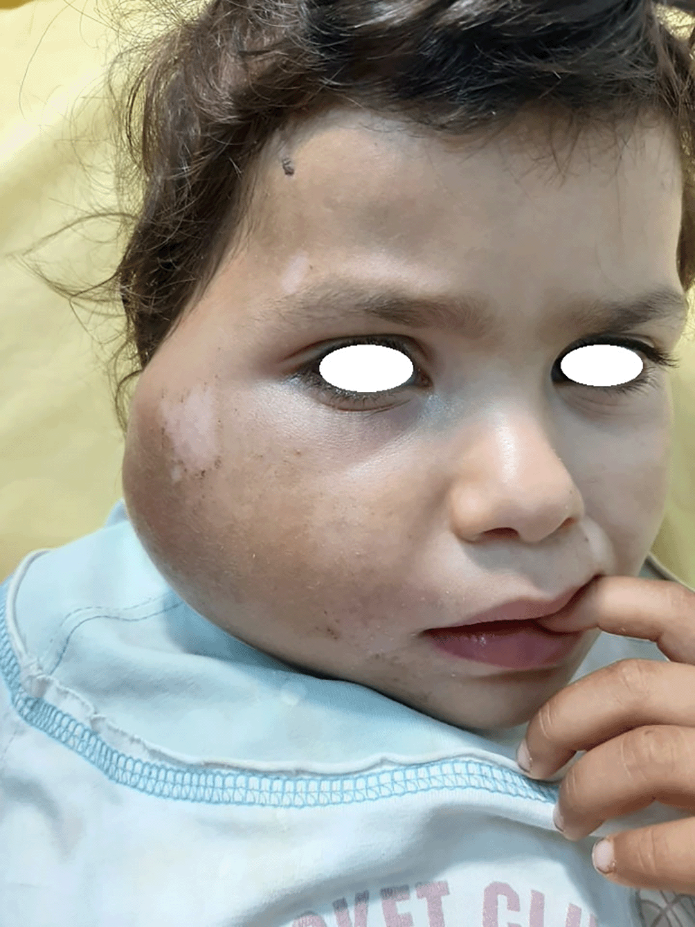
Following her admission to the otorhinolaryngology department, a detailed clinical examination was conducted for the patient which revealed a right parotid swelling measuring 7 cm long. The overlying skin showed mild inflammatory changes, but no fistula or discharge were observed. On palpation, the swelling was warm, painful and indurated, with central fluctuation. Grade II right-sided peripheral facial nerve palsy was also identified. No cervical lymphadenopathy was palpable upon examination, and the rest of the clinical assessment was unremarkable ( Figure 1).
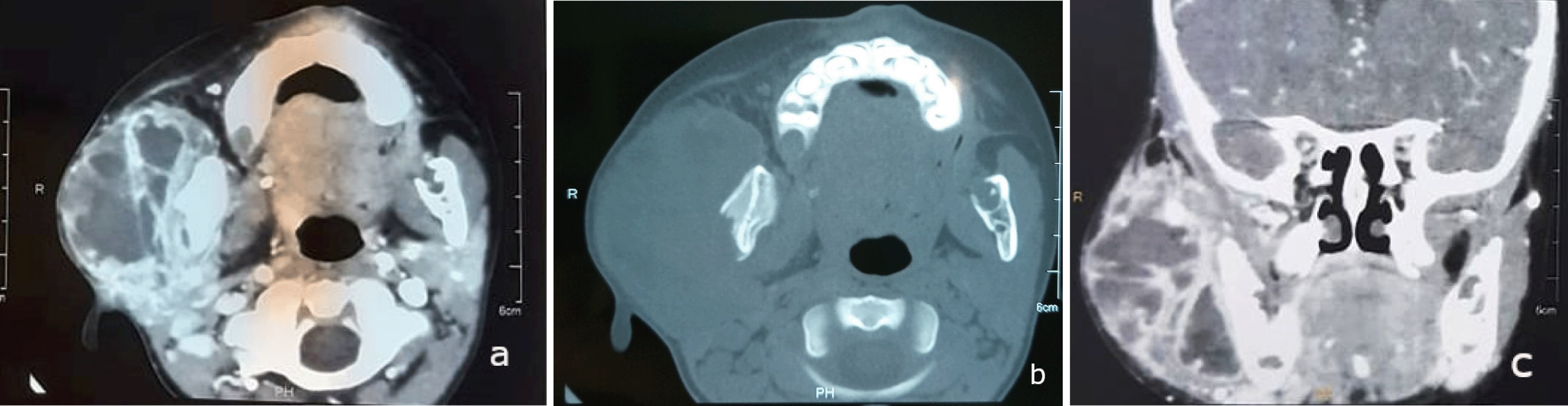
Initial laboratory tests indicated systemic inflammation. Within 24 hours, a contrast-enhanced CT scan revealed a solid cystic mass in the right parotid gland, measuring 44 x 66 mm, poorly defined with heterogeneous contrast. Partial lysis of the right mandibular condyle was observed, without extension to the deep cervico-facial spaces ( Figure 2: a,b,c).
The induration of the swelling, the facial paralysis and the bone lysis observed on CT scan were raising concern for malignancy.
On the third day of hospitalization, the patient underwent a surgical incision and drainage procedure under local anesthesia. Purulent material was obtained for bacteriological culture, and a biopsy of the lesion margins was performed for histopathological evaluation.
Both samples (pus and biopsy) confirmed the tuberculous origin of the swelling, with no evidence of malignancy. A chest X-Ray was subsequently conducted, which ruled out pulmonary involvement.
On hospital day seven, following baseline assessments that included liver and renal function tests and an ophthalmological examination, a standard anti-tuberculosis treatment regimen was initiated, adjusted according to the patient’s weight and age.
The protocol consisted of two months of quadruple therapy (Rifampicin, Isoniazid, Pyrazinamide, and Ethambutol) followed by four months of dual therapy (Rifampicin and Isoniazid).
During hospitalization, the patient was monitored for treatment tolerance and exhibited no adverse reactions. Over the subsequent weeks, clinical improvements were observed, including a reduction in parotid swelling and progressive recovery of facial nerve function.
Seven days after starting treatment, the patient was discharged with monthly follow-up appointments and successfully completed the full six-month course of therapy, resulting in favorable outcomes.
The handling of this rare pediatric case highlights effective clinical management through a systematic, multidisciplinary diagnostic approach that emphasized histopathological confirmation before treatment. The team avoided premature surgery despite radiological signs of malignancy, leading to full recovery and preserved facial nerve function. However, limitations included the lack of advanced microbiological diagnostics (PCR and GeneXpert) which could have expedited confirmation. Also, the absence of long-term follow-up, hindering the evaluation of recurrence or late complications. As with all single-case reports, the findings should be viewed carefully regarding their extensive application.
Although tuberculosis is an infectious disease that mainly affects the lungs, it can affect any organ in the body.4 However, even in endemic countries where the disease is widespread, tuberculous involvement of the parotid gland remains extremely rare.5
In children, this location is even more unusual, which can delay the diagnosis.6
In our patient’s case, the clinical presentation initially evoked a suppurative infectious etiology. However, the chronic course over the last 1 month did not support an acute infectious suppurative etiology. This suggests an inflammatory or tumoral etiology.7
It is wise to rule out a tumoral pathology of the parotid gland, due to the unilaterality, induration of the swelling, facial paralysis, and bone lysis observed on CT. Indeed, malignant tumors of the parotid gland, such as mucoepidermoid or cystic adenoid carcinomas, can present in a similar manner.8
Imaging plays an important role in diagnosis guidance. CT scans provide a detailed exploration of the parotid gland, with greater sensitivity than ultrasound. However, the radiological features observed are not specific to tuberculosis. According to the literature, the most frequently observed appearance is: thick-walled, contrast-enhanced lesion with central necrosis, characteristic of tuberculosis. Nevertheless, different aspects can be observed, such as homogeneous enhancement of the parotid gland, homogeneous enhancement with a non-enhanced microcyst, or isolated or confluent hypodense nodular lesions.9
Bacteriological examination and histopathological study were essential to confirm tuberculosis. The presence of acid-fast bacilli in the specimens, as well as epithelioid granulomas with caseous necrosis observed histologically, are characteristic of tuberculosis.10
For parotid tuberculosis, the treatment regimen is the standard extra-pulmonary tuberculosis regimen: two months of quadruple therapy (Rifampicin, Isoniazid, Pyrazinamide and Ethambutol) followed by dual therapy (Rifampicin and Isoniazid).11
Treatment lasts 6 months. Regular monitoring is essential to assess the efficiency of the anti-tuberculosis treatment, ensure that the patient is following it correctly, and take action rapidly if side effects appear. Patient education is therefore crucial.11
The effectiveness of the treatment is assessed on the basis of the clinical course and various additional tests (biology, imaging, etc.) deemed necessary by the treating physician.11
Despite the lack of formal staging system for extrapulmonary tuberculosis of the parotid gland. But several clinical features in this case were important for predicting how the disease would progress. A positive outcome was the result of early diagnosis, absence of immunosuppression and good adherence to therapy.12
From the family’s perspective, the uncertainty and fear of malignancy were a major source of anxiety. They felt a sense of relief upon receiving a clear and treatable diagnosis. Opting for a non-surgical approach, along with progressive recovery of facial function, not only alleviated their concerns but also strengthened their confidence in the healthcare team.
This case highlights the importance of considering a wide range of differential diagnosis, including tuberculosis, when faced with parotid swelling in children, especially in endemic regions. Multidisciplinary management, including appropriate investigations and well-administered anti-tuberculosis treatment, is essential to ensure a favorable prognosis. Although parotid tuberculosis is rare, it should be considered in order to avoid delays in diagnosis and treatment, which could have serious consequences for the patient.
| Views | Downloads | |
|---|---|---|
| F1000Research | - | - |
|
PubMed Central
Data from PMC are received and updated monthly.
|
- | - |
Provide sufficient details of any financial or non-financial competing interests to enable users to assess whether your comments might lead a reasonable person to question your impartiality. Consider the following examples, but note that this is not an exhaustive list:
Sign up for content alerts and receive a weekly or monthly email with all newly published articles
Already registered? Sign in
The email address should be the one you originally registered with F1000.
You registered with F1000 via Google, so we cannot reset your password.
To sign in, please click here.
If you still need help with your Google account password, please click here.
You registered with F1000 via Facebook, so we cannot reset your password.
To sign in, please click here.
If you still need help with your Facebook account password, please click here.
If your email address is registered with us, we will email you instructions to reset your password.
If you think you should have received this email but it has not arrived, please check your spam filters and/or contact for further assistance.
Comments on this article Comments (0)