Keywords
Perforator; Arterial; Continuous wave; Pulsed Dopper; Colour Doppler Perforator
This article is included in the Manipal Academy of Higher Education gateway.
This article is included in the Global Public Health gateway.
Perforator; Arterial; Continuous wave; Pulsed Dopper; Colour Doppler Perforator
An arterial perforator is a vessel that arises from a main (parent) artery and passes through the intermuscular/septocutaneous/myocutaneous plane to supply blood to superficial structures.
Defining the vascular territories of the lower extremities has greatly helped in safe flap design. Fasciocutaneous flaps based on random pedicles for lower extremity reconstruction demonstrated high necrosis rates of up to 25%. Careful anatomical study of cutaneous arteries has resulted in the emergence of the “angiosome” concept.1
Presently arterial perforators have become the mainstay of perforator flap reconstructive surgeries after trophic or superficial soft tissue defects secondary to insults such as burns, trauma, or gangrene secondary to peripheral vaso-occlusive pathology.
Despite voluminous and ever-increasing knowledge of anatomy, there is no systematic approach to locate perforating arteries in the lower limb. This is mainly due to the presence of anatomical variation among individuals, with asymmetry being the general rule in every individual. There is asymmetry in the anatomical spread of arterial perforators between the two lower limbs of one individual.
Therefore, pre-operative planning before surgical flap surgery cannot be based on any previously established set of rules. Every individual must be evaluated independently and anatomical variations in perforator mapping must be considered.
CW Doppler is a simpler configuration of Doppler imaging and consists of two different types of transducer crystals: receiving and transmitting transducer crystals. The main role of the transmitting transducer crystal was to produce a continuous ultrasound beam output at a predetermined frequency. This transmitted signal was then received by the receiving crystal. The received and transmitted signals are added, which leads to the formation of a waveform consisting of a beat frequency. The beat frequency is equal to the Doppler-shift frequency. There is no information or data provided for CWDoppler in terms of the depth of the point of analysis or at which the analyzed motion occurs, which causes the Doppler shift.2
The main aim of the development of pulse-wave Doppler was to obtain primary information regarding depth perception of the region of interest, which leads to the Doppler shift phenomenon. In addition, the PW Doppler system comprises of only one transducer crystal. The same transducer crystal does the job of transmit and receive signals. There is a substantial difference in the pulse used for pulse wave Doppler compared to the pulse in B-mode imaging. Electronic gating was used to determine the time interval between transmitted and received pulses. This allows the operator to analyze a specific point along the long axis of the transmitted beam. The main output from PW Doppler is primarily an audible signal. When combined, PW Doppler and B-mode imaging are referred to as duplex ultrasound imaging. This allows the operator to analyze both entities together and to focus on a specific point in the B-mode image for Doppler analysis.2
The recommended sites for sampling of continuous wave Doppler data in the lower limb are the dorsalis pedis artery, anterior tibial artery, distal posterior tibial artery, common femoral artery, and popliteal artery. Superficial and distal peroneal arteries can also be included sites. To obtain the best signal, the probe must be positioned along the longitudinal axis of the vessel to be analyzed and pointed cephalad. The Doppler signal should be optimized through manual adjustment of the orientation and angle until the maximum waveform amplitude is displayed on the grid and the clearest possible audible output is obtained.3
To identify and locate arterial perforators in the lower extremity using pulse wave color Doppler, and continuous-wave Doppler.
1. Mapping arterial perforators in lower limb using Continuous Wave Doppler and Pulse Wave Doppler.
2. Comparing outcomes of mapping of lower limb arterial perforators using the two doppler techniques.
3. Spectral analysis of waveforms of continuous wave doppler.
4. Achieving maximum accuracy in scanning methods of continuous wave Doppler in lower limb.
5. Collecting data for further spectral wave analysis.
6. Observing physiological and anatomical variations in lower limb arterial perforators and their effect on spectral waves.
Study settings: Department of Radiodiagnosis.
Machines used:
Samsung HS-40
Huntleigh Doppler DMX continuous wave hand held Doppler
Study design: Cross sectional study
Study participants: Patients referred to the Department of Radiodiagnosis for lower-limb arterial perforator identification and assessment.
Inclusion criteria:
Exclusion criteria:
1. Smokers
2. Known case of peripheral vascular disease.
3. Hypertensive patients/patients on anti-hypertensive therapy.
4. Diabetes with microvascular complications.
5. Recent orthopedic/surgical intervention in lower limb.
6. Any pathological data collected on Doppler study will lead to exclusion of the sample.
Study duration: November 2018 to September 2020. Sample size:
Calculated using prevalence and sensitivity.
Where Sn = 94%
The minimum number of patients required for the study was 90.
Sampling Method: Non-probability convenience sampling.
The study was conducted after obtaining permission from the institutional ethic Committee-IEC KMC MLR 10-18/382). The study was conducted at Government Hospital. This was an observational study, with data collected prospectively over a period of 2 years.
sample size was estimated based on a 5% alpha error and 95% confidence level, sample size of 90 was obtained. A total of 100 healthy volunteers underwent Doppler evaluation.
Continuous wave Doppler was performed using a Huntleigh Doppler DMX handheld device, while pulse wave and color Doppler evaluations were performed using a Samsung HS 40 machine.
Outcome variables:
7. PW Doppler: Distance from nearest anatomical landmark, depth from skin, diameter and velocity of blood flow in arterial perforator.
8. CW Doppler: Identification and site of arterial perforator with respect to nearest anatomical landmark.
In this study, we localized arterial perforators using pulse-wave Doppler and performed anatomical mapping of these perforators for ATA and PTA. Later at the localized sites, we performed a continuous wave Doppler study and attempted to record the spectral wave and sound from the continuous wave Doppler. Data were also recorded from the pulse wave Doppler data in the form of sound output using a 3.5 mm jack and an indigenous in-house amplifier. This amplifier removed the background Doppler noise and improved efficacy of data analysis.
Tabular data were obtained from the duplex ultrasound machine using a gray-scale display. The data provided were the depth of the vessel from the skin surface, maximum luminal caliber of the vessel, site of origin of the vessel from the nearest anatomical landmark, and peak systolic velocity of the wave.
Any person with diseased vessels, biphasic or monophasic waveform on pulse Doppler, or extensive atherosclerotic changes was excluded from the data set.
Tabular data and sound recordings from both continuous wave and pulse wave doppler were sent for for analysis. Sound data were analyzed using MATLAB software, and an algorithm was derived to extrapolate sound and tabular data from pulse and color Doppler onto sound recordings from continuous wave Doppler.
Data Analysis: By Chi-square test. SPSS version 22.0 was used for the analysis. p value of <0.05 was considered at statistically significant after assuming all the rules of statistical tests.
Data collection Tool: Pretested semi-structured data extraction sheet was developed based on a literature review and after consultation with experts. The study participants were informed about the objectives, and written informed consent was obtained.
We Collected 3 sets of data for each sample
• Color Doppler: recorded using a cable and mobile phone.
• Handheld Doppler, recorded using a cable and mobile phone.
• Handheld Doppler in raw data formatwas copied from the memory card of the handheld Doppler, and converted to wave. This was done to confirm that the mobile recording was proper and that there was no erroneous recoding.
In our study, out of a total of 100 participants, 77 were male and 23 were female participants ( Table 1).
The participants were predominantly residents, interns, and the volunteering public with no prior comorbidities.
Owing to maximum participation in the study by interns and postgraduate residents working in the hospital, the most common age group of participants was between 21 and 30 years, making up >40 percent of the total study participants ( Table 2).
| Frequency | Percent | |
|---|---|---|
| 20 and below | 4 | 4.0 |
| 21-30 | 42 | 42.0 |
| 31-40 | 36 | 36.0 |
| >50 | 18 | 18.0 |
| Total | 100 | 100.0 |
Most commonly, four perforators were picked up by the pulse wave and color Doppler machine while performing below-knee arterial perforator assessments. This was true for the anterior tibial artery and posterior tibial artery, individually ( Table 3).
| Parent artery | |||
|---|---|---|---|
| ATA | PTA | ||
| Perforator | 2 | 4.0% | 1.5% |
| 3 | 33.0% | 18.5% | |
| 4 | 52.5% | 46.5% | |
| 5 | 10.5% | 27.5% | |
| 6 | 0% | 6.0% | |
| Total | 100.0% | 100.0% | |
There were no cases where ATA demonstrated 6 or more perforators.
The Figures & tables depict the maximum number of perforators in a single vessel and its anatomical mapping based on the perforator count (Figure 1).
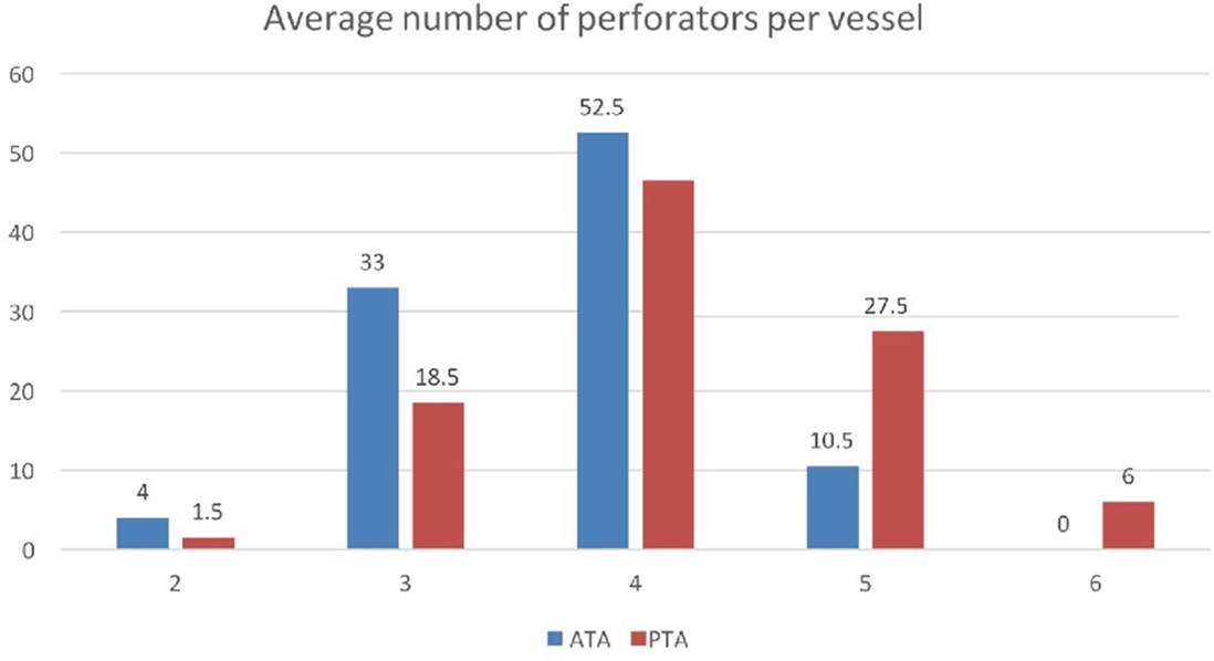
Blue bar for ATA and red bar for PTA.
Average caliber of arterial perforators was 1.49 mm for ATA, 1.53 mm for PTA and 1.51 mm overall for ATA and PTA combined (Figure 2).
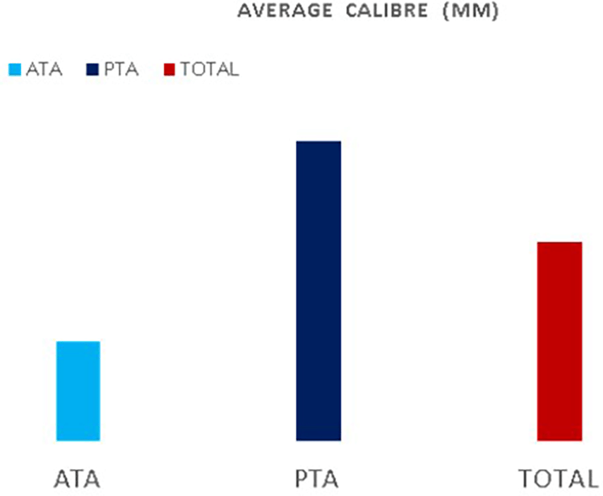
Light Blue bar ATA and Dark blue bar PTA.
For the anterior tibial artery, when only two dominant perforators were selected, both were within 15 cm of the lateral malleolus. In PTA, approximately 63 percent of the perforators were within the 11-15 cm distance from the medial malleolus. This was applied when the perforator count was a mere two for PTA.
When three perforators were picked, nearly 45 percent were within 15 cm of the lateral malleolus for the ATA. For PTA, nearly 50 percent of perforators were > 15 cm away from the medial malleolus when the total perforator count was 3. When five or more perforators were selected, the perforator that was farthest from the lateral malleolus for ATA was > 15 cm in all cases. The same is true for the posterior tibial artery from medial malleolus ( Tables 3 and 4).
| Parent artery | Total | ||||||
|---|---|---|---|---|---|---|---|
| ATA | PTA | ||||||
| Count | % | Count | % | Count | % | ||
| Perforator | 2 | 8 | 4.0% | 3 | 1.5% | 11 | 2.8% |
| 3 | 66 | 33.0% | 37 | 18.5% | 103 | 25.8% | |
| 4 | 105 | 52.5% | 93 | 46.5% | 198 | 49.5% | |
| 5 | 21 | 10.5% | 55 | 27.5% | 76 | 19.0% | |
| 6 | 0 | .0% | 12 | 6.0% | 12 | 3.0% | |
| Total | 200 | 100.0% | 200 | 100.0% | 400 | 100.0% | |
The average distance for an arterial perforator from lateral malleolus for the leg was 11 cm for ATA, with a standard deviation of 5.24 cm and a median value of 11 cm ( Table 5).
For PTA, the mean was 11.9 cm from medial malleolus with a standard deviation of 5.89 cm and median value of 12 cm (Figure 3).
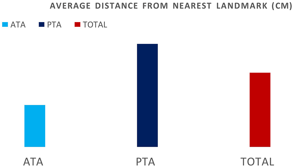
Light Blue ATA and Dark blue PTA.
The average depth from the skin surface for the anterior tibial artery perforators was 14.4 mm with a standard deviation of 3.4 mm and a median value of 14 mm (Figure 4).
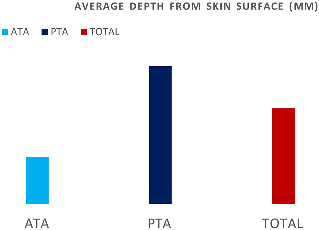
Light blue ATA and Dark blue PTA.
The average depth from the skin surface for the posterior tibial artery perforators was 14.9 mm with a standard deviation of 3.7 mm and a medial of 15 mm.
The average peak systolic velocity was 32.5 cm/s for the ATA, with a standard deviation of 5.24 cm/s and a median value of 32 cm/s ( Table 6).
For the posterior tibial artery, the mean PSV was 32.3 cm/s with a standard deviation of 8.4 cm/s and a median of 32 cm/s.
Overall, the average PSV for both ATA and PTA was 32.4 cm/s with a standard deviation of 8.3 cm/s ( Table 7).
Preliminary analysis was provided by NITK Surathkal using MATLAB software (Figure 5) ( Tables 8 and 9).
Spectral pattern for analysis.
| Frequency | Percent | |
|---|---|---|
| No | 239 | 15.2 |
| Yes | 1336 | 84.8 |
| Total | 1575 | 100.0 |
| Parent artery | Total | ||||||
|---|---|---|---|---|---|---|---|
| ATA | PTA | ||||||
| Count | % | Count | % | Count | % | ||
| Continuous wave doppler (Y/N) | N | 97 | 13.1% | 142 | 17.0% | 239 | 15.2% |
| Y | 642 | 86.9% | 694 | 83.0% | 1336 | 84.8% | |
| Total | 739 | 100.0% | 836 | 100.0% | 1575 | 100.0% | |
Colour Doppler with pulse doppler and Continuous wave doppler Image recordings are shown in Figure 6.
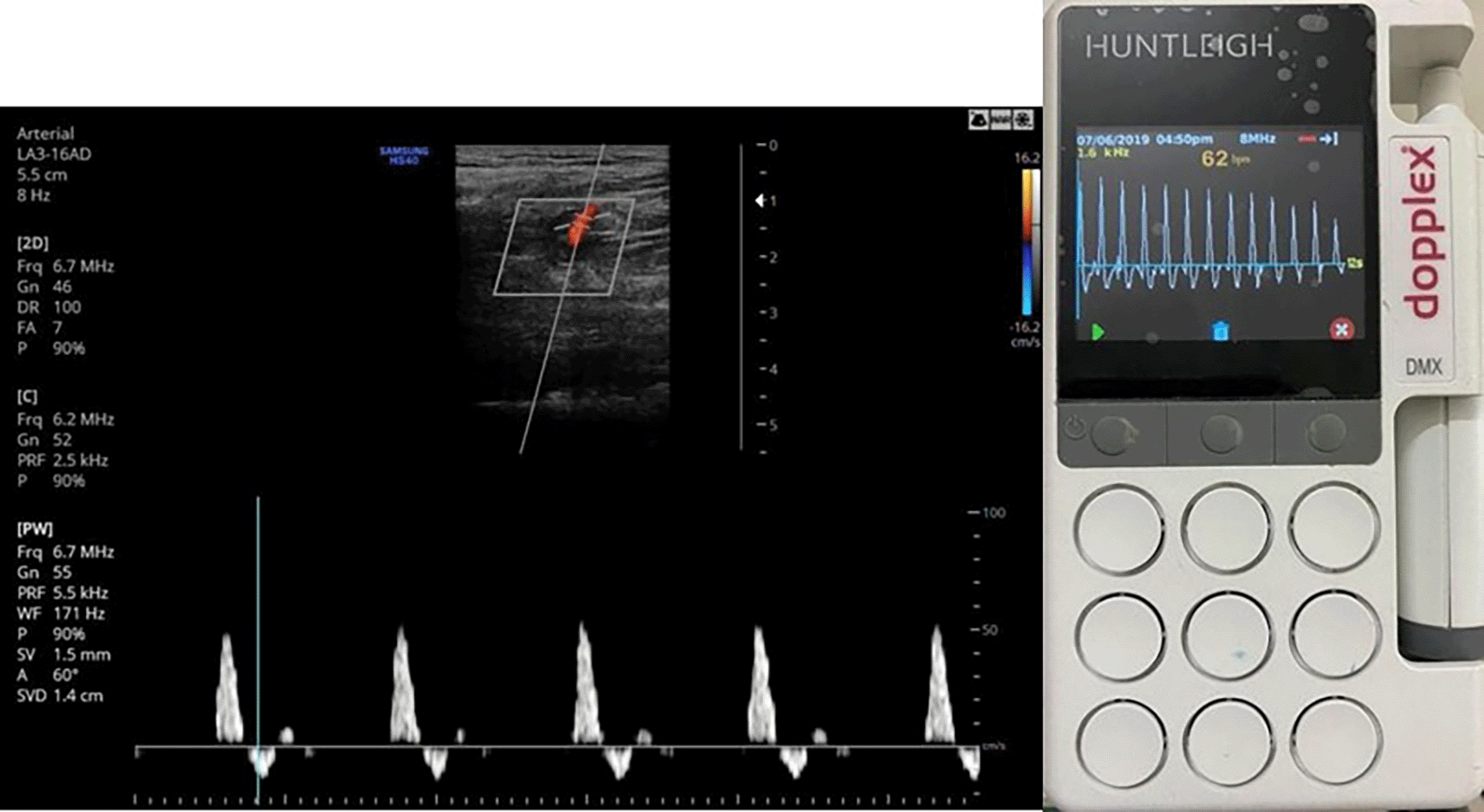
Colour and pulse wave doppler on left and continuous wave spectrum on right side.
Tables 10 & 11 shows that sample number 2 was graded as the best one by a radiologist based on the color Doppler image, perforator anatomy, and sound quality from the pulse wave Doppler.
Grading was performed by the doctor based on data from color Doppler =>2>3>4>7>(5,6,8,9/10) (Blinded study).
The average beat energy values of best 5 systolic beats (energy column in the table) almost correctly follow this trend. These energy values were obtained after the analysis of continuous wave sound recordings using an in-house algorithm for sound analysis to calculate the energy flow.
Algorithm development was carried out using MATLAB.
1) Heart rate detection algorithm
2) Best five beat extraction
• We found that the perforators that medical experts deemed “best“for surgery were those corresponding to recordings with the highest energy in the systolic beat.
• Beat extraction-
• A 10 ms moving-window-based technique was followed for extracting only the beats from the data.
• Selection of best 5 beats-
• The total energy for each extracted beat bk is calculated as, where n is the number of samples in bk
The beat energies were then sorted in descending order, and five beats with the highest energy values were selected. The average of these five energy values was calculated to determine the parameter for judging quality of the perforators.
3) Maximum frequency detection
• The extracted beat is windowed using a 40ms Hanning window, with an overlap of 20ms.
• The Short-Time Fourier Transform(FFT) is calculated for each window and stored in memory.
• The Energy in the FFT of each window was calculated, and a scaled version of these energy values was set as the threshold to find prominent frequencies.
• Out of all the frequencies with an FFT magnitude greater than the threshold, the maximum frequency was chosen.
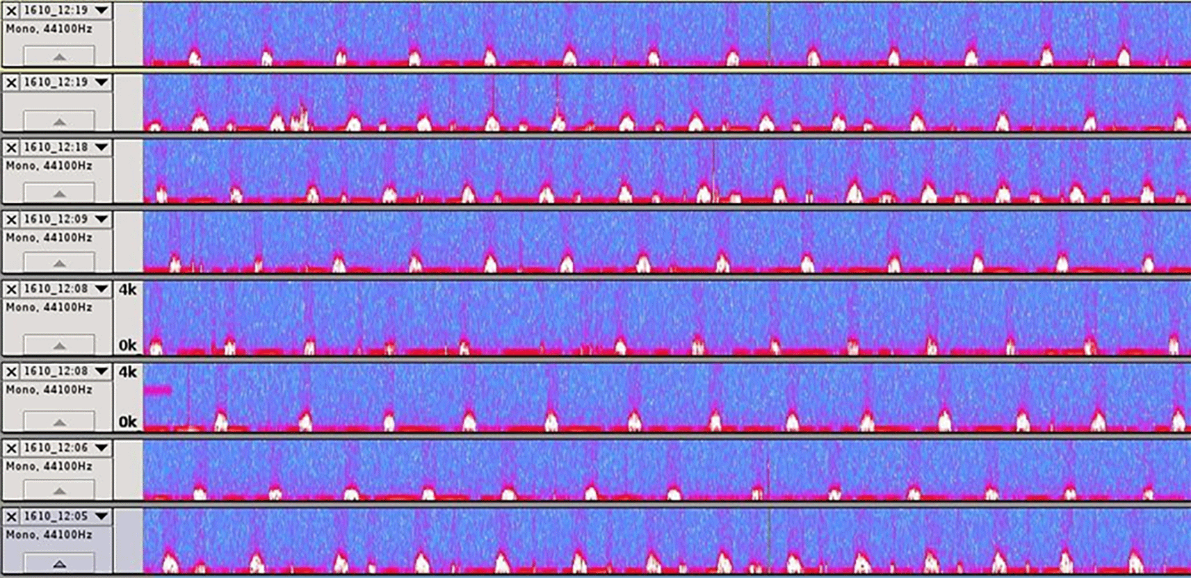
Data were recorded for analysis by sound engineers using a custom-made recording cable to meet the requirements of the audio output port of both Doppler Machines. A total of 189 sets of recordings were obtained.
After analysis of data by the engineering department, the following was noted:
a) It was surprising to note that multiple recordings of the same perforator using color Doppler machines showed huge variations in loudness as well as in peak systolic velocity (for example, in some cases, it varies from 45 cm/s to 80 cm/s) between two recordings taken within a short span of 1 min by the same practitioner. This was likely due to the change in the angle of intonation and the angle at which the Doppler probe was held.
b) The perforator, which gave better quantitative numbers, was not always the right one chosen by an experienced doctor based on the anatomy for the surgical application. Approximately 70 percent of the recordings were matched with the best perforator (based on energy of blood flow) when taking an experienced radiologist’s opinion into account (radiologist opinion based on gray scale images and anatomical course of perforator).
c) Perforators at right angles gave less quality sound, and those that were deeper from the skin showed less sound quality in the handheld Doppler.
d) In most cases, handheld Doppler correctly identified the best perforator, as suggested by the doctor, based on a blinded study. The energy numbers obtained from handheld Doppler support the quality of the perforators.
We commonly study venous perforators that become incompetent in varicose veins; arteries also have major perforators in the limbs that pass through the fasciocutaneous and septocutaneous planes to supply blood to different angiosomes. These perforators are responsible for supplying blood to tissues distant from the major arteries of the limb.
The posterior tibial artery is a branch beyond the popliteal artery, as it exits the popliteal fossa and is the largest terminal branch of the tibioperoneal trunk. It extends behind the tendinous arch of the soleus, with the majority of its course behind the tibialis posterior after the origin of the peroneal artery.4 The ankle is posterior to the medial malleolus and branches into the medial and lateral plantar arteries. The posterior tibial artery supplies the posterior compartment of the leg.
The perforators from the posterior tibial artery are most commonly septocutaneous and emerge adjacent to the flexor digitorum longus and soleus muscle.5 These perforators, located in the middle third of the lower leg, are some of the largest of the entire lower extremity.6 They are most readily identifiable 10-12 cm above the medial malleolus. A flap can be designed anywhere up to 10 cm distal to the popliteal crease.6
Peroneal artery perforators: The peroneal artery is a branch of the tibioperoneal trunk. It descends through the posterior compartment of the leg, next to the posterior intermuscular septum. Peroneal perforators, which are fewer in number can supply a perforator flap. The perforators in the middle third of the fibula are observed on Doppler. The musculocutaneous perforators emerge proximally from the soleus or peroneus longus muscles, and septocutaneous perforators emerge between the flexor hallucis longus and peroneus brevis muscles. Most perforators emerged 13-18 cm proximal to the lateral malleolus. Distally, these perforators often emerge superficial to the Achilles tendon.7
Anterior tibial perforators: The lower extremity is supplied by the anterior tibial artery. The anterior tibial artery begins at the inferior border of the popliteus muscle. It passes anteriorly through a gap in the interosseous membrane and descends on the anterior surface of the membrane between the tibialis anterior and extensor digitorum longus.8 Blood is supplied to the anterior compartment of the leg. The major perforators from the anterior tibial artery are located proximally - documented 21-26 cm proximal to the intermalleolar line between the tibia and tibialis anterior muscle. These are the largest perforators. Smaller perforators emerge 4-9 cm above the intermalleolar line between the tendons of the anterior compartment and supply the skin over both malleoli.9
Arterial perforators have important clinical applications in planning flap reconstruction surgeries of the leg in cases such as diabetic foot, trauma, and trophic ulcers.
These flap reconstructive surgeries are performed in cases of high-velocity trauma. Multiple studies have shown that selecting a suitable dominant perforator is key to the success of any flap surgery and the uneventful postoperative healing of the patient. Preoperative determination of the location and number of septocutaneous perforators provides multiple benefits for flap planning. The main benefit of preoperative flap planning is that any unplanned alteration in the operation theatre during flap reconstruction is avoided when there are no suitable dominant perforators available at the surgical site. In addition, pre-operative planning and assessment reduce the operating time and improve the outcome of the dissection. Another advantage of performing preoperative assessment via Doppler is that based on the location of the perforator, a flap may be preoperatively customized to meet all surgical requirements.
Multiple authors have previously described the distribution pattern of arterial perforators and proved that achieving a systematic approach for mapping the precise location of the dominant septo-cutaneous perforator is quite difficult. This is primarily due to the fact that there is high degree of variation in the origin and course of these vessels between multiple individuals and also between different limbs of the same individual. In our study, we identified that an individual can have as few as two dominant arterial perforators below the knee and as many as six perforators arising from a parent artery.
Color Doppler analysis associated with pulse wave Doppler allows analysis of the anatomical distribution and flow of superficial vessels in the limbs. However, this is not possible with the use of handheld continuous wave Doppler, which does not provide any information regarding the anatomical distribution of the perforators.
The main disadvantage of a color Doppler machine is that the investment and economic costs are too high to procure the machine and the machine and probe footprint large for portable ultrasound usage, especially in places like the operation theatre due to logistical problems and problems related to the sterilization of equipment. In addition, pulse wave and color Doppler require skilled expertise in operating an ultrasound machine.
Previously, many authors have recommended the use of pre-operative mapping of arterial perforators using acoustic handheld Doppler. However, the literature is limited to evidence-based evidence showing the reliability of this technique.
Studies have demonstrated variable reproducibility and success in pre-operative localization of arterial perforators, while comparing handheld data with intraoperative findings.10
In comparing intra-operative and handheld Doppler findings large underestimation (30 %) to an overestimation of 150 % for hand held Doppler findings.11
Sound data were analyzed using MATLAB software, and an algorithm was derived to extrapolate sound and tabular data from pulse and color Doppler onto sound recordings from continuous wave Doppler.
From a preliminary small dataset analysis of 45 recordings, it was found that the expected peak systolic velocity (expected PSV) from sound analysis of continuous wave Doppler was very close or nearly equal to the pulse wave Doppler actual peak systolic velocity (actual PSV) in 41 recordings. Only four recordings showed vast differences in the datasets of both ultrasound Doppler machines.
The main aim of this study was to evaluate whether the sound output of a basic continuous-wave Doppler machine could reproduce the data provided by a technologically advanced handheld Doppler machine.
While collecting data using the continuous wave handheld Doppler device, we found that the Doppler signal in the form of a spectral waveform and sound output varied drastically between different recordings of the same perforator by the same investigator (high intra-observer variation). This can be attributed to the fact that there is no way to fix the angle of insonation of the handheld Doppler ultrasound beam and that the output is dependent on the angle at which the probe is held. To counter this problem, we took multiple recordings of the same perforator and analyzed the recordings with the highest spectral peak and maximum sound output.
By having multiple recordings of the same perforator using continuous wave Doppler and performing preliminary marking of the perforator site using pulse wave doppler, we were able to set basic scanning guidelines on using continuous wave Doppler for arterial perforator analysis.
Another objective of the study was to conduct a never done before kind of pilot study for anatomical mapping of dominant arterial perforators of the below leg arteries (anterior tibial artery and posterior tibial artery).
After analyzing the results, it was found that arterial perforators in the leg can be divided into clusters with 5 cm interval, with maximum perforators arising between 5 and 10 cm, 10–15 cm, and 15–20 cm from the nearest anatomical landmarks, that is, medial and lateral malleolus or intermalleolar line. This was consistent with other studies in which clusters of 4–9 cm, 13–18 cm, and 21–26 cm were used for the origin of lower limb arterial perforators. They followed the course of the perforators in the intermuscular septae, which was also noted in our study.12
CW and PW interpretations based on years of sonographic experience analyzing 122 arterial perforators. Of all perforators, 102 originated from PTA, 4 originated from ATA, and 16 originated from the medial sural artery.13
The existence of reliable levels of septocutaneous perforators of the lower limb allows one to do reconstruction of soft tissue defects of the lower limb, especially its foot and distal third, in a much easier manner.14
1. Continuous wave Doppler cannot be used as a stand-alone modality to analyze arterial perforators of the lower limb owing to the lack of grayscale display and B mode to identify the artery of origin of the spectral waveform.
2. Spectral data obtained from continuous wave Doppler vary drastically between recordings (intra-observer variation) due to the difference in the angle of insonation during each recording.
3. Significant inter-observer variation while using continuous wave Doppler due to difference in expertise and technique of doing Doppler between 2 radiologists.
4. Spectral data cannot be used as stand-alone to analyze perforators and must be used in conjunction with B-mode display to identify a perforator with better blood flow dynamics based on its course.
5. Intraoperative and post-operative samples were not analyzed during the study.
Similar to the venous perforator anatomy, the arterial perforator anatomy has an important role and clinical application in perforator flap planning. Presently, there is a paucity of literature regarding the use of continuous wave Doppler for the assessment of arterial anatomy in the extremities. Their main advantage, in comparison to pulse wave and color Doppler, lies in their portability, ease of access, and economically cheaper devices. Another advantage is the presence of a metallic intra- operative probe in the CW Doppler, which can be sterilized. This may allow intra-operative Doppler imaging to confirm the dominant perforator during flap planning.
However, the major limitation of using continuous wave Doppler lies in its dependence on pulse wave Doppler to first identify arterial perforators in the limb, due to the lack of gray-scale imaging in CW Doppler. Without prior anatomical mapping, there is no way to confirm whether the sound output arises from a perforator or parent artery.
Analysis of the anatomical distribution of arterial perforators demonstrated that most commonly, there are 4 to 5 perforators below the knee arising from the anterior tibial artery and posterior tibial artery, with an average peak systolic velocity of ~ 30-35 cm/s.
After performing cluster analysis for the distribution of arterial perforators arising from the anterior and posterior tibial arteries in the leg, we concluded that the perforators were present in the intermuscular septa and plane. Perforator clusters were found within distinct 5 cm intervals at approximately 5–10, 10–15, and 15–20 cm from the medial malleolus, lateral malleolus, or intermalleolar line.
In conclusion, spectral analysis of a perforator could aid a surgeon in deciding a dominant arterial perforator, which will further improve the outcomes of flap surgeries and improve its planning.
The study was conducted after obtaining permission from the institutional ethic Committee- approval committee name-Institutional Ethics Committee Kasturba medical College Reg No.ECR/541/Inst/KA/2014/RR.17 study approved on Wednesday 17 th October 2018 as study no. IEC KMC MLR 10-18/382. The study participants were informed about the objectives, and written informed consent was obtained.
| Views | Downloads | |
|---|---|---|
| F1000Research | - | - |
|
PubMed Central
Data from PMC are received and updated monthly.
|
- | - |
Provide sufficient details of any financial or non-financial competing interests to enable users to assess whether your comments might lead a reasonable person to question your impartiality. Consider the following examples, but note that this is not an exhaustive list:
Sign up for content alerts and receive a weekly or monthly email with all newly published articles
Already registered? Sign in
The email address should be the one you originally registered with F1000.
You registered with F1000 via Google, so we cannot reset your password.
To sign in, please click here.
If you still need help with your Google account password, please click here.
You registered with F1000 via Facebook, so we cannot reset your password.
To sign in, please click here.
If you still need help with your Facebook account password, please click here.
If your email address is registered with us, we will email you instructions to reset your password.
If you think you should have received this email but it has not arrived, please check your spam filters and/or contact for further assistance.
Comments on this article Comments (0)