Keywords
mycotic aneurysm, rupture, repair, thoracic aorta, zone 2, TEVAR
Mycotic thoracic aortic aneurysms are rare but life-threatening, and are associated with high morbidity and mortality after operative repair. They frequently present at a late stage with sepsis and rupture; therefore, emergency management is paramount. Repair options, particularly in the thoracic aorta, are complex and include open, endovascular, and hybrid surgical approaches. We present the case of a 72-year-old woman with a ruptured mycotic thoracic aortic aneurysm who underwent successful endovascular repair with thoracic stent graft placement and left subclavian artery embolization. This case highlights the successful use of endovascular repair for the management of a ruptured mycotic thoracic aortic aneurysm in zone 2, emphasizing the importance of early diagnosis, timely intervention, and potential benefits of minimally invasive repair in these rare complex cases.
mycotic aneurysm, rupture, repair, thoracic aorta, zone 2, TEVAR
Mycotic aortic aneurysms are very rare and account for only 0.6%–2.6% of aortic aneurysms in Western countries.1–3 Mycotic thoracic aortic aneurysms (MTAA) are aneurysms of the thoracic aorta caused predominantly by bacterial or fungal infection.2,3 These are less common than their abdominal aortic counterparts and account for 30% of mycotic aortic aneurysms.3 Traditional open surgical repair carries significant perioperative morbidity and mortality, especially in comorbid and critically ill patients, making endovascular intervention an increasingly favored approach with promising outcomes. However, a consensus for optimal repair has not yet been reached due to the rarity and lack of data on MTAA in the current literature.2,3 We present a case of successful emergent endovascular repair of an MTAA in a challenging and uncommon area of the aortic arch.
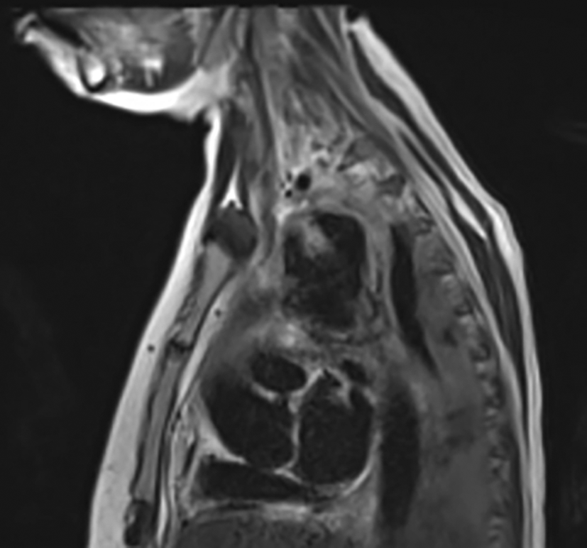
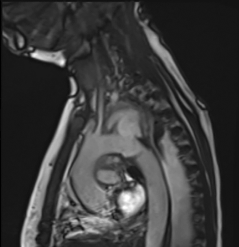
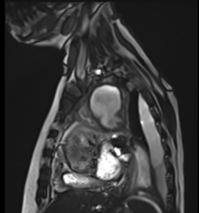
A 72-year-old woman on a bridging visa to Australia (originally from South India) presented to the emergency department with a fall and delirium secondary to recurrent blood culture-positive ESBL E. Coli urosepsis (C-reactive protein 85 mg/L, white cell count 7.4×10⁹/L) and was admitted under geriatric medicine. This was in the context of a recent discharge 1 month prior to ESBL E. coli urosepsis treated with a 2-week course of IV meropenem. She was from home with her family, independent of personal activities of daily living and mobility, and had a history of cirrhosis secondary to metabolic-associated fatty liver disease, stage IV chronic kidney disease (CKD), type 2 diabetes mellitus, hypertension, recurrent urinary tract infections, and gout. On admission, it was noted to have chronic anemia likely in the context of liver cirrhosis (haemoglobin 50 g/L, Ferritin 92 ug/L, transferrin saturation, 5% on presentation) without acute signs of bleeding, and was transfused with 2 units of red blood cells (hemoglobin 92 g/L). Urosepsis is complicated by the development of left-sided psoas abscesses, with two separate small cystic collections measuring up to 13-40 mm in diameter. These were drained under interventional radiological guidance. She developed worsening left lateral chest wall and back pain, as well as hoarseness of voice, 4 days after admission. She maintained normal hemodynamics (systolic blood pressure ranging 110-140 mmHg, heart rate 70-90 beats per minute), and no murmurs or bruits were detected on physical examination. Chest computed tomography (CT) revealed a mediastinal lesion ( Figure 1) that was further delineated with an MRI thorax ( Figure 2), suggesting a possible abscess that was closely associated with the aortic arch measuring 4.5 × 4.6 × 5.6 cm ( Figure 3). An adjacent flow void is also present along the lateral margin of the arch at the base of the mediastinal lesion. Due to an acute kidney injury in the background of her CKD, imaging with contrast CT was rationalized and minimized. Finally, an MR aortogram confirmed a communication between the cavity and the aortic arch ( Figure 4), with findings consistent with a thoracic aortic arch mycotic aneurysm involving the origin of the left subclavian artery and a left-sided pleural effusion ( Figure 5). The patient was deemed a poor candidate for open aortic arch replacement owing to extensive comorbidities. During her admission, she continued to experience recurrent severe chest and back pain, with an episode of acute drop in hemoglobin level to 66 g/L. She was urgently referred to our vascular surgery department for the management of a ruptured aortic arch mycotic aneurysm ( Figure 6). Emergent management in theatre involved thoracic endovascular aortic repair (TEVAR) with a 26 mm × 26 mm × 10 cm GORE TAG Conformable thoracic stent graft (W. L. Gore & Associates, Newark, DE) and embolization for occlusion of the left subclavian artery via a 14 × 8 mm Amplatzer Vascular Plug (Abbott Vascular, Abbott Park, IL) with uneventful deployment with a proximal landing zone just proximal to the left subclavian artery origin and a distal landing zone well above the celiac axis ( Figure 7). The patient remained hemodynamically stable throughout the case. Postoperative monitoring was performed in the intensive care unit with regular neurovascular observations of her left upper limb and hand, which remained perfused throughout this period, and her voice returned to normal on postoperative review. A follow-up CT aortogram demonstrated excellent graft placement, embolized left subclavian artery, and exclusion of the aneurysm sac without an endoleak ( Figures 8 & 9). She was then transferred back under geriatrics to continue discharge planning, and consultation for infectious diseases was obtained for lifelong antibiotic suppressive therapy. The patient was discharged uneventfully after this period.
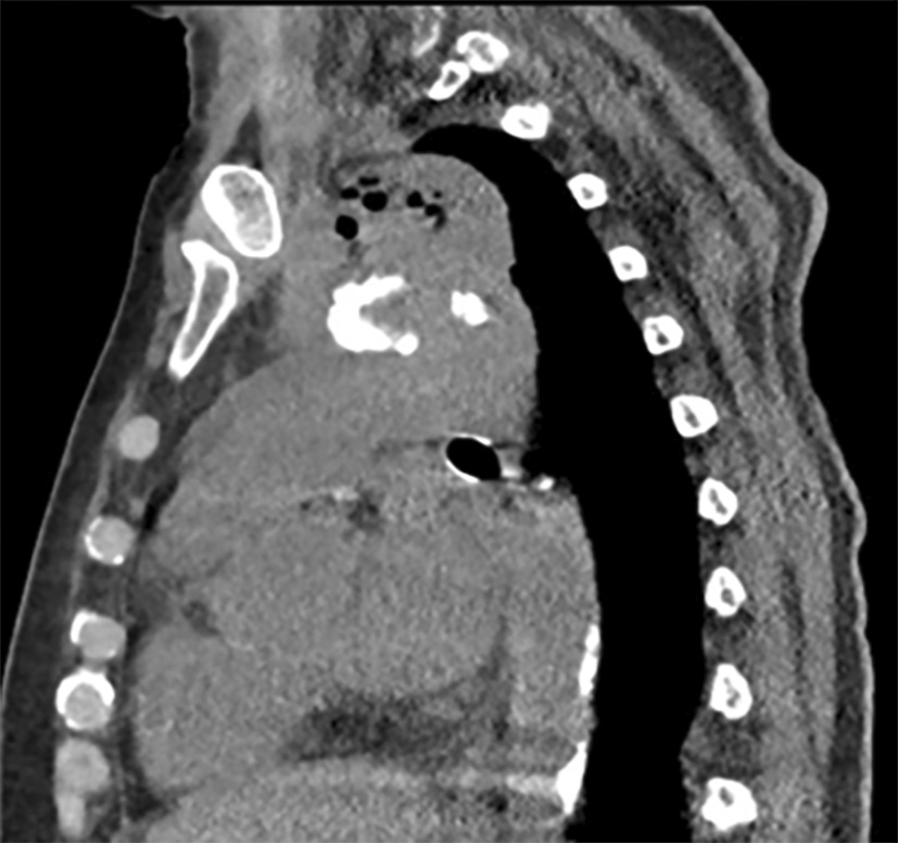
Cavitating superior mediastinal lesion with gas locules favouring infective cause.

Single sagittal slice demonstrating the position of the superior mediastinal/aortic arch lesion at the level of the left subclavian origin.
Single sagittal slice showing the large cavitating superior mediastinal/aortic arch lesion.
Confirmation of mycotic thoracic aortic arch aneurysm with contrast-filling of the aneurysm sac at the level of the left subclavian artery origin.
Confirmation of mycotic thoracic aortic arch aneurysm with a large contrast-filling aneurysm sac.
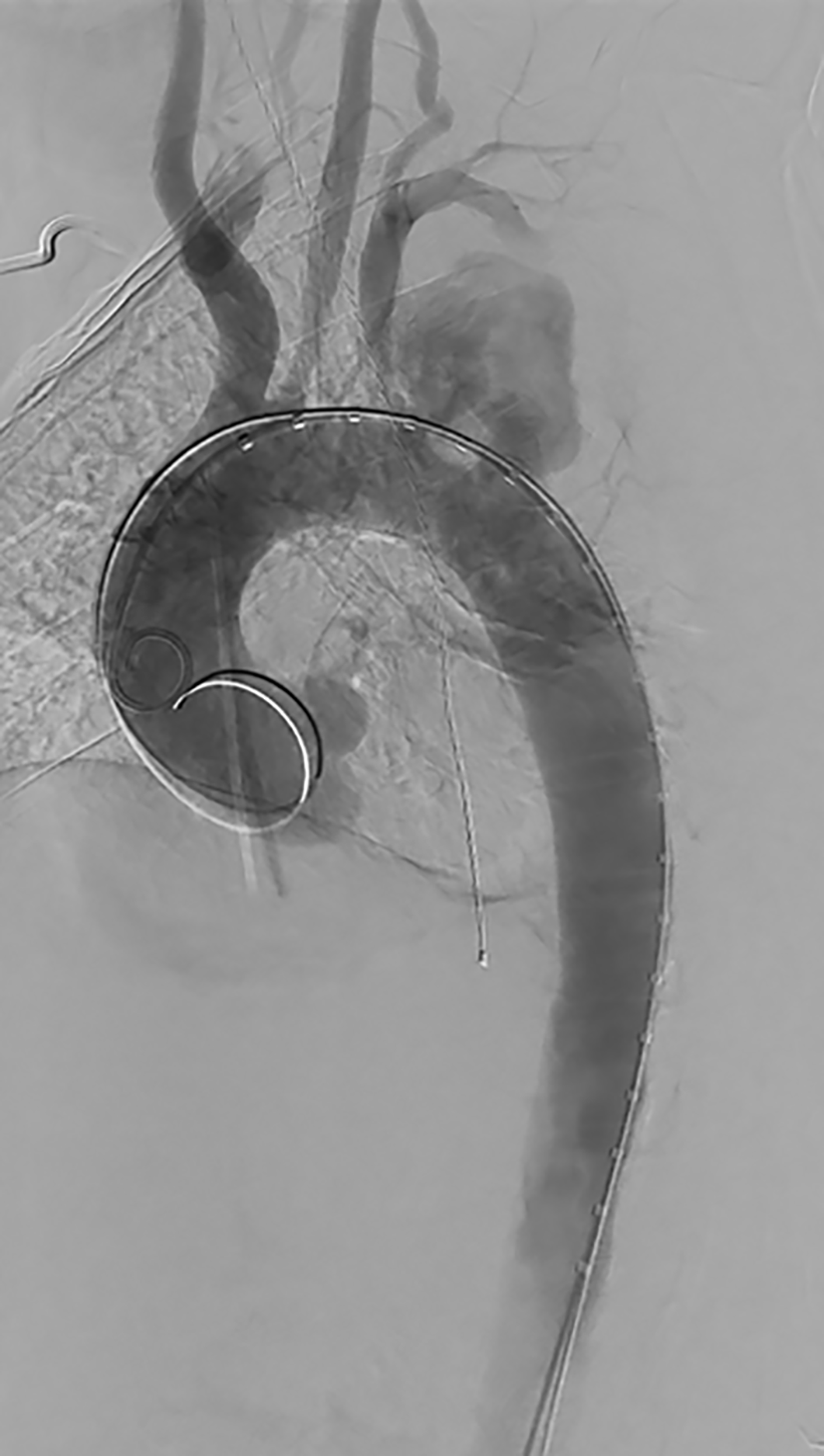
Large mycotic thoracic aortic arch aneurysm demonstrated at the level of the left subclavian artery origin.
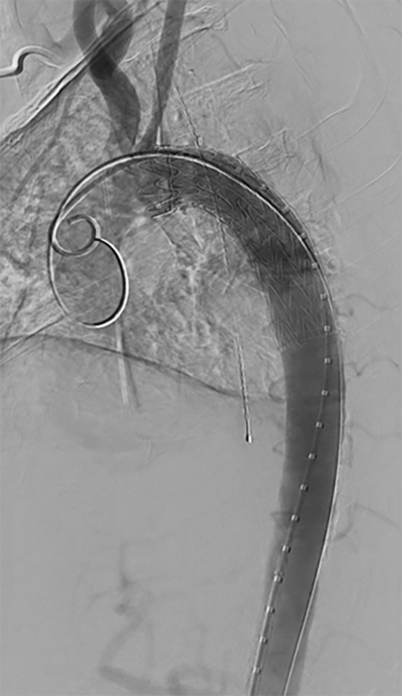
Patent thoracic stent graft with vascular plug at left subclavian artery origin with excluded aneurysm sac without endoleak.
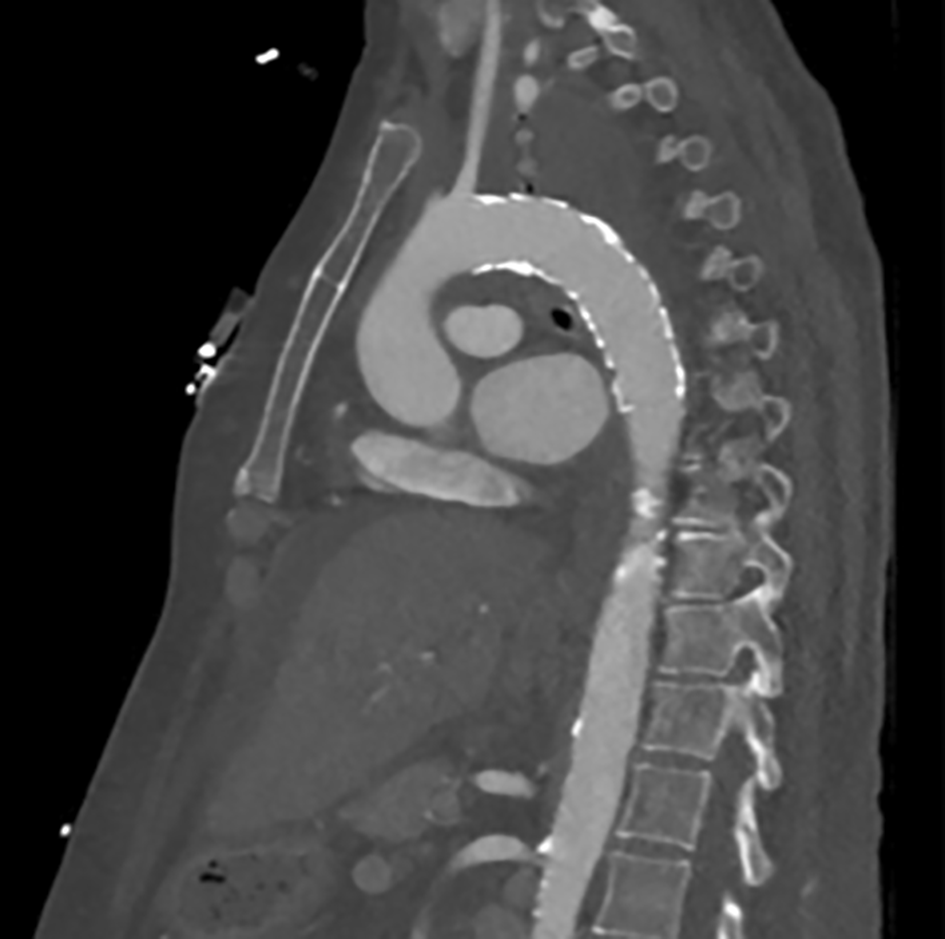
Patent thoracic endovascular stent graft with exclusion of the aneurysm sac without endoleak and successful embolization of the left subclavian artery origin with a vascular plug.
MTAA is associated with rapid expansion and rupture of the aorta,1 with the thoracic aorta and aortic arch being particularly critical sites because of their proximity to vital structures of the head, neck, spine, and upper extremities.
The management of ruptured MTAA is complex and challenging, with early diagnosis and timely operation being the key factors for patient survival in aortic sepsis.4–6 The mainstay of treatment is antibiotics and surgery.1 Traditional surgical management comprises open repair with local debridement and in situ or extra-anatomic revascularization in combination with sternotomy, circulatory arrest, or cardiopulmonary bypass, which carries significant perioperative morbidity and mortality (10-60% mortality at 30-90 days)2 in patients undergoing open surgical repair involving the aortic arch. Due to its minimally invasive nature, especially in critically ill patients at a higher risk for MTAA, TEVAR is increasingly being utilized in its management. Initially used as a temporary measure for definitive repair, the main disadvantage was prosthetic graft placement in an infected field. However, with lifelong antibiotic therapy7 the literature suggests promising long-term outcomes with low re-infection rates after TEVAR, with 81.2% freedom from re-infection at 2 years2 and lower mortality (25% mortality at 30-90 days) compared to open surgery.2 In patients with significant comorbidities, endovascular repair may be the only option available.
In our case, the MTAA was located in zone 2 of the aortic arch, presenting unique challenges for emergent repair. In these thoracic aortic arch aneurysms, signs of Ortner’s syndrome, a rare cardiovocal compressive syndrome, may develop, which can lead to vocal cord paralysis, hoarseness and finally haemoptysis.8 This is due to progressive aneurysmal growth compressing the left recurrent laryngeal nerve between the left pulmonary artery and aorta8 as in this case. She required coverage of the left subclavian artery origin and Amplatzer Vascular Plug (Abbott Vascular, Abbott Park, IL) embolization of the left subclavian artery to ensure augmentation of the proximal landing zone, as well as a decreased risk of type Ia endoleak. She did not require revascularization despite zone 2 coverage concurrently or postoperatively, and some data suggest that TEVAR and left subclavian artery coverage without bypass are associated with lower rates of complications and mortality.9
TEVAR can be successfully and safely utilized in the emergent and definitive management of a ruptured zone 2 MTAA, especially in comorbid and critically unwell patient populations, with special consideration given to the unique anatomy of aneurysms involving the aortic arch.
Written informed consent for publication of the case report and any associated images was obtained from the patient.
| Views | Downloads | |
|---|---|---|
| F1000Research | - | - |
|
PubMed Central
Data from PMC are received and updated monthly.
|
- | - |
Is the background of the case’s history and progression described in sufficient detail?
Yes
Are enough details provided of any physical examination and diagnostic tests, treatment given and outcomes?
Yes
Is sufficient discussion included of the importance of the findings and their relevance to future understanding of disease processes, diagnosis or treatment?
Yes
Is the case presented with sufficient detail to be useful for other practitioners?
Yes
Competing Interests: No competing interests were disclosed.
Reviewer Expertise: Vascular disease
Is the background of the case’s history and progression described in sufficient detail?
Yes
Are enough details provided of any physical examination and diagnostic tests, treatment given and outcomes?
Yes
Is sufficient discussion included of the importance of the findings and their relevance to future understanding of disease processes, diagnosis or treatment?
Yes
Is the case presented with sufficient detail to be useful for other practitioners?
No
References
1. Smolock CJ, Chen G, Anaya-Ayala JE, Martinez K, et al.: Successful endovascular repair of two ruptured thoracic aortic aneurysms in nonagenarians.Ann Vasc Surg. 2011; 25 (5): 697.e9-12 PubMed Abstract | Publisher Full TextCompeting Interests: No competing interests were disclosed.
Reviewer Expertise: Neuroimaging and neurointervention
Alongside their report, reviewers assign a status to the article:
| Invited Reviewers | ||
|---|---|---|
| 1 | 2 | |
|
Version 1 28 Feb 25 |
read | read |
Provide sufficient details of any financial or non-financial competing interests to enable users to assess whether your comments might lead a reasonable person to question your impartiality. Consider the following examples, but note that this is not an exhaustive list:
Sign up for content alerts and receive a weekly or monthly email with all newly published articles
Already registered? Sign in
The email address should be the one you originally registered with F1000.
You registered with F1000 via Google, so we cannot reset your password.
To sign in, please click here.
If you still need help with your Google account password, please click here.
You registered with F1000 via Facebook, so we cannot reset your password.
To sign in, please click here.
If you still need help with your Facebook account password, please click here.
If your email address is registered with us, we will email you instructions to reset your password.
If you think you should have received this email but it has not arrived, please check your spam filters and/or contact for further assistance.
Comments on this article Comments (0)