Keywords
Abdominal pain, Appendicitis, Child, Complication, Diagnosis, Differential, Ovarian Neoplasms, Teratoma
Abdominal pain, Appendicitis, Child, Complication, Diagnosis, Differential, Ovarian Neoplasms, Teratoma
Mature teratomas are a common type of ovarian tumor in children1 and are often diagnosed incidentally on imaging, which demonstrates specific findings.2 Ovarian teratomas are characterized by slow and subtle growth patterns, frequently resulting in delayed diagnosis.3 Symptomatic cases may involve complicated or large masses.3 Some forms of presentation, particularly in prepubescent girls and in the case of large lesions, make diagnosis more difficult. Computed tomography and magnetic resonance imaging (MRI) scans can be beneficial.3
Teratoma complications may arise, especially in cases of large tumors.2 Spontaneous rupture occurs in only 3.8% of the cases.2 This low rate is attributed to the presence of a thick protective capsule.2 Rupture may occur in the peritoneal cavity or a hollow organ in the surrounding area. Potential predisposing factors for such accidents include ischemia and necrosis of the mass wall, infection, trauma, and malignant transformation.4 It can present with an acute abdomen or chronic granulomatous peritonitis.5
We report a case of a ruptured mature right ovarian teratoma causing an acute abdomen that was misdiagnosed as appendicular abscess. Here, we discuss the clinical and radiologic non-specificity of these tumors in the context of complications and aspects of confusion to help surgeons anticipate such an intraoperative surprise. We also support the possibility of conservative treatment, even in the presence of such complications.
A 12-year-old previously healthy girl presented to our emergency room with a 3-day history of worsening abdominal pain predominantly located in the right iliac fossa. There was no family history of the evaluation. Her menarche occurred at 11 years of age, her menstrual cycles were regular, and her last menstrual period was one week ago.
The patient described acute onset of abdominal pain with no concept of trauma, accompanied by fever, vomiting, and diarrhea.
On physical examination, normal hemodynamic parameters were maintained, the patient was febrile at 38.8°C and had generalized tenderness, especially over the right iliac fossa. There was a palpable, firm, painful mass in the right iliac fossa that extended for at least 130 mm, with no signs of inflammation at this site. Examination of the genitals revealed a pubescent girl with patent hymen.
An emergency biology test showed that the white blood cell count (WBC) and C-reactive protein (CRP) level were significantly elevated at 16800/mm3 (4000–10000/mm3) and 140 mg/L (normal value <5 mg/L), respectively.
Given this clinical context, palpable mass, and biological anomalies, we suspected acute appendicitis complicated by an abscess. Immediate abdominal ultrasound performed by a non-experimented radiologist showed poorly limited collection with a heterogeneous echogenic content seat of echogenic spots related to extra-digestive air bubbles ( Figure 1). The collection was 123 × 154 mm in size, occupying the right lower hemiabdomen. Intraperitoneal fat infiltration and a finely echogenic, moderately abundant effusion were also observed. Given the size of the collection, identification of the appendix or the right ovary was not obvious.

The patient was perfused, monitored, and administered parenteral antibiotic therapy. The patient was then operated on via laparotomy using a McBurney incision. Intraoperatively, effusion was widespread and consisted of serous fluid, sebum, and calcium debris. The patient also had a perforated right ovarian teratoma. The mass was large, measuring 150 × 120 mm, with a unique small capsule perforation ( Figure 2). The tumor displaced the surrounding intact ovarian parenchyma, and there were no signs of adnexal torsion. The appendix appeared normal and hidden by the mass. The contralateral ovary was completely normal, and there were no metastases or peritoneal granulations.

Extemporaneous histological examinations were unavailable; however, there was no evidence of malignancy. Based on the patient’s age and aforementioned operative findings, we opted for conservative treatment. The mass was removed ( Figure 3), leaving the surrounding normal ovarian tissue in place, and peritoneal cleaning was performed.

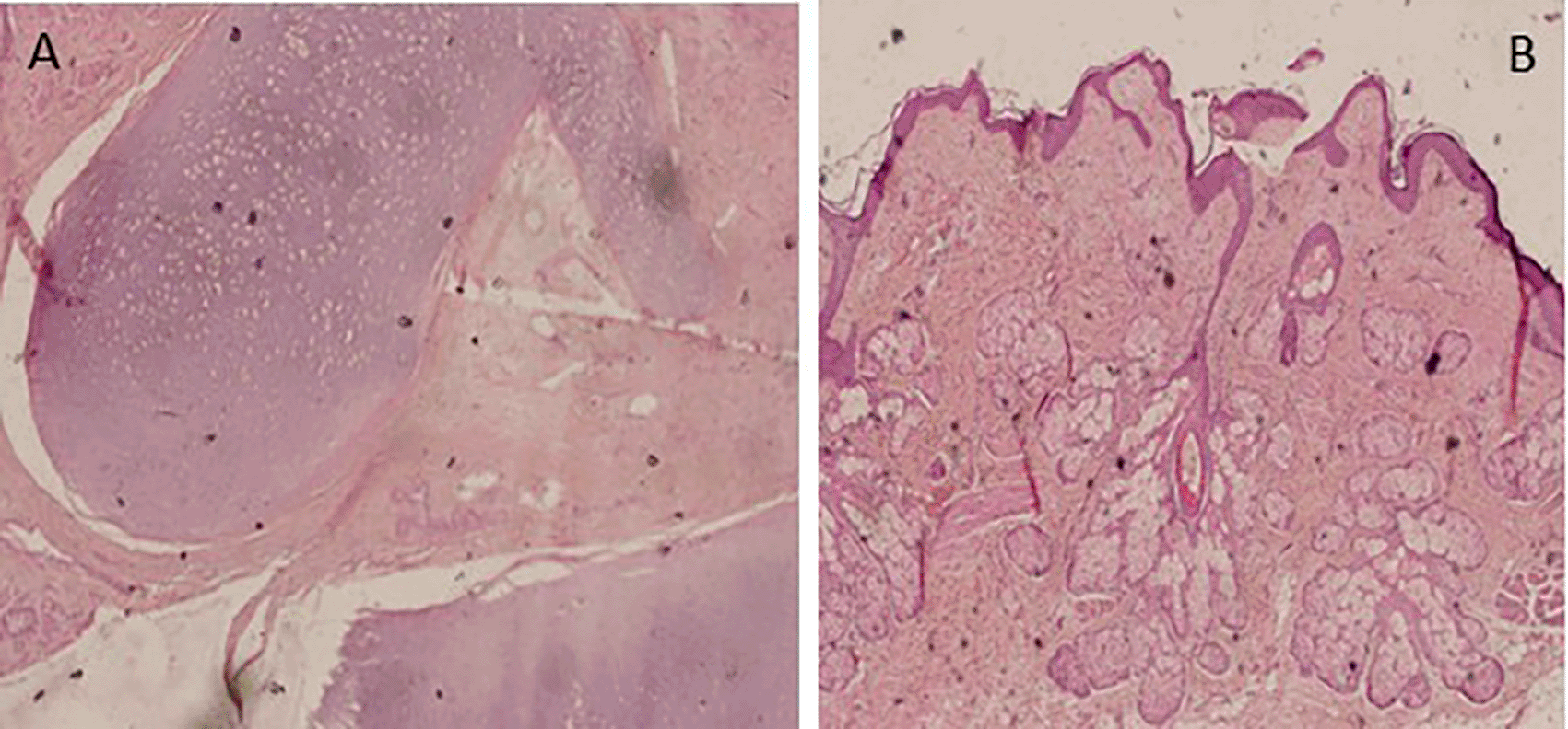
The postoperative course was uneventful, and the patient was discharged three days later. Tumor markers (AFP, HCG, CA19-9, CA-125, LDH, and Inhibin) tested immediately after surgery were negative. Histology ( Figure 4) and peritoneal lavage samples confirmed the benign nature of teratoma containing a mixture of mature benign tissues (e.g., cartilage, sebaceous glands, pancreatic tissue), as well as the complete removal of the mass. Clinical and ultrasound checkups were free of any homo-or contralateral recurrence, with a follow-up period of twenty-three months.
Rupture of an ovarian teratoma is not a common cause of acute abdomen in females. Herein, we discuss the contribution of thorough clinical and radiological investigations to the assessment of this rare condition. The distinction between acute appendicitis and teratoma ovarian rupture is the focus of this case report. Our second concern was to advocate for conservative ovarian surgery, even when faced with this complication.
One of the most common pediatric surgical emergencies is acute appendicitis, including complicated cases.6–9 In prepubertal girls, gynecologic emergencies are the most important differential diagnosis.7 Complicated right ovarian teratoma may be an imitator of acute appendicitis, as in the case of our patient and others reported in the literature, due to the nearness of the appendix and the ovary.4,7 Several factors may contribute to this clinical-radiological confusion. From a clinical point of view, a collection in the right iliac fossa in a child, even in a girl, is initially suggestive of appendicular abscess.10 However, apyrexia, the patient’s general and hemodynamic stability, the absence of vomiting, and local signs of inflammation contradict this diagnosis.10
Pain in the right iliac fossa is the first sign of appendicitis. This pain is usually tolerated at onset and is continuous and progressively worse,11 as described by our patient. Nevertheless, it is crucial to investigate the concept of intermittent and mild chronic abdominal pain characterized by a feeling of abdominal heaviness, generally associated with ovarian mature teratoma.12,13 This pain can be attributed to the compressive effect of the large mass.7 Although the acute, sudden appearance of abdominal pain in a girl should draw attention to the possibility of ovarian torsion,12 a more frequent condition, any modification in the characteristics of the sub-chronic right-sided abdominal pain, which becomes intense and accompanied by vomiting or nausea, is suggestive of rupture of an ovarian teratoma.14 These circumstances may have a confusing clinical presentation, particularly when mimicking an acute abdominal situation. In such cases, imaging is either inconclusive or not allowed by the state of emergency. Fever and transit disorders may also occur in these complicated teratomas owing to inflammatory and peritoneal irritation processes.7
On imaging, abdominal plain radiography can have an informative role as it may show calcifications and deviation of the digestive structures in relation to the mass.6 However, this type of imaging is not routinely performed for acute right iliac fossa pain. Abdominal ultrasound is the first imaging modality requested for girls with acute febrile abdominal pain, given its common gynecological etiology.9 For acute teratoma rupture, the detection of discontinuity in the tumor wall is a diagnostic key5,15; however, this was not obvious in our case. Another radiologic feature that deserves more attention is the non-visibility of the appendix, which may be located outside the right iliac fossa or even obscured by the intestine or a large mass, as in our case. A gynecological cause is still expected in such cases.
If this condition is allowed, further investigations are required and will be more useful for diagnosis. Computed tomography (CT) is a more sensitive imaging modality, revealing acute ruptured hemoperitoneum with floating fat globules.2,5,15 Tumors with only a small focus rupture may cause granulomatous chronic peritonitis, which appears to be tuberculosis or peritoneal carcinomatosis on imaging.5,14,15 Pelvic Magnetic Resonance Imaging is used to better characterize the mass and detect fat16; however, it is not systematically available and may require general anesthesia in children. Laparoscopy is considered a useful diagnostic tool for cases in which imaging is not accurate. Regardless of the diagnostic tool, when evaluating right iliac fossa pain in females, especially those of prepubertal age, clinicians should consider gynecologic causes to improve diagnosis precision.
In most cases, mature ovarian tumors are treated conservatively; the surgical approach may be either laparotomy or laparoscopy, and both approaches have comparable complication rates.13,17 Single-incision laparoscopic surgery has also proven to be effective in pelvic tumor surgery18; however, the choice of approach depends on the patient and the characteristics of the mass.
The usefulness of ovarian sparing surgery in the pediatric population has been approved considering the long-term consequences of unilateral oophorectomy and the potential risk of contralateral ovarian tumor development.17,19 If the mass is well limited and can be safely removed from the ovarian parenchyma, conservative treatment is appropriate, even in the case of large ruptured benign tumors.17,19 One review reported that ovarian-sparing surgery is associated with minimal recurrence and repeat surgery rates.19 Preoperatively, apart from complications, the benign nature of the mass is determined based on symptoms, radiologic features, and tumor markers. However, in the case of an acute presentation, the challenge remains to distinguish between a benign and malignant mass intraoperatively, as was the case with our patient. In these cases, the surgeon's estimation and pathologist's analysis will determine the therapeutic option.
Our case report may not present a complete review of all possible outcomes, which may limit the ability to draw definitive conclusions. Nevertheless, it provides a detailed example of an unusual presentation of an ovarian teratoma. This may help clinicians to better recognize the diagnostic challenges of these tumors and guide their treatment decisions when encountering similar situations.
Numerous incidents are associated with ovarian teratomas, especially when they are large. These circumstances may have a confusing clinical presentation, particularly when mimicking an acute abdominal situation. In such cases, imaging is either inconclusive or not allowed by the state of emergency. In young girls, acute abdominal pain should be given more attention because of the common occurrence of gynecological pathologies. Conservative treatment for benign complicated teratomas should be considered even with an intraoperative diagnosis.
The patient’s perspective was documented in the form of a video (available if requested). The translated text is as follows: The ailment in question afflicted me three years ago, and I am now fully recovered. I recall experiencing severe right abdominal discomfort accompanied by other symptoms including fever and vomiting. On arrival at the emergency room, the physician suspected acute appendicitis and recommended surgical intervention. I initially experienced trepidation, but as the recommended analgesic treatment took effect, my condition began to improve, thereby providing me with greater reassurance. The patient’s condition was satisfactory following the surgical procedure. We did not anticipate the existence of such a sizable cyst. I was fortunate to have been discharged from the hospital in a timely manner so that I could return to my academic duties.
| Views | Downloads | |
|---|---|---|
| F1000Research | - | - |
|
PubMed Central
Data from PMC are received and updated monthly.
|
- | - |
Provide sufficient details of any financial or non-financial competing interests to enable users to assess whether your comments might lead a reasonable person to question your impartiality. Consider the following examples, but note that this is not an exhaustive list:
Sign up for content alerts and receive a weekly or monthly email with all newly published articles
Already registered? Sign in
The email address should be the one you originally registered with F1000.
You registered with F1000 via Google, so we cannot reset your password.
To sign in, please click here.
If you still need help with your Google account password, please click here.
You registered with F1000 via Facebook, so we cannot reset your password.
To sign in, please click here.
If you still need help with your Facebook account password, please click here.
If your email address is registered with us, we will email you instructions to reset your password.
If you think you should have received this email but it has not arrived, please check your spam filters and/or contact for further assistance.
Comments on this article Comments (0)