Keywords
Osteopetrosis, Fracture, Fracture management
Osteopetrosis (OP) is a rare genetic disorder characterized by increased bone density. Monitoring is the only alternative in these patients because of bone fragility, which is a source of frequent complications.
This study aimed to describe the fracture profile, possible complications, and management in this group of patients.
We conducted a retrospective, descriptive study including 21 fractures in 8 OP patients managed in our department between 1996 and 2022, with a minimum follow-up of 2 years. Patient data included age, sex, history of surgery, fractures, intraoperative difficulties encountered, and complications.
All of our patients were young adults with a mean age of 28.4 years and an M/F ratio of 3:1. A total of 21 fractures (8 patients with OP) were managed in our department. The femur was the most frequent fracture site. The management of these fractures is surgical. Plate osteosynthesis is the most common indication. Three fractures were treated by orthopaedics. There were high rates of intraoperative and post-operative complications.
Fractures in patients with OP often involve the long bones. As this is a rare disease, there are few studies on the appropriate management of fractures in this population. Most studies are case series with a small number of cases. Osteosynthesis is the recommended treatment for these fractures despite the risk of failure. Therefore, effective preoperative planning is essential. Great care must be taken when synthesizing these fractures to avoid intraoperative complications.
Fractures in patients with osteopetrosis present a challenge to orthopaedic surgeons. Planned surgery enables the appropriate synthesis of fractures. Long-term follow-up is essential for these patients to detect complications at an early stage.
Osteopetrosis, Fracture, Fracture management
Osteopetrosis (OP), or marble bone disease, is a rare inherited genetic disorder characterized by increased bone density.1 This is due to defects in the development and function of osteoclasts.1 Osteopetrosis occurs in approximately 1 in 300.000 births.2
To date, there has been no definitive cure for OP.3 For most patients, the treatment involves complications, such as fractures, which are observed in 75% of patients.3 Treatment of these fractures is difficult and associated with a high rate of complications.4
We aimed to describe the fracture profile, possible complications, and difficulty of management in patients with osteopetrosis.
This was a retrospective, descriptive study including patients followed for OP and managed for fractures at our institute over a 27-year period between 1996 and 2022, with a minimum 2-year follow-up. We excluded patients with a follow-up of less than 2 years and those with missing data.
The diagnosis of OP was either already known and for which patients were regularly followed up or made following multiple fracture consultations and confirmed by radiological assessment. Patient data included age, sex, history of surgery, and fracture characteristics (type, site, and number).
Indications for the management of patients with osteopetrosis and intraoperative challenges have been reported. We also recorded all the complications encountered.
Clinical and radiological follow-up was carried out systematically in all patients at the second, sixth, and twelfth weeks postoperatively and at the final follow-up.
Data were analyzed using SPSS software version 26.0.
Patient anonymity was maintained during data collection. Written informed consent was obtained from all the participants.
The various parties involved in this work declare that they have no conflict of interest.
Based on these criteria, 21 fractures in eight patients with OP were included in our study. All patients were young adults in their second to fourth decade of life, with a mean age of 28.4 years, 17–42 years). They were predominantly male with an M/F ratio of 3:1. Four patients had a history of orthopaedic treatment for childhood fractures.
A kinship was present in our series. Two patients had brothers. For the others, we found no family ties, but on questioning, four patients reported a fairly high rate of fractures in certain members of their families and were not followed up at our institute.
The majority of fractures involved the femur (17 fractures), followed by both leg bones (3 fractures). Upper-limb involvement is rare. We report a single case of supra- and intercondylar fracture of the left elbow.
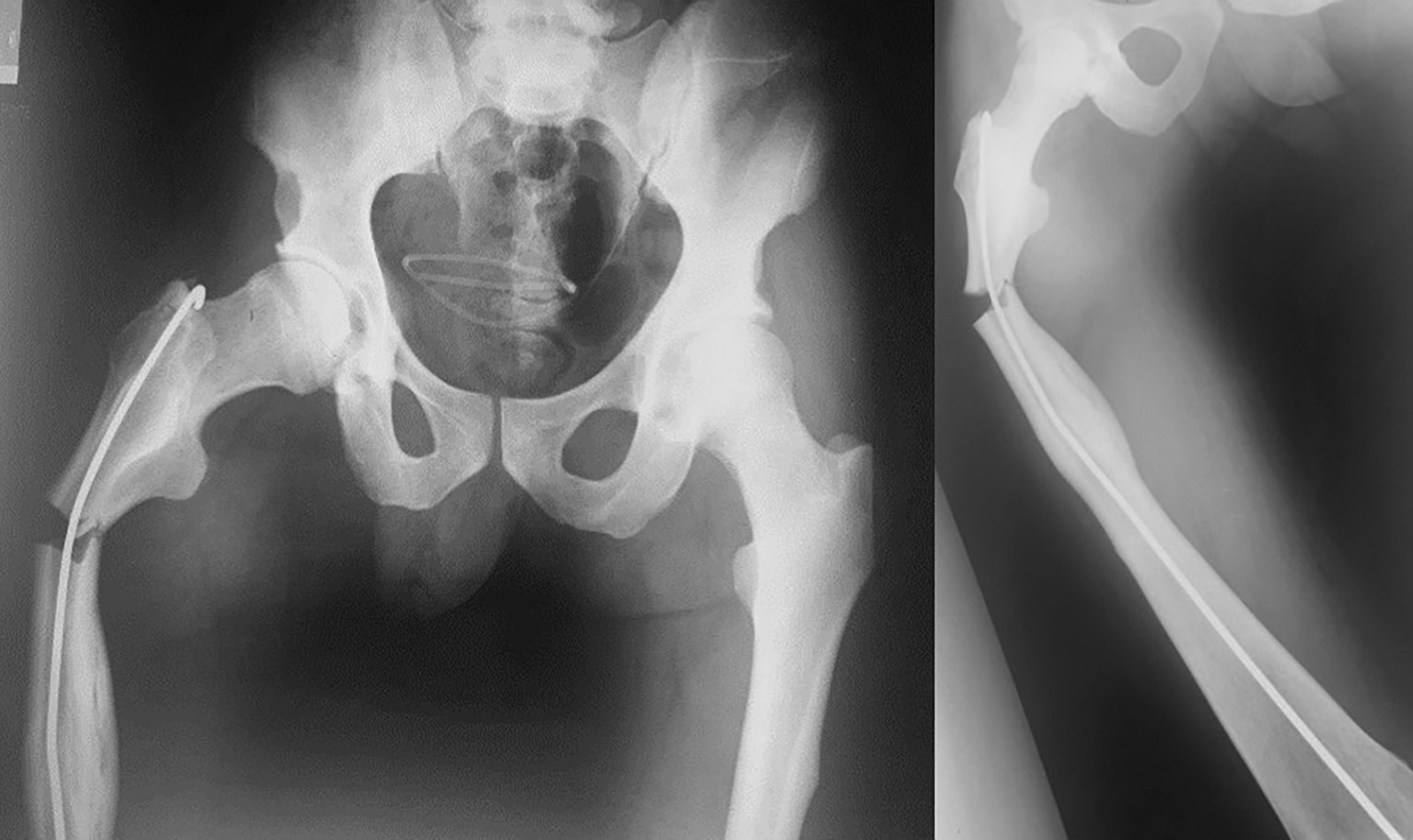
The management of these fractures is surgical. Initial plate osteosynthesis was the most common indication (16 fractures), with a Dynamic Compression Plate (DCP) of 4.5 mm for diaphyseal fractures and a Dynamic Hip Screw (DHS) for subtrochanteric fractures ( Figure 1 and Figure 2).
Only one patient was treated with an external fixator for an open fracture of the two leg bones, and another patient was treated with intramedullary pinning with a poor radiological result, necessitating repeat surgery ( Figure 3). Three fractures (one femoral and two 2-leg fractures) were initially treated orthopaedically.

Owing to the hardness of the bone, there was a high rate of intraoperative incidents involving broken instruments and implants, especially drill bits. However, tapping is difficult. The tap broke twice. We also reported breakage of intraosseous screws ( Figure 4).
Given the hardness of the bone, we used certain devices to perforate the bone, such as tungsten drill bits, square points, burs, and taps.
Intraoperative bleeding was significant in most patients. Surgery in one patient was complicated by hemorrhagic shock, requiring transfusion of 6 packed red blood cells and a stay in the surgical intensive care unit.
Post-operative complications were mainly represented by:

A summary of the management of each patient is given in Table 1.
| Gender | Age at fracture | Fracture | Treatment | Evolution/Complications |
|---|---|---|---|---|
| 1st case Male ( Figure 1,3,5) | 23 | Fracture of proximal 1/3 of right femur | Intramedullary pinning with a single pin, given the impossibility of inserting another pin | Poor radiological result; Revision by removal of the pin and insertion of a 7-hole plate |
| Subplate fracture at 4 months post-op; 9-hole anterior plate inserted without removing lateral plate; consolidation after 6 months | ||||
| 30 | Mid-diaphyseal fracture of left femur | Synthesis with 7-hole lateral plate | Fracture under plate at 2 years post-op; Removal of old plate and synthesis with longer plate (9 holes) | |
| Plate fracture at 4 months post-op with no notion of trauma; Removal of plate and synthesis with 12-hole plate | ||||
| Pseudarthrosis; Cancellous contribution and insertion of a new plate; consolidation after 9 months | ||||
| 2nd case Male ( Figure 2) | 25 | Right subtrochanteric fracture | 4-hole nail-plate | Early sepsis; Drainage washed 2 times; Consolidation with hip ankylosis after 4 mouths. |
| 33 | Left subtrochanteric fracture | DHS 4-hole | Pseudarthrosis and plate fracture; therapeutic abstention | |
| 42 | Left supra- and inter-condylar elbow fracture | 2 external and internal plates | Consolidation after 3 months with elbow stiffness and mobility of 20/90 | |
| 3rd case male | 21 | Isolated tibia fracture | Cast | Iterative fracture at 10 months post-trauma; synthesis with 9-hole external plate; consolidation after 4 months |
| 4th case female | 17 | Right femur fracture | Orthopaedic treatment with splint | Consolidation after 4 months |
| 41 | Subtrochanteric fracture | DHS 6 holes | Significant bleeding with hemorrhagic shock and stay in intensive care unit; consolidation after 6 months | |
| 5th case female ( Figure 4) | 21 | Fracture 1/3 proximal right femur | 8-hole external plate | Breakage of 2 screws proximally Fracture under the plate at 2 years post-operatively; Synthesis with 12-hole anterior plate with consolidation at 9 months post-operatively |
| 27 | Mid-diaphyseal fracture of left femur | Synthesis with 12-hole lateral plate | Delayed consolidation + plate breakage; Functional treatment with consolidation at 1 year postoperatively | |
| 29 | Open fracture of 2 leg bones | External tibio-tibial fixator | Delayed consolidation; Walking boot cast then consolidation at 6 months post-op. | |
| 6th case male | 18 | Mid-diaphyseal fracture of left femur | 10-hole lateral plate fixation | Consolidation at 9 months post-op. |
| 24 | Mid-diaphyseal fracture of right femur | Synthesis with lateral plate | Fracture above plate at 4 years post-operatively following mild trauma; plate removed and synthesis with 12-hole plate; consolidation at 6 months post-operatively | |
| 7th case female | 34 | Diaphyseal leg fracture | Cast | Consolidation at 6 months with callus vicus |
| 8th case male | 29 | Mid-diaphyseal fracture of the left femur | 8-hole lateral plate fixation | Consolidation at 1-year post-op |
| 31 | Mid-diaphyseal fracture of right femur | Synthesis with 12-hole lateral plate | Late sepsis at 18 months post-op; Drainage and plate removal |
Osteopetrosis is a rare disease characterized by increased bone deposition in unresorbed calcified cartilage or primary spongiosis.5 Its incidence is approximately 1 in 300.000 births.2 The condition is diagnosed based on radiographic findings of generalized osteosclerosis, mainly affecting the axial skeleton and long bones without involvement of the medullary canal.5 Brittle sclerotic bones are susceptible to severe fractures following relatively minor trauma, which would not result in fractures in healthy individuals.5
Few studies have focused on this subject. Indeed, the majority of publications were case reports.6
A personal history of fractures, as well as a family history, must be carefully considered. The diagnosis is often not made during the first consultation.7 Standard X-rays can guide the diagnosis, showing sclerosis in the long bones, skull, pelvis, and spine, associated with a characteristic “Erlenmeyer flask” appearance of the distal femur, or a “bone-in-bone” appearance in the spine and phalanges.8
Orthopaedic treatment is part of the therapeutic arsenal for these fractures. Indeed, cases of femoral fractures treated with casts and traction have been reported in the literature.9 However, longer recovery periods are required, with immobilization averaging three months and reduction difficult to maintain.5 This treatment can only be considered for diaphyseal fractures of long bones that are only slightly displaced, particularly in children.9
Osteosynthesis is the recommended treatment, despite the risk of failure; however, good preoperative planning and great care during surgery are required to avoid intraoperative incidents.6
Conventional plate fixation has shown limited success.5,6 This could be explained by two factors.
Screw holes and plate ends create stress zones, increasing the risk of fractures.
Plates are prone to fracture because of the high stress they are subjected to during consolidation, which is generally delayed.
Locked titanium plates are a good alternative, as they are less rigid implant systems and therefore less susceptible to damage.5
With regard to drilling, some studies have recommended the use of a high-density metal drill bit, a diamond drill bit or a tungsten carbide drill bit considered to be reliable high-density drill.10 Dawar et al.11 reported the use of multiple drill bits of progressive sizes and introduced the use of powerful high-speed motors to avoid back-and-forth drilling with continuous saline irrigation, thus preventing the problem of thermal necrosis. In addition, self-tapping screws minimize the risk of instrument breakage, eliminating the need for an additional tapping step.5
Plate fractures are generally caused by a short plate applied to the proximal femur.6,12 Because stresses through the proximal femur are very high, any implant that terminates in this region creates stresses at the end of the plate.12 Most authors recommend the use of longer plates to cover the entire bone.6 Table 2 summarizes the difficulties encountered in managing patients with OP and their solutions.
Few authors have described the results of intramedullary pinning in the literature, but they are associated with a high rate of revision surgery.7 Ding et al.4 recommended predrilling the pin under fluoroscopic control.
Intramedullary nailing is recommended to achieve long-term strength. However, it is very difficult to locate the medullary canal in long osteopetrotic bone.13 The procedure is laborious and involves opening the canal using drills and reamers adapted to this bone.13
Bone consolidation takes longer in patients with OP; therefore, the ban on weight-bearing must be prolonged. The time required for bone consolidation on the femoral shaft in these patients is approximately one year.7
Complications associated with fixation in patients with osteopetrosis should be considered when managing these fractures. Early complications consist mainly of significant blood loss, which can lead to hemorrhagic shock due to laborious synthesis and early sepsis of the material due to prolonged operating time.14 Late complications are fairly frequent, mainly delayed consolidation or pseudarthrosis due to the nature of the bone,15 fracture above or below the plate due to the biomechanical constraints imposed by the implant,5 and plate fracture due mainly to pseudarthrosis.12 A less frequent complication is chronic sepsis, explained by some authors as a result of frequent haematological disorders in these patients, leading to disruption of the local vascularization of the bone and, hence, to poor resistance to infection.7 The reoperation rate reported in the literature is 29%.5,16
The limitations of our study lie in its retrospective nature and small sample size; however, this is a rare pathology, with a small series in the literature. A multicenter study would enable better generalization of the results and could potentially lead to a consensus regarding fracture management in patients with OP.
Currently, there is no specific curative treatment for fractures in OP.1 However, a better understanding of the pathology could eventually lead to the development of less invasive treatments with fewer complications.
OP fractures are a challenge for orthopaedic surgeons. Long-term follow-up is essential for the early detection of possible complications.
Orthopaedic treatment may be indicated for fractures of the long bones that are only slightly displaced, particularly in children. In all other cases, surgical treatment with plates remains the gold standard treatment. However, this surgery is lengthy and difficult. The prolonged duration of the operation and risk of infection must be clearly explained to the patient.
Good preoperative planning is essential to anticipate intraoperative technical difficulties. The risks and benefits of each fixation modality must be considered during planning.
Written informed consent was obtained from all participants (or their legal guardians) for publication of this study, including relevant clinical details and accompanying images.
| Views | Downloads | |
|---|---|---|
| F1000Research | - | - |
|
PubMed Central
Data from PMC are received and updated monthly.
|
- | - |
Is the background of the cases’ history and progression described in sufficient detail?
Partly
Are enough details provided of any physical examination and diagnostic tests, treatment given and outcomes?
Yes
Is sufficient discussion included of the importance of the findings and their relevance to future understanding of disease processes, diagnosis or treatment?
Yes
Is the conclusion balanced and justified on the basis of the findings?
Yes
References
1. Funck-Brentano T, Zillikens M, Clunie G, Siggelkow H, et al.: Osteopetrosis and related osteoclast disorders in adults: A review and knowledge gaps On behalf of the European calcified tissue society and ERN BOND. European Journal of Medical Genetics. 2024; 69. Publisher Full TextCompeting Interests: No competing interests were disclosed.
Reviewer Expertise: Pathophysiology and clinical research in bone diseases
Alongside their report, reviewers assign a status to the article:
| Invited Reviewers | |
|---|---|
| 1 | |
|
Version 1 19 Mar 25 |
read |
Provide sufficient details of any financial or non-financial competing interests to enable users to assess whether your comments might lead a reasonable person to question your impartiality. Consider the following examples, but note that this is not an exhaustive list:
Sign up for content alerts and receive a weekly or monthly email with all newly published articles
Already registered? Sign in
The email address should be the one you originally registered with F1000.
You registered with F1000 via Google, so we cannot reset your password.
To sign in, please click here.
If you still need help with your Google account password, please click here.
You registered with F1000 via Facebook, so we cannot reset your password.
To sign in, please click here.
If you still need help with your Facebook account password, please click here.
If your email address is registered with us, we will email you instructions to reset your password.
If you think you should have received this email but it has not arrived, please check your spam filters and/or contact for further assistance.
Comments on this article Comments (0)