Keywords
Periosteal distraction; stroke; neurorehabilitation; case report
Periosteal distraction; stroke; neurorehabilitation; case report
Stroke alters the brain's structure, affecting various systems. Despite improved survival rates, it remains a leading cause of long-term disability worldwide.1 Post-stroke deficits include cognitive, sensory, visual, and language impairments, with motor impairments being the most common.2 Most patients experience some spontaneous recovery, defined as time-dependent improvement in body function and activity.3 However, this recovery is often incomplete, and neurological recovery rates vary. Motor impairments are the most frequently diagnosed and studied, with significant gains occurring within the first 3 months.3 Recovery of visuospatial neglect and orientation typically occurs 5-6 months post-stroke, while improvements in cognition, memory, and language can extend over months to years.3 The optimal restorative strategy is controversial.4 For patients who miss the optimal treatment window, conservative treatment approaches are frequently insufficient, underscoring the critical importance of early and aggressive intervention.4
Periosteal traction (PD) technique has recently been widely used to treat chronic lower limb-threatening ischemia, including diabetic foot5 and arteriosclerosis obliterans.6 It is speculated that PD surgery can effectively promote angiogenesis and tissue regeneration. In clinical practice, we also found that PD postoperative angiography showed significant new blood vessels on the surgical side. Therefore, a reasonable and highly attractive hypothesis is that applying PD technology for stroke while balancing safety and convenience, which would benefit the patient as well as lowering the budget impact. Here we present 3 cases of post-stroke patients that were managed with PD technology in the late chronic stage.
This retrospective study included post-stroke patients who were more than six months post-injury and received PD surgery between June 2024 and February 2025. The diagnosis of stroke was established based on clinical presentations and imaging findings. To thoroughly assess the potential therapeutic effects of PD, we specifically selected patients whose neurological recovery had reached a plateau. We excluded individuals lacking detailed information about the nature of their stroke and those with a history of potential additional brain injuries, such as traumatic brain injury, anoxic brain injury, or subarachnoid hemorrhage. This study received approval from our Institutional Review Board, and written informed consent was obtained from all participants.
The surgical site was located contralateral to the hemiplegic limb. The center of the marked point was positioned 2 cm below the junction of the ear canal and the parietal tubercle. A 3-cm longitudinal scalp incision was made parallel to the parietal tubercle. A vertical incision was then performed through the temporal region and the periosteum, extending approximately 1 cm to the skull. A subperiosteal channel measuring 3 cm in length and 1 cm in width was created in the skull. Two microfractures were drilled into the outer table of the skull, and a periosteal stretch system (Kehui's periosteal stretch system) was inserted into the subperiosteal channel. The stretch screw was advanced into the skull using a screw fixation needle until it reached the inner table. A second screw fixation needle was then inserted parallel to secure the periosteum and the cap-like fascia of the temporal muscle. Refer to Figure 1 for a schematic representation of the surgical procedure.

a-c: After the skin incision, the peroneal aponeurosis and the cranial membrane were transected, exposing the parietal bone; d-f: subperiosteal implantation of the plate. Drill a Φ1.5mm Kirschner wire in the distal hole of distraction plate. Insert a special locking hollow screw along the Kirschner wire and lock it to distal hole, leaving part of the screw exposed outside the skin for periodic postoperative adjustments. Drill another K-wire to a depth of 5 mm into the single-layer cortex in the most-distal hole of distraction plate to prevent lateral rotation of the plate; g-i: skin sutures were performed. Intraoperative X-rays confirmed the accurate placement of the instrument.
Considering the compliance of the periosteum of the skull, we then stretched the plate at a rate of 0.5 mm per day (0.25 mm in the morning, and 0.25 mm in the evening). After 3 weeks of stretching, the distraction plates, and screws were removed. During the distraction period, attention was paid to nursing the wound at the distractor, to prevent complications such as nail tract infection and periosteal infection, and we kept the nail tract and surrounding skin tissue under close observation.
Patient 1 was a 62-year-old male with a history of hypertension and diabetes, diagnosed with a left hemisphere ischemic stroke two years prior. He was receiving long-term treatment with clopidogrel, rosuvastatin, amlodipine, metformin, and inglitazone. Preoperative evaluation revealed right limb dysfunction (right shoulder abduction at 60 degrees, muscle strength graded at 3, and hypertonia), central facial palsy, and speech dysfunction. On the second postoperative day, these symptoms showed significant improvement, with the most notable recovery in facial nerve function and speech. By the 18th postoperative day, right shoulder abduction had improved to 80 degrees, with muscle strength rated at 5. However, a follow-up three months after surgery indicated a deterioration in the patient's symptoms compared to postoperative status, suggesting that PD surgery may need to be performed regularly to maintain efficacy.
Patient 2 is a 68-year-old male diagnosed with a left basal ganglia cerebral infarction six years ago. After undergoing routine rehabilitation training, he achieved partial recovery of function in the affected area. Preoperative evaluation indicated right-sided hemiplegia, characterized by limited shoulder abduction (approximately 70 degrees) and elbow extensor muscle strength rated at grade 3. Wrist extensor muscle strength was also graded at 3, and the patient was unable to extend the right thumb. Additionally, muscle strength in the quadriceps, tibialis anterior, and both long and short fibular muscles was rated at grade 3. By the 20th postoperative day, significant recovery of motor function was observed, including improved shoulder and elbow joint function, partial recovery of thumb extension, and an increase in finger extensor muscle strength to grade 4. Lower limb muscle strength reached grade 5, enabling the patient to ambulate slowly without noticeable gait abnormalities.
Patient 3 is a 54-year-old male with a history of cerebral infarction lasting over ten years, initially managed with long-term oral nifedipine for blood pressure control. Preoperative evaluation revealed reduced muscle strength in the right limbs, with a strength grade of 3 in the right upper limb and 4 in the right lower limb. The patient exhibited limited movement in the four fingers of his right hand (excluding the thumb), which impaired his ability to form a fist. After 14 days of follow-up, significant improvement in the finger function of his right hand was noted compared to its initial state. However, the patient is currently lost to follow-up. To objectively evaluate the impact of PD surgery on cerebral blood perfusion, Patient 3 underwent Computed tomography Perfusion imaging (CTP) of the head both before the surgical procedure and 60 days postoperatively. The CTP results demonstrated that the PD surgery effectively enhanced cerebral blood supply in the patient ( Figure 2).
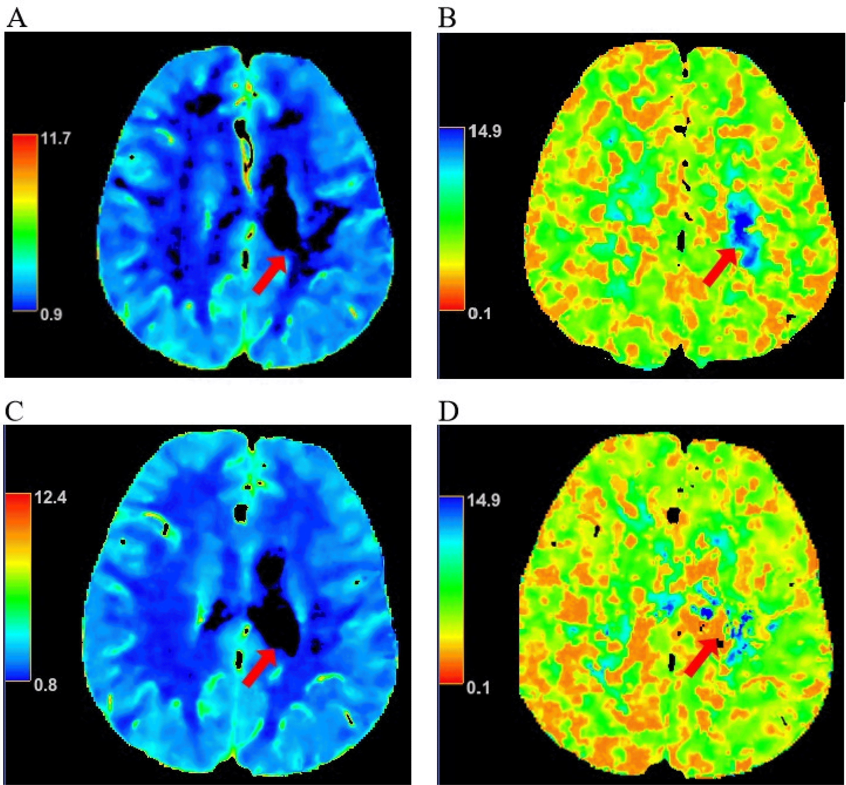
A-B: Preoperative cranial CTP showed decreased MTT and TTP , and decreased CBV and CBF in the left basal ganglia region; C-D: Postoperative cranial CTP showed MTT and TTP were shorter, and CBV and CBF were increased compared with the preoperative period in the left basal ganglia region; MTT: mean transit time, TTP: time to peak, CBV: cerebral blood volume, CBF: cerebral blood flow.
The mechanism by which PD therapy restores brain function is still unclear. However, clinical feedback indicating its rapid effects suggests several possibilities. First, the presence of cortical vessels and the periosteal vascular network may serve as the anatomical basis for the efficacy of PD treatment.7 Second, in strokes, a crucial target for recovery is the penumbra—the area surrounding the infarction. This region, characterized by electrical depletion yet preserved energy metabolism, holds potential for recovery.3 The penumbra, having survived the initial injury, can reorganize to facilitate healing. More importantly, local and remote unaffected areas may also serve as targets.3 PD may enhance the function of the penumbra and related regions by promoting neuroplastic changes. Finally, findings from the Cranial Bone Maneuver program indicate that PD may enhance nerve function in stroke patients by promoting the regeneration of meningeal lymphatic vessels, improving their drainage capacity, and effectively alleviating neuroinflammation.8
We propose that PD is a novel alternative surgical option for treating neurological dysfunction in stroke patients. Key advantages include: i) the small incision minimizes complications associated with skin incisions; ii) the traction devices needed for periosteal stretching consist only of screws and standard locking plates, thereby imposing minimal economic burden on patients; iii) using periosteal infiltration local anesthesia with PD can decrease anesthesia-related complications, reduce the need for general anesthesia, and significantly alleviate the strain on patients' vital organs.
However, the results are limited by the small sample size and short follow-up period. Additionally, the optimal settings for retractor parameters (e.g., force applied and duration) and the specific patient subgroups most likely to benefit remain unclear. The potential synergistic effects of PD combined with other treatments also warrant further exploration. Further studies are needed to answer these questions.
In conclusion, the preliminary results of our study show that the PD technique has surprising efficacy in improving functional recovery in stroke patients, with great prospects for future application. However, more in-depth basic and clinical studies are needed to optimize protocols and explore potential mechanisms, providing a stronger scientific basis for treating neurological diseases.
| Views | Downloads | |
|---|---|---|
| F1000Research | - | - |
|
PubMed Central
Data from PMC are received and updated monthly.
|
- | - |
Provide sufficient details of any financial or non-financial competing interests to enable users to assess whether your comments might lead a reasonable person to question your impartiality. Consider the following examples, but note that this is not an exhaustive list:
Sign up for content alerts and receive a weekly or monthly email with all newly published articles
Already registered? Sign in
The email address should be the one you originally registered with F1000.
You registered with F1000 via Google, so we cannot reset your password.
To sign in, please click here.
If you still need help with your Google account password, please click here.
You registered with F1000 via Facebook, so we cannot reset your password.
To sign in, please click here.
If you still need help with your Facebook account password, please click here.
If your email address is registered with us, we will email you instructions to reset your password.
If you think you should have received this email but it has not arrived, please check your spam filters and/or contact for further assistance.
Comments on this article Comments (0)