Keywords
Mycoplasma bovis, Diagnosis, Biomarker, P48 protein.
This article is included in the Pathogens gateway.
Mycoplasma bovis, Diagnosis, Biomarker, P48 protein.
Mycoplasma bovis (M. bovis) is a significant opportunistic pathogen responsible for causing various diseases and economic losses in cattle worldwide, affecting both dairy and beef industries (Ammar et al., 2021; Ohtsuka et al., 2019; James et al., 2013). This bacterium is known for its ability to cause a wide range of clinical manifestations, including bovine respiratory disease (BRD), mastitis, arthritis, and otitis media, affecting cattle of all ages. Initially, M. bovis was considered to be a harmless commensal microorganism found in the lungs of healthy cattle. However, over time, it has emerged as one of the primary pathogens contributing to multiorgan infections in cattle, with serious implications for animal welfare and farm productivity (Maunsell & Chase, 2019). The ability of the pathogen to cause chronic, often subclinical, infections and its capacity for immune evasion complicates its management and control (Buchenau et al., 2010; Rosengarten et al., 1994). This is further exacerbated by its resistance to common antimicrobial treatments and the lack of an effective vaccine, making rapid and accurate diagnostic methods essential for managing M. bovis infections (Fasogbon et al., 2024; Zengin et al., 2024; Hasoon et al., 2023). Despite the availability of diagnostic tools, current methods often fail to provide accurate, early detection, which hinders effective disease management and control measures (Shirani et al., 2020; Fu et al., 2014c).
The identification of diagnostic biomarkers is essential to improve the detection and management of M. bovis infections. Such biomarkers can be proteins, antigens, or nucleotides that indicate disease conditions. Proteins and protein antigens offer compelling advantages as diagnostic biomarkers because they directly reflect biological activity. Proteins actively participate in physiological processes, meaning that changes in their levels can provide real-time insights into the presence or progression (Lino et al., 2022). Additionally, many proteins undergo post-translational modifications, which can serve as highly specific indicators of certain conditions, offering a level of detail that is not available with DNA or RNA biomarkers (Chu et al., 2021). Moreso proteins are often more easily detectable in accessible body fluids, such as blood or urine, making them practical for routine diagnostics (Lino et al., 2022).
Various molecular methods, including polymerase chain reaction (PCR), enzyme-linked immunosorbent assay (ELISA), and proteomic approaches, have been employed to identify potential protein and gene markers specific to M. bovis (Kumar et al., 2014; Zubair et al., 2020). However, variability in these markers, such as variable surface proteins (Vsp) and nucleomodulins, has introduced complexities in achieving consistent and reliable diagnostics (Lu et al., 2024; Behrens et al., 1994).
This study aimed to consolidate the available literature on potential diagnostic proteins and protein antigens of M. bovis. By evaluating the identification methods and diagnostic efficacy of these proteins, we sought to bridge the gap between bench research and clinical applications, thereby offering insights into the most promising candidates for future diagnostic tools (Ayan et al., 2023; Dawood et al., 2023).
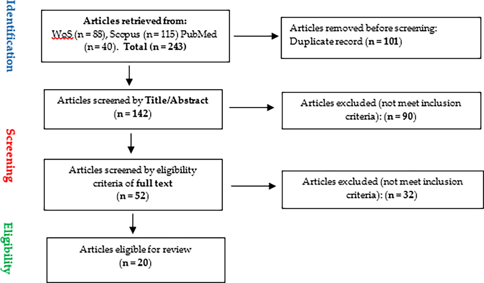
A comprehensive literature search was conducted on 31st August, 2024 to identify the diagnostic proteins relevant to M. bovis. The search targeted the major scientific databases Scopus, Web of Science (WoS), and PubMed, using the search terms Mycoplasma bovis, diagnosis/detection, and protein. The search fields were the title, abstract, and keywords. Boolean operators (AND/OR/NOT) were used to develop search strategies ( Table 1) tailored to each database (Fasogbon et al., 2023, 2024; Mitaki et al., 2024). No date limits were applied, and the search was restricted to original, peer-reviewed articles published in English. The search yielded 243 articles from the three databases. This included 115 articles from Scopus, 88 from the Web of Science, and 40 from PubMed.
The paper selection process followed the Preferred Reporting Items for Systematic Reviews and Meta-Analyses (PRISMA) 2020 guidelines (Page et al., 2021; Swase et al., 2025). The inclusion and exclusion criteria ( Table 2) were used to select articles to be reviewed in the study. A total of 101 duplicate articles were removed owing to duplicated entries from the databases, resulting in 142 unique articles for screening. The titles and abstracts of the remaining 142 articles were screened to determine their relevance to the research objective. The screening was done by two independent reviewers (Ilemobayo Victor Fasogbon and Angela Mumbua Musyoka) via an online platform, Rayyan (https://new.rayyan.ai/) (Johnson & Phillips, 2018) to ensure objectivity. As illustrated in Figure 1, after screening, 52 articles were selected for the full-text review. A thorough review of the full text of the 52 articles was conducted. After excluding irrelevant articles based on the inclusion criteria, 20 articles were deemed eligible for inclusion in the systematic review, as outlined in Table 2.
Two independent reviewers (Ilemobayo Victor Fasogbon and Angela Mumbua Musyoka) extracted the following data from each of the 20 included articles:
• Diagnostic proteins or protein antigens: Specific protein or protein antigens reported in the study were identified.
• Method of identification: Techniques involved in the identification of proteins or protein antigens, such as PCR, ELISA, proteomic analysis, or western blotting.
• Diagnostic evaluation: The study assessed the diagnostic sensitivity and specificity of the identified proteins or protein antigens.
• Author, year of study and other meta-data : The identity of the study and year of study were also identified from the 20 included records.
The primary sequence of the P48 protein was downloaded from the UniProt database with the ID A0A1S6K889. Protein properties were predicted using the Expasy ProtParam tool (Gasteiger et al., 2005). The server was used to predict the number and composition of amino acids in the sequence, molecular weight (Da), theoretical isoelectric point (pI), instability index, aliphatic index, GRAVY (Grand Average of Hydropathicity), total number of negatively charged residues (Asp + Glu), total number of positively charged residues (Arg + Lys), extinction coefficient (M−1 cm−1, assuming all pairs of Cys residues form cystines), extinction coefficient (M−1 cm−1, assuming all Cys residues are reduced), estimated half-life (mammalian reticulocytes, in vitro), estimated half-life (yeast, in vivo), and estimated half-life (Escherichia coli, in vivo) of the protein.
This systematic review analyzed 20 studies that focused on identifying diagnostic proteins and protein antigens for M. bovis proteins. These proteins have varying levels of diagnostic potential, and have been identified through diverse methods such as PCR, recombinant protein expression, proteomics, and ELISA. Table 3 presents a summary of the diagnostic molecules identified in the 20 studies along with their detection methods and evaluation of diagnostic efficacy.
| Putative diagnostic protein | Detection methods | Diagnostic evaluation | References |
|---|---|---|---|
| P48 | Recombinant protein expression, PCR, ELISA, ELAA | Yes | James et al. (2013), Fu et al. (2014a, 2014b), Robino et al. (2005) |
| Variable Surface Proteins | Monoclonal antibodies, SDS-PAGE, Western blotting, PCR | Yes | Behrens et al. (1994), Brank et al. (1999), Buchenau et al. (2010) |
| MbovP Proteins | Proteomics, 2D Gel Electrophoresis, MALDI-TOF-MS, Immunoinformatics | Yes | Zhang et al. (2021), Zubair et al. (2020), Shirani et al. (2020), Lysnyansky et al. (1996) |
| MilA Protein | Phage-based ELISA, Western Blotting | Yes | Wawegama et al. (2014), Farzaneh et al. (2022) |
| Interleukin-17A | Proteomic Analysis | Yes | Ohtsuka et al. (2019) |
| Pyruvate Dehydrogenase E1 | 2D Gel Electrophoresis, Immunoblotting, MALDI-TOF-MS | Yes | Sun et al. (2014) |
| Cyclooxygenase-2 & | Immunohistochemical Labeling | No | Rodriguez et al. (2015) |
| GAPDH Protein | RFLP, PCR | No | Perez-Casal and Prysliak (2007) |
| Alpha Enolase | Cloning, Expression, Purification, Immunofluorescence Labeling | No | Song et al. (2012) |
| Haptoglobin & Serum Amyloid A | - | Yes | Saher et al. (2024) |
| uvrC | PCR | Yes | Subramaniam et al. (1998) |
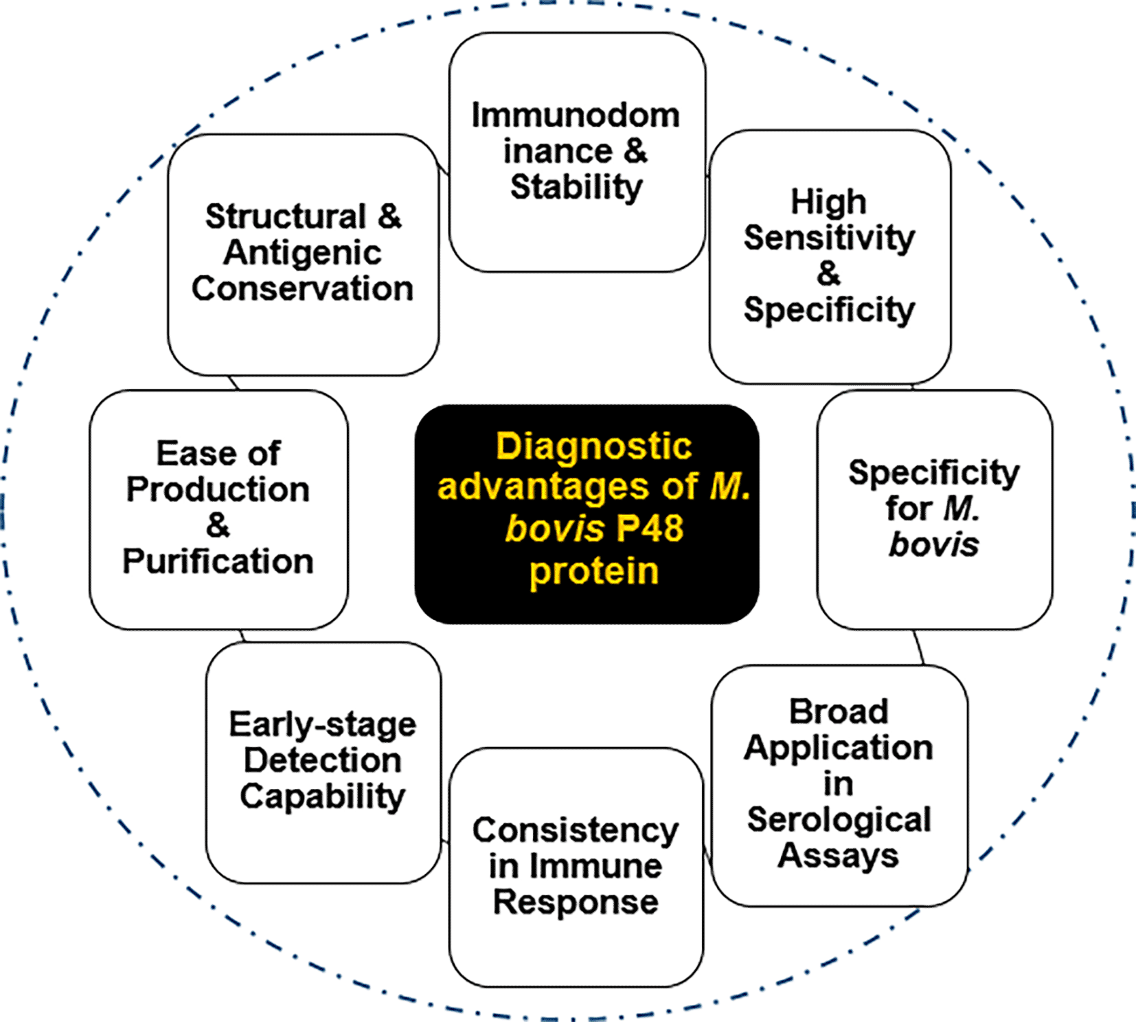
• P48 protein: The P48 protein is one of the most frequently reported diagnostic biomarkers for M. bovis. P48 is a well-established immunodominant lipoprotein in M. bovis and is used as a specific marker for infection, with consistent results showing a high sensitivity and specificity (James et al., 2013; Fu et al., 2014c). Detection methods such as PCR, competitive ELISA, and ELAA have been used to accurately identify P48 in infected cattle. These findings strongly support the inclusion of P48 in routine diagnostic assays for M. bovis because of its comparative advantages ( Figure 2).
• Variable Surface Proteins (VSPs): VSPs (including VspA, VspB, and VspC) are a group of surface-expressed proteins in M. bovis that undergo antigenic variations. They play a key role in immune evasion but are useful for diagnostics because they trigger immune responses. Their variability allows for the differentiation between strains and can aid in epidemiological tracking. They are often detected using techniques like Western blotting and SDS-PAGE (Behrens et al., 1994; Brank et al., 1999). However, their variability poses challenges for standardization of diagnostic assays.
• MbovP proteins: The MbovP proteins, including MbovP274, MbovP570, and others, were identified primarily using proteomic approaches. These proteins are surface-exposed and are involved in pathogen-host interactions. MbovP proteins are valuable biomarkers for detecting M. bovis because of their role in stimulating host immune responses, making them potential targets for serodiagnostic tools. These proteins are increasingly recognized as promising biomarkers owing to their high antigenicity and unique expression patterns in M. bovis (Zhang et al., 2021; Zubair et al., 2020). MALDI-TOF-MS and immunoinformatic approaches have significantly enhanced the identification of these proteins, leading to their potential inclusion in future diagnostic assays.
• MilA protein: The MilA protein, an immunogenic lipase, was shown to demonstrate strong reactivity in phage-based ELISA assays (Wawegama et al., 2014; Farzaneh et al., 2022). MilA is a putative virulence factor whose diagnostic relevance is tied to its immunogenicity, making it an excellent candidate for inclusion in serological diagnostics of M. bovis.
• Interleukin-17A: IL-17A is a cytokine involved in the immune response and has been identified as a putative diagnostic marker. In M. bovis infections, elevated levels of IL-17A may indicate an active inflammatory response, making it a potential marker of immune system involvement in disease progression. (Ohtsuka et al., 2019).
• Pyruvate dehydrogenase E1: Pyruvate dehydrogenase E1 (PDH E1) component beta subunit (PDHB) in M. bovis has been identified as an immunogenic protein with high immune reactivity. It shows potential for diagnostic applications owing to its ability to elicit a strong immune response, making it a candidate for assays such as indirect ELISA (Sun et al., 2014).
• Cyclooxygenase-2 (COX-2) and GAPDH protein: COX-2 are involved in the inflammatory response. Elevated COX-2 expression during M. bovis infection may indicate inflammation, whereas M. bovis GAPDH results in a strong humoral immune response; hence, it is a putative candidate for diagnostics and vaccine development (Rodriguez et al., 2015; Perez-Casal and Prysliak, 2007).
• Alpha enolase: Alpha-enolase in M. bovis diagnosis plays a crucial role as an adhesion-related factor in M. bovis diagnosis. It facilitates adherence by binding to the host plasminogen, aiding in the pathogen’s colonization and evasion of host defenses (Song et al., 2012). This protein, found on the bacterial surface and in membrane fractions, is essential for understanding the pathogenicity of M. bovis.
• Haptoglobin & Serum Amyloid A (SAA): Haptoglobin is an acute-phase protein that responds to inflammation and infection, making it useful for detecting the early stages of the disease. For an accurate diagnosis, monitoring haptoglobin levels alongside other biomarkers, such as serum amyloid A, enhances sensitivity (Saher et al., 2024). Their increase during M. bovis infection makes them non-specific but valuable indicators of ongoing inflammation and disease severity.
• uvrC product: Deoxyribodipyrimidine photolyase, encoded by uvrC, plays a crucial role in the repair of DNA damage caused by UV light in M. bovis. This enzyme is a part of the excision DNA repair system, making it a specific target for genetic detection. In diagnostic applications, the uvrC gene is utilized for PCR-based assays to detect and identify M. bovis efficiently (Subramaniam et al., 1998).
The P48 protein was found to possess excellent molecular properties, as predicted by the Expasy ProtParam tool, thus corroborating the findings of this systematic review.
The theoretical isoelectric point (pI) of the P48 protein was calculated to be 7.14 ( Table 4), suggesting that it carries no net charge at near-neutral pH, which is a common feature of proteins that interact in a neutral physiological environment. A key indicator of protein stability is its instability index, calculated as 23.64. This value categorizes the P48 protein as stable, which is advantageous for its potential use in biosensor applications, where structural integrity over time is critical. Furthermore, the aliphatic index (83.40 points to a high level of thermostability, which is particularly beneficial for a protein that may be exposed to varying environmental conditions during diagnostic assays. The grand average hydrophobicity (GRAVY) score of -0.349 indicates that the P48 protein is mildly hydrophilic, which may influence its solubility and interaction with other biomolecules in aqueous environments. The protein extinction coefficient, a critical parameter for determining protein concentration via absorbance at 280 nm, is 40,340 M−1 cm−1, assuming that all cysteine residues form disulfide bonds. This same value applies if cysteine residues are reduced, which is consistent with the minimal presence of cysteine. The calculated absorbance at 280 nm for a 0.1% solution of the protein was 0.981, providing a useful measure for quantifying proteins in laboratory settings. Regarding the protein’s turnover, the estimated half-life varies significantly across different biological systems: 5.5 hours in mammalian reticulocytes (in vitro), 3 minutes in yeast (in vivo), and 2 minutes in Escherichia coli (in vivo). These differences highlight the varying stability of the P48 protein across different cellular environments, which could influence its function and longevity during infection or when used as a diagnostic tool. The amino acid (all amino acids named in this work and their 3-letter representation are presented in supplementary file 2) (Fasogbon et al., 2025). composition of P48 ( Table 5) provides further insight into its structure and function. Alanine (Ala) and lysine (Lys) were the most abundant residues, comprising 10.6% of the total amino acids. This high lysine content may contribute to the ability of the protein to interact with negatively charged molecules such as DNA or acidic polysaccharides. The presence of cysteine (Cys), although minimal at 0.3%, is significant for the formation of disulfide bonds, which could further stabilize the structure of the protein. The atomic composition, represented by 1853 carbon, 2901 hydrogen, 485 nitrogen, 568 oxygen, and 2 sulfur atoms, contributed to the overall structural and functional attributes of the protein, resulting in a chemical formula of C1853H2901N485O568S2.
Given these detailed molecular properties, P48 has emerged as a robust candidate for diagnostic applications, particularly in the context of M. bovis infection. The stability and specific molecular characteristics of P48 make it an ideal target for the development of peptide aptamers and biosensors.
The P48 protein emerged as a highly valuable diagnostic biomarker for M. bovis infection because of its distinct immunological characteristics. One of the most significant advantages of P48 is immunodominance. Several studies, including those by Robino et al. (2005) and Fu et al. (2014c), have demonstrated that P48 elicits a strong and consistent antibody response in infected cattle, making it a reliable target for diagnostic assays, such as ELISA and Western blotting. Unlike the variable surface proteins (VSPs) of M. bovis, which are known for their high-frequency phase variation, P48 remains structurally and antigenically conserved across different field isolates of M. bovis (Robino et al., 2005). This stability makes P48 a more reported candidate for diagnostic applications than VSPs, which may evade detection due to their variability (Behrens et al., 1994). In their development of a direct competitive ELISA (Dc-ELISA), Fu et al. (2014c) confirmed that P48 showed no cross-reactivity with other pathogens such as Mycoplasma agalactiae or Mycoplasma bovirhinis, further enhancing its diagnostic specificity. The stability and immunodominance of P48 enable it to serve as a reliable biomarker for detecting M. bovis infections at various stages of the disease, from early to chronic phases. The P48 protein has been utilized in the development of diagnostic assays that offer high sensitivity and specificity. For example, Fu et al. (2014c) developed a monoclonal antibody-based competitive ELISA that specifically detected M. bovis antibodies with a cut-off inhibition value of 32%, providing significantly higher sensitivity than other commercial ELISA kits. Additionally, a study by Robino et al. (2005) showed that recombinant P48 (rP48) can detect both IgM and IgG antibodies, with IgM antibodies appearing within 6–9 days of infection. This early antibody response position, P48, is an effective marker for early-stage detection, offering a diagnostic window for many other proteins, such as IL-17A (Robino et al., 2005; Ohtsuka et al., 2019).
Specificity of the P48 protein for M. bovis is another critical advantage that distinguishes it from other proteins used in diagnostic assays. As demonstrated by Fu et al. (2014c), the P48 protein does not cross-react with other common pathogens, such as bovine viral diarrhea virus or infectious bovine rhinotracheitis virus, ensuring that diagnostic assays based on P48 can confidently distinguish M. bovis infections from other respiratory pathogens common in cattle populations. In contrast, proteins such as GAPDH and COX-2, although useful in some contexts, may lack the same level of specificity for M. bovis (Perez-Casal & Prysliak, 2007). P48 has proven to be a versatile protein for the development of various serological assays, including indirect ELISA, sandwich ELISA, and competitive ELISA (Fu et al., 2014c; Robino et al., 2005). The ability to incorporate P48 into different assay formats allows its application in both research and clinical settings. For example, Enzyme-Linked Aptamer Assays (ELAA) using P48 have demonstrated high sensitivity and have the potential to replace traditional antibody-based assays because of the ease of aptamer production and stability under various conditions (Fu et al., 2014c; Zhang et al., 2012). When compared to other diagnostic molecules, such as variable surface proteins (VSPs), MbovP proteins, or IL-17A, P48 stands out because of its consistency and robust immune response. Although variable surface proteins are valuable in distinguishing chronic infections, they are prone to antigenic variation, which can reduce the reliability of diagnostic assays based on these proteins (Behrens et al., 1994; Buchenau et al., 2010). Similarly, while MbovP proteins have shown promise in proteomic studies, they lack broad validation across multiple diagnostic platforms that P48 has achieved (Zhang et al., 2021; Zubair et al., 2020). Additionally, P48 has been shown to be highly effective when combined with recombinant protein expression systems, allowing easy production and purification for large-scale diagnostic applications.
This systematic review highlights the diagnostic potential of the P48 protein for Mycoplasma bovis infections in cattle, supported by its immunodominance, stability, and high specificity in serological assays. However, limitations include the small number of included studies (n = 20), methodological heterogeneity across diagnostic techniques, and a lack of longitudinal data assessing P48’s performance over time. The review also did not formally evaluate study bias, and its computational predictions require experimental validation. Despite these constraints, the findings advocate for P48 as a promising biomarker, encouraging the development of P48-based diagnostic tools like ELISA and lateral flow tests. Future research should focus on large-scale validation and comparative studies with other biomarkers to optimize M. bovis detection and improve cattle disease management.
This study establishes P48 as a more reported diagnostic protein for Mycoplasma bovis detection, based on systematic literature evaluation and computational analysis. Compared to alternative biomarkers, such as VSPs, MbovP proteins, and MilA, P48 consistently demonstrated higher specificity, stability, and immunodominance in infected cattle.
Computational analysis confirms P48’s structural and physicochemical advantages, including stability (instability index: 23.64), thermostability (aliphatic index: 83.40), and moderate hydrophilicity (GRAVY score: -0.349). These properties make P48 a reliable target for serological assays, biosensors, and recombinant antigen-based diagnostics.
Given these findings, we strongly recommend further experimental validation and development of P48-based diagnostic tools for early, accurate, and cost-effective M. bovis detection. Future research should explore P48’s integration into next-generation biosensors to improve their diagnostic efficiency in veterinary medicine.
| Views | Downloads | |
|---|---|---|
| F1000Research | - | - |
|
PubMed Central
Data from PMC are received and updated monthly.
|
- | - |
Provide sufficient details of any financial or non-financial competing interests to enable users to assess whether your comments might lead a reasonable person to question your impartiality. Consider the following examples, but note that this is not an exhaustive list:
Sign up for content alerts and receive a weekly or monthly email with all newly published articles
Already registered? Sign in
The email address should be the one you originally registered with F1000.
You registered with F1000 via Google, so we cannot reset your password.
To sign in, please click here.
If you still need help with your Google account password, please click here.
You registered with F1000 via Facebook, so we cannot reset your password.
To sign in, please click here.
If you still need help with your Facebook account password, please click here.
If your email address is registered with us, we will email you instructions to reset your password.
If you think you should have received this email but it has not arrived, please check your spam filters and/or contact for further assistance.
Comments on this article Comments (0)