Keywords
Hepatic tuberculoma, chronic hepatitis B, cirrhosis, liver mass, case report
Hepatic tuberculoma, chronic hepatitis B, cirrhosis, liver mass, case report
Tuberculosis remains a major global health concern, particularly in developing countries.1,2 While hepatic involvement by Mycobacterium tuberculosis typically occurs as part of miliary or disseminated disease, isolated hepatic tuberculosis (HTB) is an extremely rare entity, accounting for less than 1% of all tuberculosis cases.3,4 In cases of isolated hepatic involvement, it poses substantial diagnostic challenges due to its nonspecific clinical manifestations and radiological resemblance to primary liver malignancies, such as hepatocellular carcinoma (HCC) and intrahepatic cholangiocarcinoma (iCCA).5–7
The clinical presentation of HTB may include constitutional symptoms, hepatomegaly, or focal hepatic lesions, often indistinguishable from those seen in malignant tumors.8 In patients with underlying chronic liver disease, particularly advanced hepatitis B virus (HBV) related cirrhosis, the appearance of a new liver mass is frequently presumed to be malignant, prompting consideration of surgical or oncologic intervention. In this context, misdiagnosis can result in unnecessary or inappropriate treatment. Hence, histological confirmation remains essential when imaging findings are atypical or equivocal. Here, we present a rare case of isolated HTB mimicking a malignant liver tumor in a patient with compensated HBV related cirrhosis, highlighting the importance of liver biopsy in equivocal presentations.
The patient, an 81-year-old man, had been followed since 2013 for chronic hepatitis B at the stage of compensated advanced chronic liver disease, with liver stiffness measured at 37.2 kPa on FibroScan®. He was treated with Entecavir 0.5 mg/day, with a sustained virological response. His past medical history included type 2 diabetes mellitus, arterial hypertension, dyslipidemia, hypothyroidism, benign prostatic hyperplasia, and cardiac arrhythmia.
He was admitted for evaluation due to rapidly progressive deterioration of his general condition, marked by a 20-kg weight loss over three months, severe asthenia, anorexia, and localized right upper quadrant pain without radiation. On physical examination, general condition was preserved (WHO performance status 1), with normochromic conjunctivae, no jaundice, and no palpable lymphadenopathy. The abdomen was soft with no palpable hepatomegaly but mild tenderness in the right upper quadrant.
Laboratory investigations revealed normal liver tests: aspartate aminotransferase (AST) at 13 U/L, alanine aminotransferase (ALT) at 9 U/L, alkaline phosphatase (ALP) at 135 U/L, and gamma-glutamyl transferase (GGT) at 36 U/L. Total bilirubin was 12 μmol/L, serum albumin 35 g/L, and prothrombin time (PT) was 92%. No biological evidence of systemic inflammation or renal impairment was identified.
Abdominal ultrasound revealed a dysmorphic liver with left lobe atrophy and mildly irregular margins. A 50-mm heterogeneous hypoechoic mass with irregular margins and a Doppler signal was detected in segment IV. A complementary contrast-enhanced computed tomography (CT) scan showed a 47-mm heterogeneous, necrotic mass in segment IV. The lesion was hypodense, exhibiting progressive contrast enhancement, without significant arterial phase wash-in, and capsular retraction adjacent to the lesion, features strongly suggestive of malignancy ( Figure 1). Given the clinical and radiological profile, the primary differential diagnoses included iCCA and fibrolamellar HCC. The lesion was classified as LR-M according to the LI-RADS. Considering the clinical and radiological suspicion of malignancy, serum tumor markers, including alpha-fetoprotein (AFP), carcinoembryonic antigen (CEA), and carbohydrate antigen 19-9 (CA 19-9) were carried out, all of which yielded negative results.
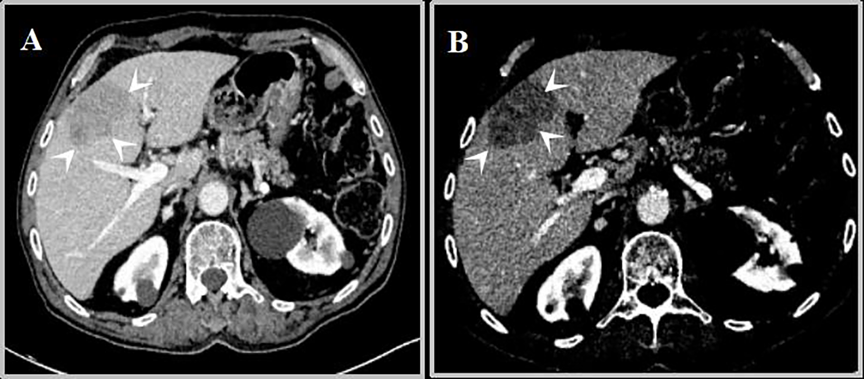
(A) Arterial phase: Subcapsular pseudomass in segment IV without arterial wash-in.
(B) Portal venous phase: Persistent hypodensity of the lesion and adjacent capsular retraction, suggesting malignancy.
Subsequently, a percutaneous liver biopsy was performed, revealing a dense inflammatory infiltrate composed of lymphocytes, plasma cells, neutrophils, and epithelioid granulomas, some centered around caseating necrosis without identification of Mycobacterium tuberculosis on staining. No tumor cells were identified. The histological findings were highly suggestive of HTB ( Figure 2).
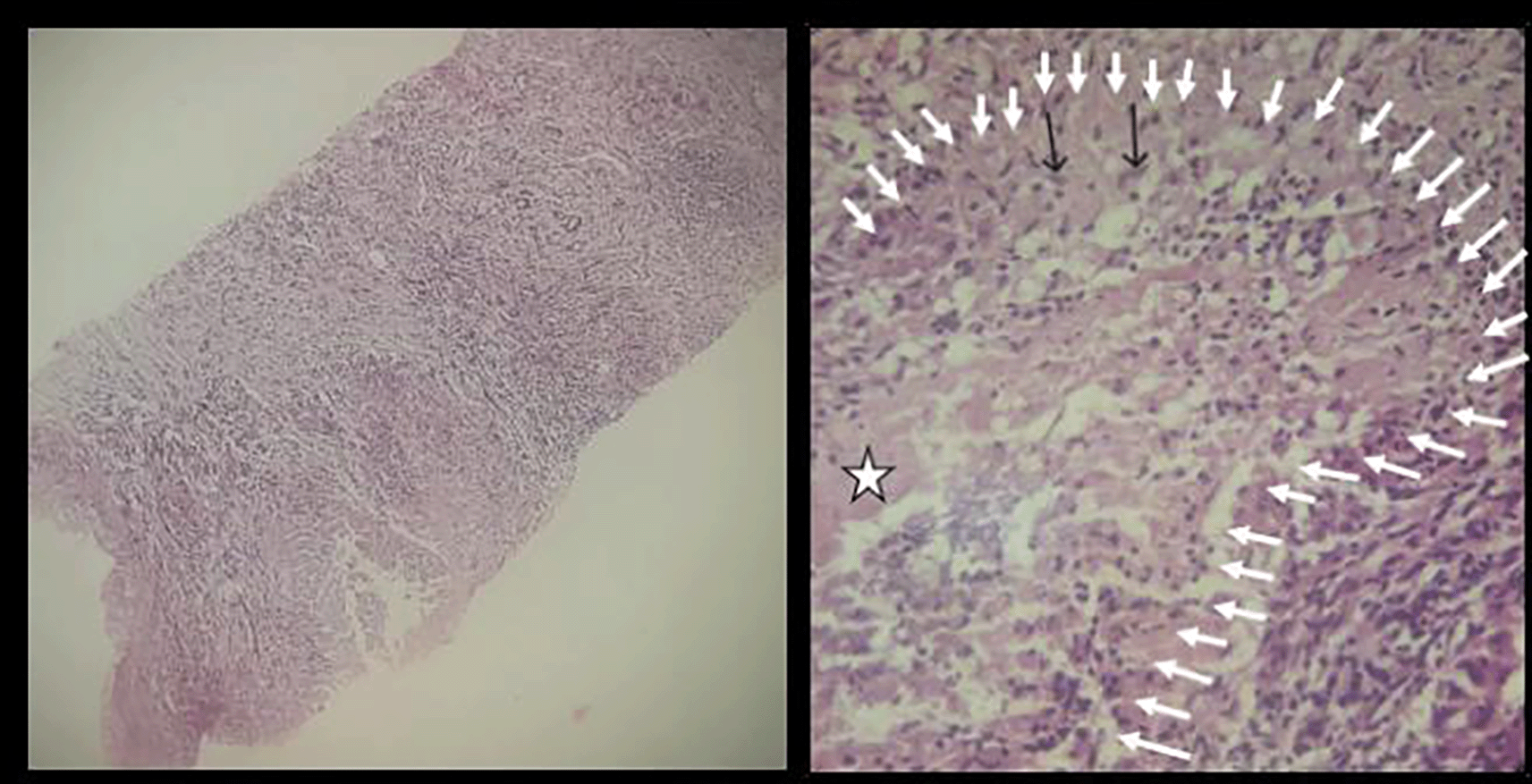
Left (Hematoxylin and Eosin, ×20): Liver parenchyma showing extensive fibrosis with dense inflammatory infiltrate and epithelioid granuloma formation.
Right (Hematoxylin and Eosin, ×40): Well-formed granuloma (white arrows) composed of epithelioid histiocytes (black arrows), centered around caseating necrosis (star).
A full workup for tuberculosis dissemination was negative: sputum examination for Mycobacterium tuberculosis, including acid-fast bacilli (AFB) smear and culture, yielded negative results, and thoracoabdominopelvic CT showed no other tuberculous involvement.
Given the constellation of clinical, biological, radiological, and histopathological findings, including a necrotic hepatic mass with caseating granulomatous inflammation, absence of malignant cells, negative tumor markers, and no evidence of disseminated tuberculosis, the diagnosis of isolated hepatic tuberculoma was retained.
A comprehensive pre-treatment evaluation was conducted, including complete blood count, liver and renal function tests, acetylation testing, and an ophthalmologic examination. The acetylation test recommended a daily isoniazid (H) dose of 150 mg. The full anti-tuberculous regimen, initiated based on histological confirmation, consisted of quadruple therapy (HRZE): rifampicin (R) at 10 mg/kg/day, pyrazinamide (Z) at 20 mg/kg/day, and ethambutol (E) at 15 mg/kg/day, in addition to isoniazid (H) at a fixed dose of 150 mg/day. Liver function reassessed on Days 3, 7, and 10 of therapy showed no abnormalities, allowing the continuation of treatment without modification.
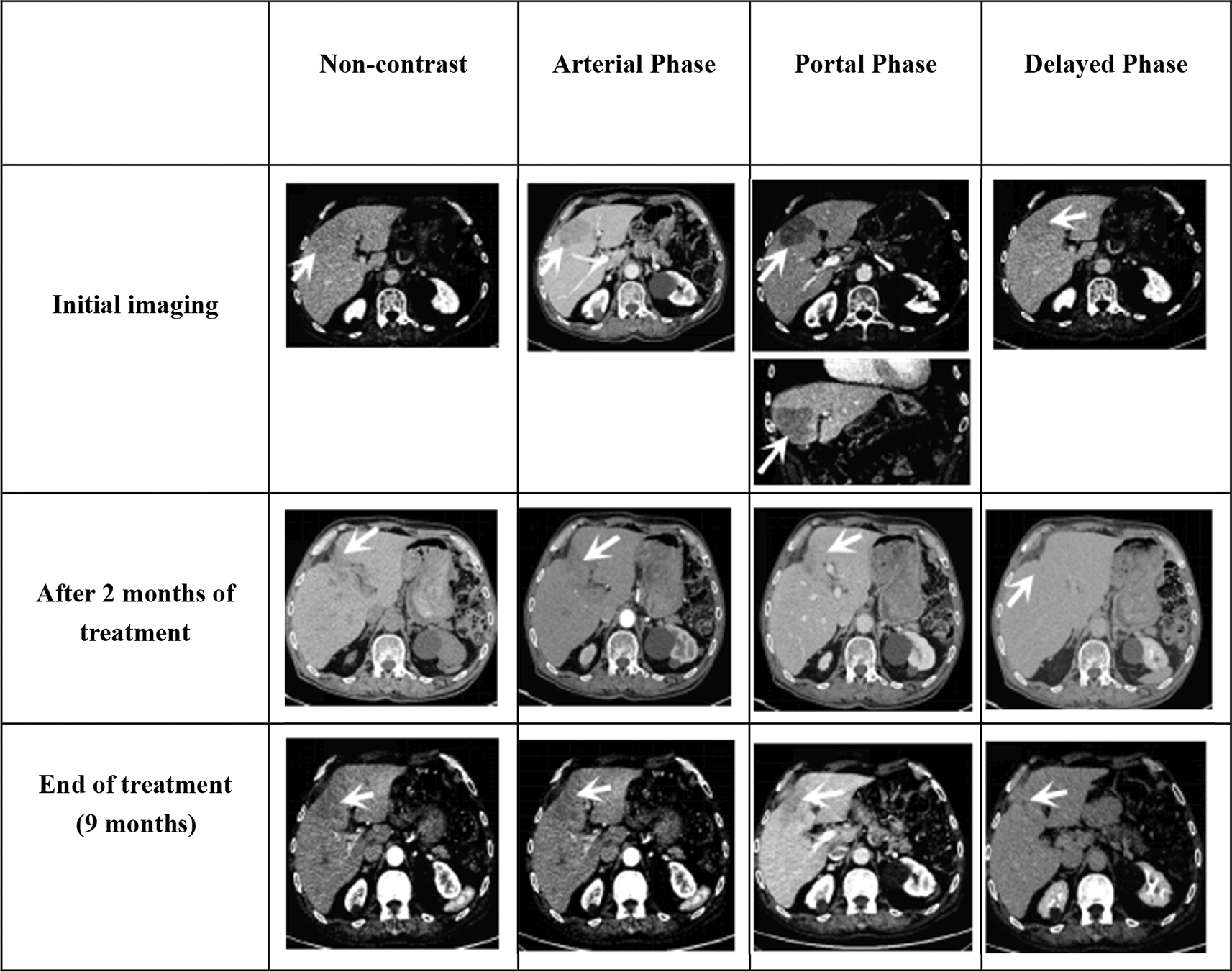
At the 2-month follow-up, the patient showed clear clinical improvement, with good tolerance to treatment and recovery of appetite. A follow-up CT scan, performed after completion of the quadruple therapy and before transitioning to dual therapy, revealed partial regression of the hepatic lesion, which had begun to recover a normal parenchymal appearance ( Table 1).
|
The patient completed a standard 9-month course of antituberculosis therapy (ATT), consisting of two months of quadruple therapy followed by seven months of dual therapy (HR). Follow-up assessments demonstrated favorable clinical and radiological outcomes. Post-treatment CT revealed complete resolution of tuberculous lesions, with only a residual hypodense fibrous scar observed. The absence of contrast enhancement confirmed successful radiological recovery without evidence of active disease ( Table 1).
HTB is a rare extrapulmonary manifestation of Mycobacterium tuberculosis, reported in 50–80% of autopsy cases involving patients who have died from pulmonary TB.9 Despite its relatively high prevalence in postmortem studies, HTB frequently remains asymptomatic and is likely underdiagnosed in clinical settings.10 In recent years, the global resurgence of TB, driven by HIV infection, multidrug-resistant strains, intravenous drug use, and inadequate public health infrastructure, has been associated with an increased incidence of hepatic involvement.1,2,11
HTB predominantly occurs in individuals aged 11 to 50 years, with the highest incidence reported in the second decade of life. Isolated forms of HTB are more frequently observed in patients in their fourth to sixth decades. The condition shows a clear male predominance, with a reported male-to-female ratio of approximately 2:1.8
The classification of HTB varies across the literature. Levine described five forms: miliary tuberculosis, pulmonary TB with hepatic involvement, primary HTB, tuberculoma or abscess, and tuberculous cholangitis.12 In contrast, Reed proposed a simplified classification comprising three forms: HTB associated with generalized miliary disease, primary miliary HTB, and isolated tuberculoma or hepatic abscess.13
The liver’s rich vascularization, abundance of reticuloendothelial cells, and anatomical position at the distal end of the portal circulation predispose it to granulomatous inflammation. However, its relatively low oxygen tension is thought to limit mycobacterial proliferation, which may explain the rarity of isolated HTB.14
Isolated HTB remains an exceptionally rare entity that presents considerable diagnostic challenges, primarily due to its nonspecific clinical manifestations and radiological features that closely mimic primary hepatic malignancies such as HCC and iCCA.5 These difficulties are further compounded in patients with advanced chronic liver disease, particularly in the setting of HBV-related cirrhosis, where space-occupying hepatic lesions are more likely to raise suspicion for HCC. To date, only one case of hepatic tuberculosis (HTB) complicating post-hepatitis B cirrhosis has been reported by Hu et al.8 To the best of our knowledge, the present case represents the second documented instance of this association. This highlights the importance of considering HTB in the differential diagnosis of liver masses in patients with HBV-related cirrhosis and emphasizes the need for histological confirmation before initiating oncologic treatment.
Clinically, HTB often manifests with systemic signs such as fever, significant weight loss, abdominal pain, and hepatomegaly. Less commonly, patients may develop obstructive jaundice or portal vein thrombosis, further complicating differential diagnosis.8,15,16 The considerable overlap between these symptoms and those of HCC frequently contributes to delayed or inaccurate diagnosis. Our patient experienced significant weight loss, asthenia, anorexia, and localized right upper quadrant pain, without fever, jaundice, or lymphadenopathy.
On imaging (ultrasound, CT, or Magnetic resonance imaging (MRI)), HTB can present as solitary or multiple lesions. These lesions often mimic malignancy in their morphology, especially in cirrhotic livers.8,17 Features such as arterial enhancement and venous washout on MRI or contrast-enhanced ultrasound can be misleading, as they are commonly associated with HCC but may also occur in HTB.17 In this case, imaging revealed a hypodense hepatic lesion with progressive contrast enhancement, absent arterial wash-in, and associated capsular retraction, features raising suspicion for iCCA or fibrolamellar HCC.
Laboratory markers may also lack diagnostic specificity. AFP levels, typically elevated in HCC, can also be increased in HTB, as demonstrated in the case reported by Hu et al.8 Therefore, sole reliance on imaging and biomarkers may be insufficient for accurate differentiation between these entities. Our patient had normal serum levels of tumor markers, including AFP, CEA, and CA 19-9.
Histopathology remains the cornerstone for diagnosing HTB. However, liver biopsy may be contraindicated in patients with advanced liver disease due to bleeding risks. When feasible, biopsy often reveals granulomatous inflammation and caseous necrosis, hallmarks of tuberculosis, though these findings are not entirely specific.15,16 AFB staining, while confirmatory, has limited sensitivity, making negative results inconclusive. Unlike diffuse hepatic tuberculosis, in which Mycobacterium tuberculosis is often detectable, hepatic tuberculoma typically lacks direct microbiological confirmation, as AFB staining and cultures are frequently negative.18 In our case, despite the presence of hallmark histopathological features, namely granulomatous inflammation with central caseating necrosis, no AFB were identified, and comprehensive microbiological investigations, including sputum cultures and cross-sectional imaging, revealed no evidence of extrahepatic involvement. This absence of bacteriological confirmation significantly complicated the diagnostic process, necessitating a multidisciplinary evaluation. Ultimately, the diagnosis was supported by a coherent constellation of clinical, laboratory, radiological, and histological findings, in the absence of alternative explanations. This case highlights the diagnostic challenges specific to hepatic tuberculoma and underscores the critical role of integrated indirect evidence when definitive microbiological proof is lacking.
When clinical suspicion is high and biopsy is not feasible, empirical ATT may be considered. A positive clinical and radiological response thereafter can serve as retrospective diagnostic confirmation.8,15 In more complex diagnostic scenarios where additional characterization is needed, PET/CT can play a complementary role by localizing metabolically active lesions and optimizing biopsy targeting. However, its utility in HTB remains limited by the metabolic overlap with malignant lesions, necessitating careful correlation with clinical and histopathological findings.19
Beyond imaging and histology, laboratory parameters like elevated Erythrocyte Sedimentation Rate (ESR) and C-Reactive Protein (CRP) can point toward an infectious etiology. Likewise, high adenosine deaminase (ADA) levels in ascitic or pleural fluid may suggest tuberculous involvement, although their specificity for HTB is limited.16,20
ATT remains the mainstay of treatment for HTB. Although the optimal duration of ATT in HTB remains controversial, it generally ranges from 6 to 12 months, depending on the patient’s response and any co-existing conditions.21 In our case, the patient received a 9-month treatment course. This treatment duration is consistent with established anti-tuberculosis chemotherapy guidelines, which have demonstrated favorable efficacy in the management of drug-susceptible tuberculosis infections.21 Most patients exhibit favorable therapeutic responses to this regimen, with longitudinal studies demonstrating sustained efficacy and low recurrence rates.22 However, in clinically complex presentations, particularly those with delayed diagnosis or diagnostic uncertainty, adjunctive surgical intervention may be required alongside antimicrobial therapy.22
In patients with underlying HBV-related liver disease, treatment must be approached cautiously due to the risk of drug-induced liver injury (DILI). Close monitoring of liver tests and appropriate adjustment of hepatotoxic agents are often necessary.23,24 Hepatotoxicity is a major concern during ATT, particularly in those with underlying chronic liver diseases. Its incidence is estimated at approximately 14.1%, highlighting the need for regular liver function monitoring throughout treatment.25
In the present case, treatment was carefully tailored based on the patient’s acetylation status, with close monitoring of hepatic function during the course of therapy. This individualized approach was well tolerated, and no hepatic complications were observed.
Co-infection with HBV poses additional risks, as patients with dual infection are more likely to experience liver decompensation or failure during treatment.24,26 Thus, vigilant HBV monitoring during ATT is essential.
In our patient, HBV DNA (PCR) remained undetectable throughout and after the ATT, indicating stable viral suppression and favorable hepatic tolerance.
Misdiagnosing hepatic tuberculoma as HCC may lead to inappropriate interventions such as surgical resection, transarterial chemoembolization (TACE), or administration of systemic therapies, all of which are ineffective against TB and may result in harm.17
Isolated HTB should be considered in the differential diagnosis of focal liver lesions, even in patients with underlying liver disease, particularly those from TB-endemic regions. Liver biopsy remains indispensable in ambiguous cases to prevent misdiagnosis and avoid unnecessary oncologic interventions.
| Views | Downloads | |
|---|---|---|
| F1000Research | - | - |
|
PubMed Central
Data from PMC are received and updated monthly.
|
- | - |
Provide sufficient details of any financial or non-financial competing interests to enable users to assess whether your comments might lead a reasonable person to question your impartiality. Consider the following examples, but note that this is not an exhaustive list:
Sign up for content alerts and receive a weekly or monthly email with all newly published articles
Already registered? Sign in
The email address should be the one you originally registered with F1000.
You registered with F1000 via Google, so we cannot reset your password.
To sign in, please click here.
If you still need help with your Google account password, please click here.
You registered with F1000 via Facebook, so we cannot reset your password.
To sign in, please click here.
If you still need help with your Facebook account password, please click here.
If your email address is registered with us, we will email you instructions to reset your password.
If you think you should have received this email but it has not arrived, please check your spam filters and/or contact for further assistance.
Comments on this article Comments (0)