Keywords
Dentigerous cyst, radiograph, dental cyst, pediatric patients, surgery
This article is included in the Global Public Health gateway.
Dentigerous cyst, radiograph, dental cyst, pediatric patients, surgery
Dentigerous cysts are considered one of the most common benign developmental lesions faced by maxillofacial surgeons during their training and practice.1,2 Historically, an early report by Gabell, James and Payne documented 84 dentigerous cysts in 1914. The authors reported that these cysts present with the following typical features: asymptomatic, accidental finding, associated with supernumerary or failure of tooth eruption in the maxilla or mandible.3 These manifestations remain the same in the current literature, with more studies reporting the hallmark of the cyst lining attached to the cementoenamel junction.2,4
The authors agree that dentigerous cysts are the second most common odontogenic cyst.5–7 The pathogenesis of dentigerous cyst formation relies on fluid accumulation between the reduced enamel epithelium and tooth, which is released from compromised follicular veins around the tooth.8,9 In terms of age and sex prevalence, Zhang et al. analyzed 2082 dentigerous cysts diagnosed in British Columbia, Canada. The authors found that dentigerous cysts are more likely to be encountered in male patients in their twenties and thirties. These observations are considered rare in younger and older age groups.2
Radiographically, dentigerous cysts in most cases show well-defined radiolucent lesions associated with an unerupted tooth. However, some instances demonstrated ill-defined borders if they get infected10,11 In terms of surgical management, enucleation of the cyst is usually the method of choice. However, marsupialization is considered a surgical modality, especially when the cysts are adjacent to vital structures. This technique induces cyst decompression, which aids total enucleation in the second surgery with fewer postoperative complications.12 We report a rare case of dentigerous cyst in a 1-year-old boy. To the best of our knowledge, this is the second documented case in the literature since the first report by Suresh et al. in 2011.13
A 1-year-old boy medically fit and pain was referred to the Oral and Maxillofacial Surgery Department of King Fahad Hospital University (KFHU), Khobar, Saudi Arabia as case right facial swelling and pain with history of 5 days. General examination showed that all vital signs were normal, with no signs of dehydration or dysphagia. Extraoral examination revealed a right buccal swelling that was consistent with good mouth opening. Intraoral examination revealed a bluish color on the right posterior mandibular ridge with no vestibular swelling.
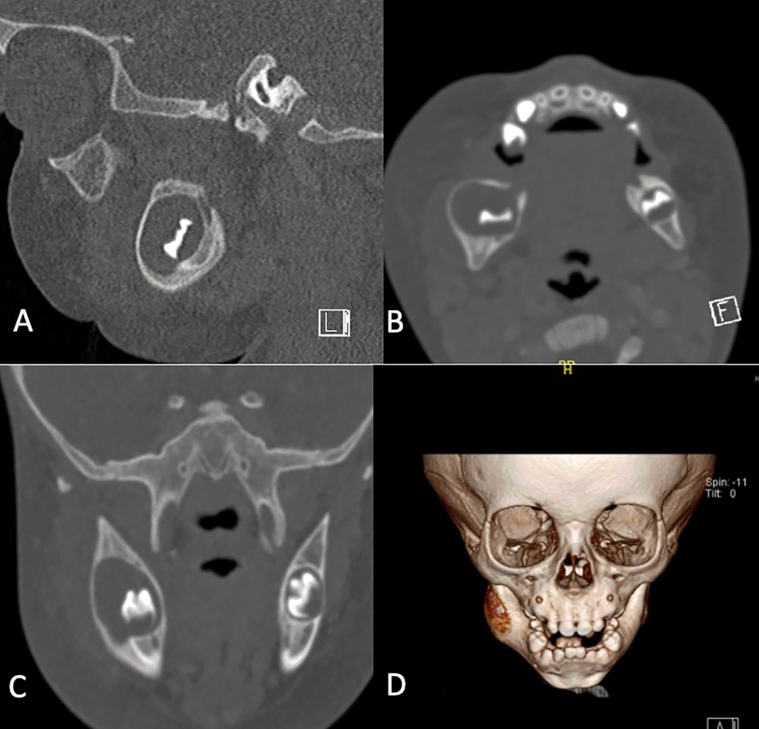
Computed tomography (CT) demonstrated a well-defined expansile lytic lesion in the posterior body of the mandible involving an unerupted tooth measuring approximately 1.6 × 1.3 cm (Figure 1). The radiographic appearance is highly suggestive of a dentigerous cyst or inflammatory cyst based on histopathological correlation.
The parents were reassured, and the treatment plan options were explained in detail. The parents agreed to proceed with surgical enucleation of the lesion under general anesthesia. The patient was admitted to the KFHU, and consent was signed. Under all aseptic conditions, the patient was prepared using povidone-iodine solution, and the patient was covered with a sterile towel. Good exposure of the lesion was achieved by raising a full mucoperiosteal flap, enucleation of the entire cyst along with the associated tooth, and bleeding from all margins of the cavity lesion. The specimen was placed in a labelled container for histopathological examination. Irrigation with normal saline and hemostasis were achieved, and the surgical site was closed with 3/0 Vicryl sutures.
A gross examination of the specimen consists of tooth and cyst lining (1.5 cm × 1.5 cm with a wall thickness of 0.2). Microscopic analysis revealed a cyst wall lined by non-keratinized squamous epithelium with intraepithelial neutrophil infiltration and pus formation. This finding is consistent with an inflamed dentigerous cyst. The patient was followed on a regular basis for up to 8 months after the surgery. The parents were informed of the outcomes of the surgical operation. The surgical site was intact, swelling was reduced significantly, and no complaints were reported. Three-month post-operative CT) scan was requested and reviewed. Compared with the previous CT, no evidence of bone destruction, pathology, or soft tissue abnormalities was observed or noted (Figure 2).

Dentigerous cysts are a major area of interest in the field of oral and maxillofacial surgery as they are the second most common developmental cysts. Therefore, a deeper understanding of different presentations can play a crucial role in providing the optimum care to the patients.14,15 Several studies have explored the pathophysiological processes that underlie dentigerous cysts. Benn and Altini (1996) suggested that three possible mechanisms could initiate the pathological process based on the source of the inflammation. First, the radicular cyst in the pathway of eruption of the successor’s tooth was left in situ following exfoliation or extraction of primary teeth. The second is the presence of a nonvital tooth. Finally, there are other sources of inflammation in the jaw.16 However, as mentioned previously, the pressure from fluid accumulation between the reduced enamel epithelium and unerupted tooth seems to be the most common pathophysiological theme of dentigerous cysts.8,9,12,17 In our case, the pathological process mirrored the generally agreed pathological pattern of a dentigerous cyst.
A dentigerous cyst is usually discovered incidentally during routine dental examinations.9–11 Our patient presented with right facial swelling and pain, which could be attributed to the fact that fluid accumulation over time results in expansion, leading to facial swelling accompanied by pain. No intervention to break the expansion pressure could result in a negative effect on the surrounding vital structures such as the inferior alveolar nerve and maxillary sinus.12,17 Authors are in agreement that dentigerous are commonly associated with unerupted mandibular third molars followed by maxillary canines.2,10,12 This finding broadly supports the current literature on the most commonly impacted teeth.12,17,18
In terms of age range, despite global population variations, the current evidence suggests that the second and third decades of life are the peak incidences of dentigerous cysts at approximately 23% and 20%, respectively. Notably, a considerable percentage of cases were reported in the fourth, fifth, and sixth cases.2 Several reports in the literature were published in the first decade. Hedge reported a case of 9 years old boy with left mandible swelling and pain,19 Dave reported two cases of 9 years old boy with asymptomatic swelling and 7 years old boy who was discovered during a routine dental examination,12 Deepa documented a case of 10 years old girl with a history of difficulty in mastication due to a dentigerous cyst in her lower right jaw20 and McKinney also reported 4 years old child with asymptomatic facial swelling related to dentigerous cyst.21 The first documented case in one a old boy was reported by Suresh et al. in 2011. The patient presented with left mandibular angle swelling associated with a history of pain, difficulty in swallowing, and an obliterated buccal sulcus.13 Our case is the second documented case after Suresh et al. at this very young age. However, the case we reported differed in some clinical manifestations, as our patient presented with a history of rapid right facial swelling and pain. No difficulty in swallowing or buccal sulcus obliteration. This difference illustrates the benefit of reporting such cases to aid and build up evidence-based practice, as this will result in positive reflections on the care that we provide for our patients.
A unilateral and single presentation is considered a classic demonstration of a dentigerous cyst, similar to our case. Nevertheless, multiple dentigerous cysts can be observed in patients with basal cell nevus, mucopolysaccharidoses, and cleidocranial dysplasia syndromes.20 However, few cases of bilateral dentigerous cysts have been reported in the literature in non-syndromic patients.22 In terms of radiographic presentation, based on a CT scan, our case showed a well-defined radiolucent lesion associated with an unerupted tooth which is in alignment with the characteristic appearance of a dentigerous cyst.12,13,20
Surgical treatment techniques for dentigerous cysts include enucleation and marsupialization. The surgical decision is formulated based on several factors, such as the patient’s medical history, location and size of the lesion, and the facilities where the surgery will take place.20 Dave et al. suggested that a multidisciplinary approach is required for such cases to establish a fully comprehensive treatment plan. The authors acknowledge that enucleation is the gold standard surgical option for most dentigerous cyst cases. However. Marsupialization followed by complete removal of the cyst should be considered in cases of close proximity to vital structures.12 The recurrence of dentigerous cysts is not a concern for most patients.11,13 Our patient underwent complete removal of the cyst under general anesthesia with the associated tooth to eliminate the need for second-stage surgery, as well as to improve the patient and family’s quality of life by reducing the required time in the hospital.
| Views | Downloads | |
|---|---|---|
| F1000Research | - | - |
|
PubMed Central
Data from PMC are received and updated monthly.
|
- | - |
Provide sufficient details of any financial or non-financial competing interests to enable users to assess whether your comments might lead a reasonable person to question your impartiality. Consider the following examples, but note that this is not an exhaustive list:
Sign up for content alerts and receive a weekly or monthly email with all newly published articles
Already registered? Sign in
The email address should be the one you originally registered with F1000.
You registered with F1000 via Google, so we cannot reset your password.
To sign in, please click here.
If you still need help with your Google account password, please click here.
You registered with F1000 via Facebook, so we cannot reset your password.
To sign in, please click here.
If you still need help with your Facebook account password, please click here.
If your email address is registered with us, we will email you instructions to reset your password.
If you think you should have received this email but it has not arrived, please check your spam filters and/or contact for further assistance.
Comments on this article Comments (0)