Keywords
Port catheter, catheter rupture, migration, pulmonary artery, sternotomy
Port catheters are commonly used in oncology for chemotherapy, but they can sometimes rupture and migrate, leading to serious complications. Early detection and management are crucial, and surgical intervention may be required when less invasive approaches fail. This report highlights these rare but significant complications.
A 57-year-old male, with no significant medical history, underwent surgery for sigmoid colon adenocarcinoma and planned chemotherapy. His port catheter became nonfunctional, and on presentation, he was hemodynamically stable with normal vital signs. Chest radiography revealed catheter rupture, and thoracic angio-CT confirmed the fragment’s migration into the left pulmonary artery. After a failed percutaneous attempt, emergency surgery was performed via median sternotomy, successfully retrieving the catheter fragment.
Catheter rupture and migration are rare complications, with an incidence of less than 1%. The clinical presentation can vary from asymptomatic to severe, requiring a high index of suspicion. Imaging, including chest radiography and thoracic angio-CT, is essential for accurate diagnosis and treatment planning. Management options include percutaneous retrieval, but surgery may be necessary when complications arise, as in this case. Preventive strategies, such as proper insertion and regular surveillance, are key in minimizing risks.
Catheter rupture with migration is a life-threatening complication that requires urgent diagnosis and intervention. This case underscores the importance of vigilance in monitoring oncology patients with implantable devices and emphasizes the critical role of surgical intervention when less invasive approaches fail.
Port catheter, catheter rupture, migration, pulmonary artery, sternotomy
Port catheters are widely used in oncology to ensure reliable venous access for long-term treatments, such as chemotherapy.1 While generally safe, these devices are not without risks, and complications such as infections, thrombosis, and mechanical failures can occur.1,2 Among these, catheter rupture and migration are rare but potentially life-threatening events, often requiring urgent diagnosis and intervention.1,3 Migrated fragments can lead to serious consequences, including vascular injury, pulmonary embolism, and hemodynamic instability, necessitating a multidisciplinary approach to management.1,2
This report emphasizes the importance of early detection and prompt management of port catheter complications. It highlights the role of surgical intervention when minimally invasive techniques fail and underscores the need for vigilance in managing rare but serious events associated with implantable vascular devices. This case report has been prepared in line with the SCARE criteria.4
A 57-year-old male, with no significant past medical history, underwent surgery one month prior for a sigmoid colon adenocarcinoma classified as pT4N3M0. A low anterior resection with colorectal anastomosis was performed, and the postoperative course was uneventful. Adjuvant chemotherapy was indicated, but the implanted port catheter became nonfunctional.
On presentation, the patient was hemodynamically stable, with a blood pressure of 120/75 mmHg, a heart rate of 78 beats per minute, and he was afebrile at 36.8°C. Physical examination revealed no abnormalities: heart sounds were regular without murmurs, lung auscultation was clear bilaterally, and the abdomen was soft and non-tender. There were no signs of localized swelling, tenderness, or infection at the catheter insertion site.
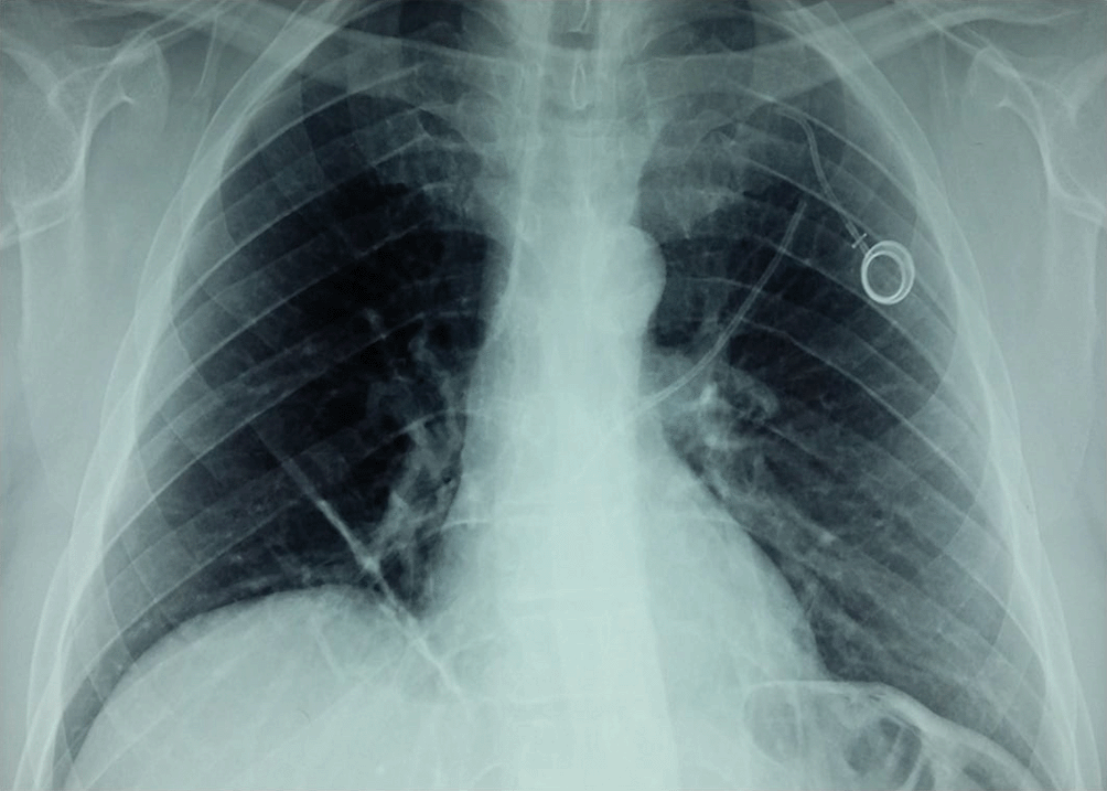
A chest radiograph revealed catheter rupture, with the distal fragment migrating away from its original position ( Figure 1). The exact location of the fragment was further delineated by thoracic angio-CT, which identified it lodged in the left pulmonary artery, extending into the left upper lobar branch. The imaging showed no evidence of vascular thrombosis, embolism, or pulmonary infarction. The surrounding lung parenchyma appeared unremarkable, with no signs of atelectasis or consolidation. The major thoracic vessels, including the aorta and superior vena cava, were intact, and there was no associated pleural effusion or mediastinal shift. This precise localization was crucial in planning the subsequent management approach.

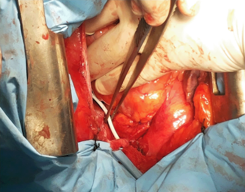
An initial attempt at percutaneous retrieval resulted in pulmonary artery injury, leading to tamponade, necessitating immediate intervention. Emergency surgery was performed through a median sternotomy, providing optimal access to the thoracic cavity. The pericardium was opened carefully to relieve the tamponade and allow direct visualization of the heart and great vessels. After systemic heparinization to prevent thrombosis, the pulmonary artery was meticulously dissected to expose the area of interest. A 1 cm longitudinal incision was made just proximal to the bifurcation of the pulmonary artery, precisely at the location of the lodged catheter fragment as identified on preoperative imaging ( Figure 2). The fragment was successfully retrieved using fine vascular forceps under direct visualization to avoid further injury to the vessel wall ( Figure 3). Hemostasis was ensured by securing the incision site with a 5-0 Prolene purse-string suture, placed in a concentric fashion to reinforce the arterial closure. The pericardium was loosely re-approximated, and chest drains were placed to monitor for potential bleeding or effusion. The sternotomy was closed layer by layer using standard surgical techniques, and the patient was transferred to the intensive care unit for close postoperative monitoring. The recovery was uneventful, with no signs of recurrent bleeding or pulmonary complications, and the patient was discharged in stable condition on postoperative day 5.

Port catheters are essential devices in oncology, providing reliable venous access for chemotherapy and other long-term treatments.1,5 Despite their routine use, complications occur in approximately 5–10% of patients, with infection, thrombosis, and mechanical failures being the most common.1,5 Catheter rupture and migration, as in this case, are rare, with an incidence of less than 1%.5,6 Migrated catheter fragments can embolize to critical vascular sites, such as the pulmonary artery, causing complications ranging from asymptomatic presentations to life-threatening events like pulmonary infarction or vascular injury.1 Understanding the epidemiology of these complications helps clinicians remain vigilant and adopt preventive measures, such as proper catheter insertion techniques and routine monitoring.1,5
The clinical presentation of port catheter rupture can vary widely, ranging from asymptomatic cases to severe systemic symptoms.6 According to a retrospective analysis of 41 patients with centrally dislocated catheter fragments, 53.7% of cases were found incidentally, while 39% presented with catheter malfunction.1 Only 7.3% of patients with fragments in the right atrium, right ventricle, or pulmonary artery exhibited cardiac symptoms.1 A more comprehensive review of 215 cases of intravenous catheter embolization revealed that catheter malfunction was the most common clinical sign, occurring in 56.3% of cases.7 Other presentations included arrhythmia, pulmonary symptoms, and septic syndromes.7
The time between catheter rupture and symptom onset can vary significantly, with presentations occurring between 0- and 60-days post-procedure in some cases.1,7 In conclusion, the clinical presentation of port catheter rupture is diverse and often non-specific. Healthcare providers should maintain a high index of suspicion, particularly in patients with a history of catheter use who present with unexplained symptoms or catheter malfunction.5
Diagnostic workup begins with a chest radiograph, which is often sufficient to confirm catheter rupture and identify the location of the migrated fragment, as seen in our case.8 However, advanced imaging, such as thoracic angio-CT, is essential for precise localization and assessing potential complications, including pulmonary embolism, thrombosis, or vascular injury.8,9 In this case, thoracic angio-CT clearly demonstrated the fragment lodged in the left pulmonary artery and ruled out additional complications, guiding subsequent management. Routine surveillance imaging of port catheters in high-risk patients, particularly those undergoing prolonged treatment, may aid in early detection of such complications.8,9
Management of catheter rupture and migration depends on the clinical presentation, fragment location, and associated complications.1 Asymptomatic cases with accessible fragments can often be managed with percutaneous retrieval, which is considered the first-line treatment.7,10 Percutaneous techniques have a high success rate, but complications such as vascular injury, as observed in this case, may occur.10 Pulmonary artery rupture, although rare, is a recognized complication that requires immediate intervention.1,10
When minimally invasive approaches fail or lead to complications, surgical retrieval becomes necessary.1,11 Sternotomy provides direct access to the pulmonary artery, allowing precise localization and safe removal of the fragment. In our case, surgical intervention was lifesaving, with the fragment retrieved via a controlled pulmonary artery incision and closure using a purse-string suture to minimize bleeding risk.1,10,11 Multidisciplinary coordination among interventional radiologists, thoracic surgeons, and anesthesiologists is essential to optimize outcomes in such scenarios.7,11
Preventive strategies play a crucial role in minimizing catheter-related complications.5,12 These include proper insertion techniques, regular device maintenance, and patient education regarding early signs of catheter dysfunction.9,12,13 Additionally, the use of improved catheter materials may reduce the risk of mechanical failure.1,12
In conclusion, catheter rupture with migration, although rare, represents a potentially life-threatening complication that demands prompt diagnosis and a personalized management strategy.5,7 This case emphasizes the necessity of heightened vigilance in the monitoring and care of oncology patients with implantable devices.5,13 It also highlights the pivotal role of timely surgical intervention in managing such severe complications, ultimately ensuring optimal patient outcomes.7,12
Written informed consent to publish this case and associated images was obtained from the patient.
The completed CARE checklist for this case report is available via the Zenodo repository under a CC0 license:
Title: CARE Checklist – Case Report on Choledochal Cyst in an Elderly Patient
Repository: Zenodo
DOI: 10.5281/zenodo.1568500314
License: CC0 1.0 Universal (CC0 1.0) Public Domain Dedication.
| Views | Downloads | |
|---|---|---|
| F1000Research | - | - |
|
PubMed Central
Data from PMC are received and updated monthly.
|
- | - |
Is the background of the case’s history and progression described in sufficient detail?
No
Are enough details provided of any physical examination and diagnostic tests, treatment given and outcomes?
No
Is sufficient discussion included of the importance of the findings and their relevance to future understanding of disease processes, diagnosis or treatment?
No
Is the case presented with sufficient detail to be useful for other practitioners?
No
Competing Interests: No competing interests were disclosed.
Reviewer Expertise: Vascular access device, hereditary angioedema, internal medicine
Alongside their report, reviewers assign a status to the article:
| Invited Reviewers | |
|---|---|
| 1 | |
|
Version 1 02 Jul 25 |
read |
Provide sufficient details of any financial or non-financial competing interests to enable users to assess whether your comments might lead a reasonable person to question your impartiality. Consider the following examples, but note that this is not an exhaustive list:
Sign up for content alerts and receive a weekly or monthly email with all newly published articles
Already registered? Sign in
The email address should be the one you originally registered with F1000.
You registered with F1000 via Google, so we cannot reset your password.
To sign in, please click here.
If you still need help with your Google account password, please click here.
You registered with F1000 via Facebook, so we cannot reset your password.
To sign in, please click here.
If you still need help with your Facebook account password, please click here.
If your email address is registered with us, we will email you instructions to reset your password.
If you think you should have received this email but it has not arrived, please check your spam filters and/or contact for further assistance.
Comments on this article Comments (0)