Keywords
Brain Imaging, Diagnosis, Magnetic Resonance Image, Medulla Oblongata, Stroke
Bilateral medial medullary infarction (BMMI) is a rare ischemic lesion that typically presents as dysarthria, dysphagia, tetraplegia, and respiratory failure. It is known for its characteristic « heart-shaped » appearance on magnetic resonance imaging (MRI). The etiological profile of this disease has not been studied previously. Considering the unfavorable outcome of BMMI, it is crucial to investigate the clinical and etiological profile to optimize the management of these patients.
A 49-year-old north-african smoker male patient was admitted to the neurology department of the military hospital of Tunis-Tunisia 24 hours after acute-onset slurred speech, weakness and impaired sensation in all four limbs, predominantly in the lower limb. His family history was remarkable for young-onset brainstem ischemic stroke (IS) in a brother. Neurological examination a right hemiparesis with tactile hemihypoesthesia and right astereognosia (modified Rankin scale of 2). Cerebral MRI showed radiological aspect compatible with diagnosis of « heart-shaped » BMMI. A heterozygous mutation R506 of Leiden Factor V was identified. The patient’s condition remained stable at 15 months follow-up (Modified Rankins Scale at 2) and no recurrence of IS was noted under anticoagulation to date.
BMMI is a rare case of medial medullary infarction, accounting for less than 1% of IS cases. To our knowledge, this is the first case of « heart-shaped » BMMI linked to Leiden Factor V mutation.
Brain Imaging, Diagnosis, Magnetic Resonance Image, Medulla Oblongata, Stroke
Bilateral medial medullary infarction (BMMI) is a rare ischemic lesion that typically presents as dysarthria, dysphagia, tetraplegia, and respiratory failure. It is known for its characteristic « heart-shaped » appearance on MRI scans. Its prognosis is usually poor, with major mortality and morbidity.1 The etiological profile of this disease has not been studied previously. Considering the unfavorable outcome of BMMI, it is crucial to investigate the clinical and etiological profile to optimize the management of these patients.
A 49-year-old male patient was admitted to the Neurology Department of the Military Hospital of Tunis-Tunisia 24 h after acute-onset slurred speech, weakness, and impaired sensation in all four limbs, predominantly in the lower limb. His family history was remarkable for young-onset brainstem ischemic stroke (IS) in a brother. His medical history was unremarkable.
General physical examination revealed an unremarkable blood pressure of 110/65 mmHg and heart rate of 70 beats per minute. Neurological examination revealed right hemiparesis with tactile hemihypoesthesia and right astereognosia. Examination of the cranial nerves yielded normal results. His National Institutes of Health Stroke Scale (NIHSS) score was 6 and the modified Rankin scale score was 2.
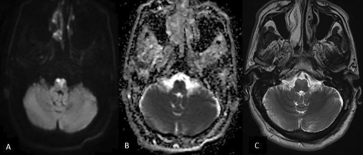
Brain MRI performed 24 h after symptom onset showed bilateral symmetrical T2/FLAIR hyperintense lesions in the bilateral pyramids and medial lemniscus of the middle medulla oblongata with restricted DWI ( Figure 1a and b). No visible artery occlusion was found on MR ( Figure 1c). Given this radiological aspect, the diagnosis of « heart-shaped » BMMI was confirmed.

Trans-thoracic echocardiogram showed no intracardial thrombus, permeable foramen ovale, diastolic or systolic abnormalities, and left ventricular ejection fraction was normal at 82%. No valvular lesions were detected. The cardiac cavity was not dilated.
Laboratory investigations revealed total cholesterol, triglycerides, HDL-cholesterol, and LDL-cholesterol levels. The glycated hemoglobin levels were normal in our patient. serum cyanocobalamin level was normal (831pg/ml). Antinuclear antibodies and antineutrophil cytoplasmic antibodies were negative. Thrombophilia workup showed free protein S within normal limits at 125.5% and protein C within normal limits at 89%, as was the case with liquid antithrombine at 85%. The patient tested negative for both G2021A Factor II and C677T methyl tetrahydrofolate reductase mutations. A heterozygous mutation, R506 of Leiden Factor V was identified. Etiological laboratory investigations of IS in our patient were normal ( Table 1).
Based on these elements, genetic thrombophilia due to a heterozygous R506 mutation of Leiden factor V was considered an etiology of IS in our patient.
Treatment with acenocoumarol 4mg/day was initiated as well as physical therapy.
The patient’s condition remained stable at 15 months follow-up (Modified Rankin Scale at 2), and no recurrence of IS has been noted under anticoagulation therapy to date (Figure 2).
We describe a case of BMMI that is original in many respects. First, our patient had a relatively less severe clinical presentation than previously reported cases. Moreover, inherited thrombophilia as an etiology of BMMI has not been previously documented.
A BMMI is a peculiar case of medial medullary infarction. According to a systematic review published in 2012, it accounted for a total of 38 cases published until March 2011.2 As shown in Table 2 (Refer: https://doi.org/10.6084/m9.figshare.29082734.v1), 21 BMMI cases were published between March 2011 and December 2024.
As was the case in our patient, the clinical presentation is explained by anatomical structures affected by infarction, such as the medial lemniscus and medullary pyramids, which contain sensory spinothalamic tract of conscious proprioception, vibration, fine touch, and the contralateral corticospinal tracts. Hypoglossal nerve is rarely affected in BMMI, this is explained by the fact that the hypoglossal nucleus is located close to the border between the anteromedial and lateral territories in thedorsal portion of the medulla oblongata.3 However, six out of the 21 cases we reviewed displayed either ipsilateral or bilateral hypolglossal paresis.
BMMI are a diagnostic challenge. Cases with unusual presentations such as stepwise flaccid tetraparesis and respiratory failure mimicked Guillain-Barré syndrome (GBS), leading to misdiagnosis (Table 2).4 GBS was the first cause of misdiagnosis of BMMI in the literature, with four cases to date. Vomitting, nausea, bulbar affect, and tetraparesis can be misleading clinical features, leading to a misdiagnosed and treated case of botulism.5 Misdiagnosis of myasthenic crisis leading to late diagnosis and severe outcome.6
The only pathognomonic feature of BMMI is the radiological « heart-shaped » sign, highlighted in our case. DWI hyperintense lesions with reduced ADC values are the core features that confirm the diagnosis of IS. Another variable aspect of BMMI is the occluded artery. In fact, the characteristic « heart-shaped » appearence is linked to the anteromedial arterial territory of the medulla oblongata. This territory can be supplied by the vertebral artery, anterior spinal artery, or basilar artery, depending on anatomical variation (Table 2). A variation of the « heart-shaped » sign in BMMI is the « airpod sign » which occurs in case of BMMI in a patient with Type 3a anatomical variant of anterior spinal artery coming from a single verterbal artery.7 Visualization of artery occlusion is not constant on MR, especially in small vessel disease-related IS cases. MR angiography is also used to detect the dominant vertebral artery or agenesis of one vertebral artery. In fact, BMMI may be induced by occlusion of the non-dominant VA that supplies the bilateral anteromedial territories of the medulla.3 In as many as 60% of cases, the DWI hypersignal with restricted ADC may not be present if MRI is performed in the first 24 h after symptom onset.8 This pleads in favor of iterating the MRI after 24 hours if the diagnosis is suspected and the initial MRI is normal, as false-negative precocious DWI is a known feature of posterior circulation IS and most specifically in brainstem IS cases.
The causes of BMMI are diverse, with large artery atherosclerosis and small vessel disease being the most frequent. Rarer causes, such as aneurysm, dolichoectasia, and Fabry disease, have been reported.9–11 To our knowledge, this is the first case of « heart-shaped » BMMI linked to Leiden Factor V mutation.
The imputability of IS to Leiden Factor V mutations has been well documented and proven in children, young adults, and older adults in several previously published studies.12,13 Although inherited thrombophilia was first linked to venous thrombosis, its involvement in arterial thrombosis pathogenesis, including IS, was confirmed by several studies with early onset IS even in the absence of a left-right cardiac shunt.14
Mortality and morbidity in BMMI are considerably high, with severe residual handicap (Modified Rankin Scale score of 5 in most cases).1 In our patient, the residual handicap was relatively lower than that in previously described cases (Table 2).
This study has some limitations need to be highlighted. The family history of young-onset brainstem IS could be related to inherited thrombophilia, but etiological work-up for IS was not performed in our patient’s brother, who was also affected by young-onset IS.
Written informed consent for the publication of clinical details and clinical images was obtained from the patient.
Mohamed Slim Majoul MD,
Contribution: Writing – Original Draft/Visualization
Meriam Messelmani MD,
Contribution: Writing – Review & Editing/Methodology
Moncef Aloui MD,
Contribution: Resources
Fekih Nejiba Mrissa MD,
Contribution: Project Administration
Jamel Zaouali MD,
Contribution: Validation
Ridha Mrissa MD
Contribution: Supervision
Title: CARE guidelines for Case Report: Heart-shaped sign in bilateral medial medullary infarction: A case report and review of the literature.
Data Repository: Figshare
Licence: CC0 1.0 Universal
All data underlying the results are available as part of the article.
| Views | Downloads | |
|---|---|---|
| F1000Research | - | - |
|
PubMed Central
Data from PMC are received and updated monthly.
|
- | - |
Provide sufficient details of any financial or non-financial competing interests to enable users to assess whether your comments might lead a reasonable person to question your impartiality. Consider the following examples, but note that this is not an exhaustive list:
Sign up for content alerts and receive a weekly or monthly email with all newly published articles
Already registered? Sign in
The email address should be the one you originally registered with F1000.
You registered with F1000 via Google, so we cannot reset your password.
To sign in, please click here.
If you still need help with your Google account password, please click here.
You registered with F1000 via Facebook, so we cannot reset your password.
To sign in, please click here.
If you still need help with your Facebook account password, please click here.
If your email address is registered with us, we will email you instructions to reset your password.
If you think you should have received this email but it has not arrived, please check your spam filters and/or contact for further assistance.
Comments on this article Comments (0)