Keywords
Neuromuscular fatigability, children, Sex differences, Surface electromyography, Muscle fiber recruitment, Fatigue resistance
Neuromuscular fatigability, children, Sex differences, Surface electromyography, Muscle fiber recruitment, Fatigue resistance
Strength is a fundamental determinant of both athletic performance and overall health across the lifespan.1 Next to maximal strength, the level of strength that can be sustained over a certain time or until fatigue occurs is a key component of physical performance However, differences between children and adults as well between sexes in both, maximal strength and fatiguability have been observed, and the mechanisms still lack clarity.2
To interpret fatigue-related findings accurately, it is necessary to distinguish between related but distinct physiological concepts: muscular endurance, fatigue, and fatigability. Muscular endurance refers to the duration for which a given force can be maintained and is typically quantified as time to task failure (TTF). On the other hand, neuromuscular fatigue describes an objective decline in maximal voluntary contraction torque (MVC) following a fatiguing task. Fatigability, on the other hand, is understood as the susceptibility to fatigue and can also be objectively quantified.3,4 The process of fatigue is influenced by multiple factors at both the individual level (e.g., voluntary activation, maximal strength, fiber type distribution) and the exercise mode (e.g., intermittent vs. continuous, dynamic vs. isometric). These interactions create a complex interplay that has led to varying findings regarding fatigue-related differences across age groups and sexes.5
Generally, children tend to exhibit greater resistance to fatigue,6–11 and the underling mechanisms have been summarized in the differential motor unit activation hypothesis. Dotan et al.12 suggested that children either lack or are not able to use type II muscle fibers to the extent that adults can. Support for this theory comes from studies showing that children have reduced voluntary activation, lower anaerobic capacity13 and reduced type II fibre content.14,15 However, higher muscle mass occluding blood flow, mixed findings regarding type II muscle fibers and inconsistent findings in isometric sustained tasks between children and adults still contribute to uncertainty regarding physiological explanations.
In adults, females often demonstrate greater resistance to fatigue than males due to a higher proportion of type I fibers, enhanced oxidative capacity, and more efficient blood flow regulation.16 However, the extent to which these differences emerge in prepubertal populations remains unclear, as studies on sex-related differences in fatigue before puberty are limited.11
In sustained exercises, where recovery plays a minor role, studies have reported negligible differences in fatigue between children and adults during sustained contractions of the plantar flexors,17,18 knee extensors,19 and back extensors.20 These studies employed surface electromyography (EMG) to quantify muscle activity and frequency changes, which are indicative of fiber type recruitment.21 However, these studies had methodological limitations, including very small sample sizes or exclusion of female participants. Furthermore, they assessed muscle groups that, based on literature, were expected to have a high proportion of type I fibres, potentially limiting their generalisability.22
Given the inconsistencies and limitations in current research, the present study aims to investigate differences in neuromuscular fatigability between male and female prepubertal children and young adults during a sustained isometric contraction of the quadriceps femoris muscle, which is predominantly composed of type II fibers.22 We hypothesized that children would be less fatigable than adults and that sex differences would be minor in children but more pronounced in adults.
The piloting cross-sectional study comprised a sample of 24 prepubertal children (12 males and 12 females aged 7-10 years) and 36 young adults (18 males and 18 females aged 20-30 years). Sex was self-reported. They were recruited from various sports clubs (mostly soccer or tennis), or by a selection from the sport student community by means of personal advertising and flyers. Inclusion criteria, based on self-report, were participation in individual or organized physical activity between 4-9 h a week, and absence of any acute or chronic diseases or injuries. Adults volunteered to participate in this study after providing written informed consent. For children informed consent was obtained verbally from all participants and written informed consent was provided by their legal guardians. Prior to participation, children were informed about the study using age-appropriate videos and image materials, and the procedures were explained to them in a clear and understandable manner. Ethical approval was granted by the local ethics committee (Ethikkommission Nordwest- und Zentralschweiz (EKNZ), Project-ID: 2023-00181) and the procedures were conducted according to the Declaration of Helsinki.
In this cross-sectional study, the participants performed two maximal voluntary isometric contractions (MVCpre), followed by a failure-task, and another MVCpost in knee extensor muscles. At the end perceived effort was asked, and anthropometric measurements were taken. During the failure-task, EMG signals were obtained. The participants were advised to avoid intensive exercise for 48 hours before the test day. This study was not preregistered as it represents a pilot investigation designed to explore age- and sex-related differences in neuromuscular fatigability. This study followed the STROBE (Strengthening the Reporting of Observational Studies in Epidemiology) guidelines. A completed STROBE checklist has been uploaded to OSF and is available at: https://doi.org/10.17605/OSF.IO/9A42S. The checklist is available under the Creative Commons CC0 1.0 Public domain dedication.
Strength testing
After a 5-min-warm-up on the treadmill at a self-perceived moderate intensity (children: 7.2 ± 1.1 km/h, adults: 8.6 ± 1.2 km/h), the familiarization tests, MVCs and the failure-task were performed on the isokinetic dynamometer (Isomed 2000, D. & R. Ferstl GmbH, Hemau, Germany). All isometric tests were conducted in knee extensor muscles of the dominant leg in an upright sitting position with the back inclination set at 75° with a knee angle of 60°. The lever arm length of the dynamometer was placed at 2/3 of tibia length. The familiarization included submaximal and maximal isometric contractions prior to the actual MVC, which ensured that participants understood the instructions.
The MVCpre test was performed twice with a rest of 1 min in between. According to Maffiuletti et al.23 the test instruction was to “push as fast and hard as possible” to achieve a rapid force increase to measure rate of torque development (RTD, 150 mx) as well as reach a force plateau of 3-4 s to obtain the peak torque. High reliability for isometric peak torque of knee extensors have been reported in prepubertal and early pubertal children (ICC = 0.95)24 as well as in adults (ICC > 0.967; SEM < 3.8%).25 The peak torque of the best trial and the highest RTD were considered as MVCpre and used in further analysis.
In the failure-task, participants were instructed to sustain an isometric contraction at 60% of their individual MVCpre for as long as possible. The required level was visualized on a computer for the participants and feedback from the test instructor was given as well. When 50% of MVCpre could not be reached for more than 2 seconds despite strong encouragement from the test instructor, the test was ended (=failure). TTF was used for further analysis. 10 seconds after termination, another MVCpost was performed in the same way as MVCpre in order to quantify MVC reduction after the failure-task. Rate of perceived effort during the failure-task was asked after MVCpost using the modified Borg’s scale.1–10,26
Electromyography
To obtain electromyographic activity during the strength measurements, surface EMG was applied on vastus lateralis (VL), vastus medialis (VM), rectus femoris (RF), and biceps femoris muscles (BF) using bipolar silver chloride surface electrodes (Blue Sensor N-00-S, 30x22 mm; Ambu, Denmark). Skin preparation and placement of the electrodes was done according to SENIAM guidelines.27 For data collection, the Myon system 320 (Myon AG, Schwerzenberg, Switzerland) with a sampling rate of 4 kHz per channel and 12-bit analogue to digital conversion was used. EMG data was analysed offline with proEMG 2.0 software (Prophysics AG, Kloten, Switzerland). Raw data were low-pass (400Hz) and high-pass (10Hz) filtered, rectified, and smoothed with a moving average of 100 ms. To quantify the fatigue-related changes of myoelectric signals in those intervals, muscle activity with root mean square (RMS) and median frequency (MDF) after Fast-Fourier-Transformation, respectively, were considered. Time windows of 4 s each were analysed at the beginning (0%: after 2 seconds after the start), quarter (25%), half (50%), three-quarter (75%) and end (100%: 2 seconds before end). RMS during the fatigue trial was normalized to the maximal RMS (1000 ms window) of MVCpre. An increase in RMS coupled with a decline in MDF parameters is a known characteristic in fatiguing processes.28
Anthropometry
Standing and sitting height were measured before the bioimpedance analysis on the Inbody 720 (Biospace Co., Ltd., Seoul, Korea). To account for variations in biological maturation, the Mirwald method was further used to calculate the offset from peak height velocity (PHV) in children.29
Mean and standard deviation (mean ± SD) are presented for all variables. To investigate neuromuscular fatigability, the outcome variables MVC reduction (MVCpre vs. MVCpost in %), RTD reduction (RTDpre vs. RTDpost in %), TTF, perceived effort, and RMS and MDF from EMG during the fatigue-task were considered. For MVC and RTD reduction, TTF, perceived effort as well as the initial RMS values (at 0% TTF) linear models were used to analyse differences between age and sex categories (~ age + sex), as well as age*sex interaction (~ age * sex). The linear mixed models for RMS and MDF further included the time points (0%, 25%, 50%, 75% and 100% of TTF) during the contraction as time covariates and random intercepts per participant. This means that ~ time*age, ~ time*sex and ~ time*age*sex interactions were calculated for change of activity (RMS) and frequency (MDF) of agonist muscles (VL, VM, RF) as well as antagonist co-activity (RMS BF). Normality of residuals was checked by visually inspecting the Q-Q plots of the models.
Meaningful differences are interpreted by observing the appropriate effect sizes (Cohen’s d).30 Effects below 0.2 are considered trivial, between 0.2-0.5 small, 0.5-0.8 moderate and >0.8 as large. Statistical significance was set at a predetermined alpha level (p < 0.05). All statistical analyses and figures were performed with RStudio (version 4.2.0) using the package lme4.31
We used the STROBE reporting guideline32 to draft this manuscript, and the STROBE reporting checklist33 when editing, included in supplement A. Patients and the public were not involved in the design, conduct, reporting, or dissemination plans of this research.
Descriptive characteristics of the participants with anthropometrics and maximal strength results separated for children and adults and both sexes are displayed in Table 1.
Table 2 and Figure 1 illustrate that, following the failure-task, there was a moderately greater reduction in MVC among children than adults. Additionally, there was a likely trivial sex effect observed, indicating a potentially smaller reduction in females. The differences between age groups within female and male sex were negligible. The decline in RTD was likely lower in children than in adults as well as in females compared to males (both small effects lacking significance). Further, the difference in RTD reduction between female children and adults was likely smaller than that of male children and adults. Notably, the difference in TTF between female adults and children was greater than the difference between male adults and children. Children reported a moderately lower perception of effort, while females also tend to report lower values. Combined, there was likely a moderately greater difference between female children and adults compared to male children and adults.
Statistically significant results are marked with a * (p < 0.05).
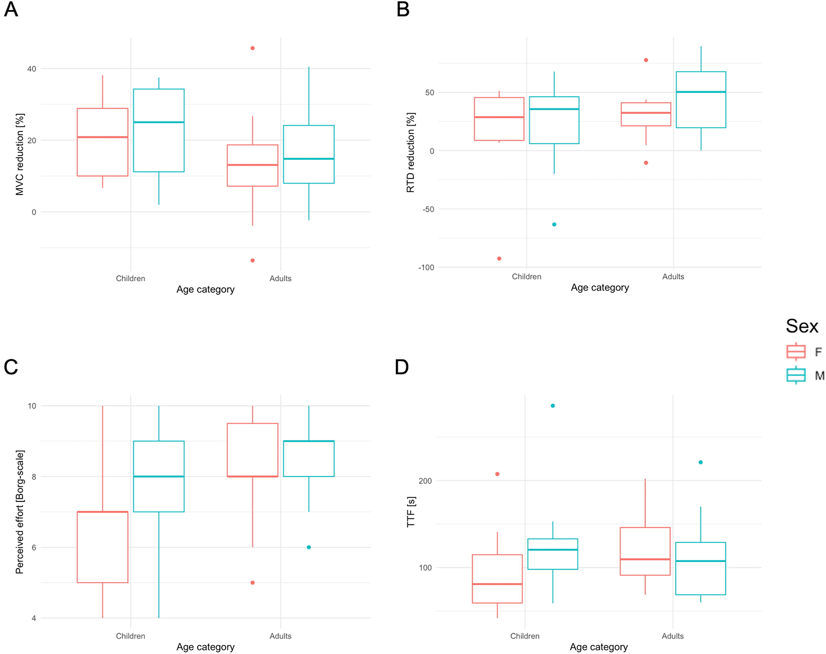
Boxplots of MVC reduction (A), RTD reduction (B), TTF (C) and perceived effort (D) separated for age categories and sex.
Electromyography
Initially, the RMS of all muscles as a percentage of RMS during the MVCpre was higher in children than in adults, with no differences observed between sexes, as depicted in Table 3 and Figure 2. Small or negligible effects were found for both the RMS and the MDF changes between age and sexes throughout the failure-task across all muscles. Statistical significance was evident for time*age differences in RMS VL, VM, RF, and MDF VM, RF indicating more change in adults.
Reference group are adults and males. Statistically significant results are marked * (model coefficients) (p < 0.05).
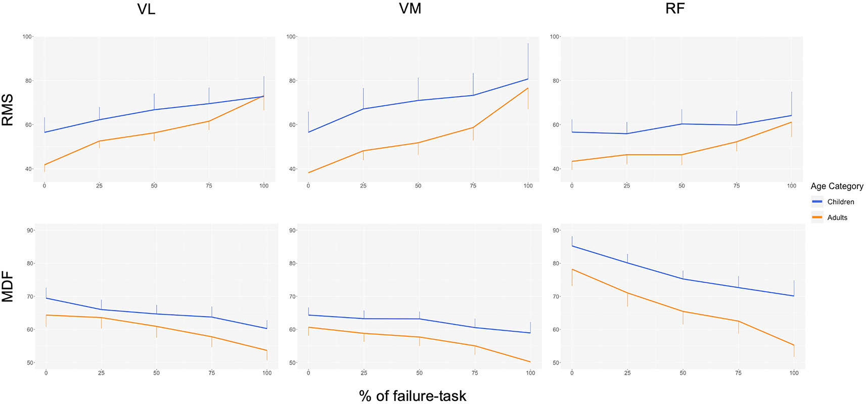
Change of root mean square (RMS) and median frequency (MDF) across the agonist muscles (rectus femoris (RF), vastus lateralis (VL) and vastus medialis muscles (VM)) visualized as mean and 95% confidence intervals (only lower or upper bound displayed) separated for age category (blue: children, orange: adults).
The aim of this study was to examine age and sex differences in neuromuscular fatigability during a sustained submaximal contraction of the knee extensor muscles until failure. In contrast to the hypothesis, MVC reduction was more pronounced in children. On the other hand, smaller RTD reduction in children and female sex is in line with the expectation of reduced fast-twitch fiber usage in these subgroups. Initial RMS was higher in children than adults, yet the changes in activity and frequency throughout the failure task was smaller in children, indicating less fatigue. To our knowledge, this is the first study to investigate such a broad spectrum of neuromuscular fatigue facets between children and adults of both sexes in the quadriceps femoris muscles with predominantly fast-twitch fiber muscles.
Our results showed higher MVC reduction in children compared to adults with negligible sex differences in the knee extensor muscles. This was surprising given previous results in plantar flexor muscles with negligible MVC reduction between male children and adults after submaximal failure-tasks.17,18 As there is no sound physiological explanation for these results, the reduced MVC in children after fatigue may be due to cognitive or motivational factors.34 Given that the MVC task was performed immediately following the demanding failure-task in this study, children might not have been mentally prepared to refocus for that maximal effort.
The trivial sex differences in MVC reduction are unexpected too, given that males were assumed to fatigue more. This was anticipated given their higher maximal force capacity, larger muscle mass, and higher proportion of fast-twitch fibers, all of which are associated with higher anaerobic metabolism and greater accumulation of fatigue-inducing by-products.22,35,36 However, the present data suggest that these factors do not meaningfully influence MVC reduction.
Contrasting the unexpected findings in MVC reduction, the decrease in RTD is physiologically plausible for each subgroup. To our knowledge, this study is the first to assess changes in explosive force after fatiguing exercise in children. Our findings appear reasonable when considering the differential motor unit activation hypothesis and the typically lower fast-twitch fiber content in females.16,37 Given that male adults are likely to have the highest proportion of those fast-fatigable muscle fibers, the most significant reduction in RTD was expected.22 Notably, a study on fatiguing dynamic isokinetic exercises at 60°, 180° and 300°/s of knee extensor muscles in adults found no sex differences in RTD reduction despite differences in maximal torque decrement, which is intriguing and warrants further investigation.38
Although there is general evidence that children are more resistant to fatigue than adults,2 the variability and inconsistency of our results align somewhat with findings on TTF during sustained contractions in earlier studies.19,20 The only study directly comparable, which examined three submaximal intensities of knee extensor muscles, reported longer TTF in prepubertal male children than in adults at 25% of MVC. This resonates with our findings for male participants. However, that study also reported considerable variability, especially among children, and did not extensively discuss the results.19 Even if different muscle groups with probably different fibre type compositions have to be compared with caution, negligible difference in TTF was seen in children and adults in the plantar flexors.18
Other studies using repeated MVCs have mostly found that children showed greater fatigue resistance in these intermittent tasks, suggesting a discrepancy with our findings, as the role of recovery may come back into play.2 Additionally, in dynamic exercises like intense running or cycling, either no differences or a shorter TTF in children were observed, which would be in line with our observation.2
We must once more take into account motivational aspects, given that TTF is determined by voluntary failure. As children, particularly female children, may be less adept at perceiving fatigue and pain, their tolerance and motivation to continue could be lower, resulting in a shorter TTF.39 Thus, our study reinforces the idea that neuromuscular fatigue mechanisms are intricately linked to the type of exercise, which could explain the controversies observed in our sustained isometric contraction results.
The results of perceived effort are consistent with previous research, which found that children generally rate their effort lower than adults do, particularly for short bouts of exercise.40 Possible explanations for this could include children’s limited experience with the range of sensations or a misunderstanding of Borg’s scale, leading to less accurate effort ratings. On the other hand, the observation might stem from lower mechanical and metabolic indicators of peripheral fatigue mechanisms. In this regard, it has been postulated that metabosensitive group III/IV muscle afferents are less responsive in children due to a smaller accumulation of H+ ions, resulting in milder sensations of effort and pain.41
The age-dependent changes in RMS and MDF are in line with our hypothesis, but not with findings of previous studies.19 Although small effects, our results could reflect the differential motor unit activation hypothesis, with children presenting higher initial agonist muscle activity. This aligns with the suggestion that larger motor units, which predominantly innervate fast-twitch fibers and exhibit higher EMG amplitudes, may be less activated in children.37 Since RMS values during the failure-task were normalized to the initial MVCs, a higher relative percentage during the task in children is expected, corroborating the hypothesis. In addition, the smaller changes in RMS and MDF in children, support this hypothesis. As those higher-threshold motor units cannot be activated in the same extent as in adults, children’s change of EMG parameters would be smaller than that of adults, as observed in our results.37 Furthermore, the potential lower voluntary activation level in children would result to a lower relative intensity in submaximal failure-tasks, and potentially allows more rotation of motor units, which both can delay fatigue.42
A similar study of knee extensor muscles in sustained contraction observed comparable changes in EMG activity between male children and adults, with generally higher RMS in children. However, their report lacked detail on the measurement and did not provide effect sizes, potentially overlooking minor differences.19 For plantar flexor muscles, no difference in the increase in activity over the duration of the task was found between male children and adults, although this study was limited by its exclusive focus on males and a fixed task duration (10 min) rather than until failure.17 When examining knee extensor muscles at 20% and 60% of MVC until failure, another study found no significant differences in EMG changes between children and adults.18
Further supporting these findings, the study on back extensor muscle endurance revealed that MDF and median power frequency slopes were steeper in male children than female children, with both sexes showing fewer changes in children than adults.20 This could potentially be explained by muscle fiber type distribution, as it has been shown in female adults that decreasing rates of MDF slopes are more associated with fast-twitch fibers.21 Their finding suggests sex differences in muscle fiber type distribution, which seem to be established before puberty. On top, it implies that adult males may have a higher content or utilization of more fatigable fast-twitch muscle fibers.37
A higher antagonist co-contraction could underestimate the resulting maximal force of agonists and its reduction during fatiguing exercise could therefore result in a higher reserve. In our results, we found the change of co-contraction to be non-significant, in line with recent studies that regard it as a minor factor in explaining neuromuscular differences in such simple tasks.42
The great variability and some contradictory results also show the difficulties and limitations of this study. First of all, no familiarization measurement on a separate day was performed. Despite thorough instruction and familiarization with the tasks on the test day, children experienced greater difficulty maintaining a steady force output during the failure task. The end criterion for the task was a force drop below 50% of the target for more than two seconds; however, if the force briefly fell below this threshold but was then quickly re-established within the acceptable range, the test was not terminated. This allowed for short periods of recovery which could have affected the outcomes. Moreover, the task’s intensity, set at 60% of MVC, was contingent upon the accuracy of the initial MVC measurement, which might have been less precise for children than for adults.
The research methods used, particularly surface EMG, has limitations in interpreting results and is incapable of distinguishing between central and peripheral fatigue mechanisms. While we only examined the muscular level, the muscle-tendinous stiffness could have been another potential factor impacting strength and fatigue. However, lower muscle-tendinous stiffness as a mechanical buffer has so far only been modelled in mice. Further, female children recruited in this study were slightly older and more mature than male children, based on chronological age and the PHV offset. Even if they were still pre-pubertal, it cannot be excluded that their biological maturation has altered the results.
The findings of this study offer valuable insights into age- and sex-related differences in neuromuscular fatigability, with implications for sports science and pediatric health. It highlights knowledge gaps that require further investigation to optimize exercise and rehabilitation programs. Notably, we observed distinctions between male and female children, contrary to previous assumptions. However, fully understanding the nuances of neuromuscular fatigue mechanisms across ages and sexes requires further research, also including motivational and cognitive influences.
Future studies should employ more complex methods such as multichannel EMG, interpolated twitch technique, and various fatiguing exercises to examine age and sex differences. Additionally, exploring how neuromuscular fatigue changes in response to resistance training or across maturation would clarify child-adult differences in performance. These steps will enhance our understanding of motor development in youth, particularly for male and female children. This research is crucial for shaping age-appropriate training strategies that improve athletic performance and support long-term musculoskeletal health.
All participants, or their legal guardians of children, provided written informed consent prior to participation in the study. The consent process adhered to ethical guidelines, and all participants were informed about the nature, purpose, and potential risks of the study.
All authors have reviewed the manuscript and approved the final version. They agree to take responsibility for all aspects of the work to ensure that any questions regarding the accuracy or integrity are properly investigated and resolved.
| Views | Downloads | |
|---|---|---|
| F1000Research | - | - |
|
PubMed Central
Data from PMC are received and updated monthly.
|
- | - |
Provide sufficient details of any financial or non-financial competing interests to enable users to assess whether your comments might lead a reasonable person to question your impartiality. Consider the following examples, but note that this is not an exhaustive list:
Sign up for content alerts and receive a weekly or monthly email with all newly published articles
Already registered? Sign in
The email address should be the one you originally registered with F1000.
You registered with F1000 via Google, so we cannot reset your password.
To sign in, please click here.
If you still need help with your Google account password, please click here.
You registered with F1000 via Facebook, so we cannot reset your password.
To sign in, please click here.
If you still need help with your Facebook account password, please click here.
If your email address is registered with us, we will email you instructions to reset your password.
If you think you should have received this email but it has not arrived, please check your spam filters and/or contact for further assistance.
Comments on this article Comments (0)