Keywords
Chronic Otitis Media, Anatomical variations, HRCT, computed tomography
Chronic Otitis Media, Anatomical variations, HRCT, computed tomography
Chronic otitis media (COM) is defined as intermittent or persistent inflammation of the middle ear cleft characterized by chronic otorrhea and hearing impairment.1 Affecting an estimated 65–330 million people worldwide and remains a major public health concern, particularly in low- and middle-income countries.2
The etiology of COM is multifactorial and includes genetic, environmental, and anatomical factors.3 Among anatomical factors, the Eustachian tube (ET) has been consistently identified as a key anatomical structure involved in the pathogenesis of COM.4 ET is responsible for ventilation of the middle ear, drainage, and protection against infections.5 Changes in its angulation with respect to the Reid’s plane or external auditory canal can disrupt these functions, thus predisposing individuals to recurrent or chronic middle ear infections.3 Moreover, anatomical changes in the nose and paranasal sinuses, such as a deviated nasal septum, have also been proposed to disrupt aeration of the middle ear cleft and contribute to the pathogenesis of COM.6,7
COM usually affects both the ears.2 Unilateral COM, a condition in which only one ear is clinically affected, offers a unique opportunity to investigate anatomical variations that may predispose one side to disease while sparing the other. It has been postulated that the contralateral ear in these cases may reveal early stage pathologies, such as retractions or tympanosclerosis, but have not progressed enough to cause symptoms.1,4
This study aimed to compare the anatomical features of the temporal bone, ET, and paranasal sinuses between the affected and unaffected sides of patients with unilateral COM. This will provide insights into the anatomical predispositions that may contribute to the pathogenesis of COM.
This within-subject case-control study was conducted in the Department of ENT at Khyber Teaching Hospital, Peshawar, Pakistan, from August 2024 to December 2024, according to the ethical standards of the Institutional Review Board of Khyber Teaching Hospital, Peshawar, Pakistan (registration no. 495/DME/KMC issued on 18/08/2023). A non-probability convenience sampling method was used. All adult patients (aged ≥18 years) with recurrent or unsettled unilateral otorrhea and hearing loss were screened. Diagnostic bilateral otoscopy and audiological investigations were performed for every patient. The diagnosis of unilateral COM was made in which one ear had a perforated tympanic membrane, while the other ear was intact with no previous history of ear discharge. Patients with a history of trauma, temporal bone or nasal surgery, or any additional anomalies affecting the skull were also excluded. Those who refused to participate were excluded from the study. Fifty patients (100 temporal bones) were included.
All patients underwent high-resolution computed tomography (HRCT) of the temporal bones with paranasal sinuses, and axial and coronal reconstructions were performed. These scans were assessed for rhinologic variants including the presence and angulation of septal deviation, concha bullosa, turbinate hypertrophy, and opacification of the paranasal sinuses. Otologic variants were also interpreted, including the pattern of mastoid pneumatization, presence and thickness of the Korner septum, and ET measurements (length, angle with respect to Reid’s plane, tubotympanic angle, and pretympanic diameter). All scans were interpreted by two radiologists independently, and significant discrepancies were resolved by discussion with a third radiologist. The COM-affected side (case) and healthy side (control) were evaluated for within-subject comparisons.
The data were analyzed using SPSS v23.0. The distribution of data was determined using the Shapiro-Wilk test. For continuous and categorical variables, means with standard deviations and frequencies with percentages were calculated, respectively. For comparison, McNemar’s test was applied for paired categorical variables, whereas paired t-tests were used to compare continuous variables between the COM and healthy sides. Odds ratios (OR) were calculated for the significant dichotomous variables. For the effect sizes, Cohen’s d was used for continuous variables to estimate the magnitude of differences. At the same time, Cramer’s V was applied to categorical variables to measure the strength of association. Statistical significance was set at P <0.05. The results of this study were selected for a presentation at the 158th meeting of the American Otological Society.8
A total of 100 temporal bones of 50 patients with a mean age of 37.72 ± 10.47 years, comprising 21 females and 29 males, were included in the study. These yielded 50 bones from the COM-affected side serving as cases and contralateral 50 healthy bones as controls. Of the 50 case-temporal bones, 29 were right sided and 21 were left sided.
Rhinologic variants ( Table 1):
1. Deviated Nasal Septum (DNS)
DNS was more frequent towards the COM side compared to the healthy side (p = 0.05, marginally significant). The OR = 2.37 showed that it was 2.37 times more likely towards the COM side.
2. Angle of Septal Deviation (ASD)
The ASD was significantly greater on the COM side compared to the healthy side (1.89 ± 2.66 vs. 0.46 ± 1.13, p = 0.003). Cohen’s d = 0.44, indicated a medium effect size for the difference between the two sides.
3. Concha Bullosa
It frequent in the COM side (18 vs. 9, p = 0.04). The OR = 3.25 suggested its presence was strongly associated with the COM side.
4. Turbinate Hypertrophy
There significant difference in the frequency of inferior or middle turbinate hypertrophy between the COM and healthy sides (p > 0.05).
5. Sinus Opacification
No significant differences were observed between the COM and healthy sides for opacification in the frontal, maxillary, or sphenoid sinuses (p > 0.05).
| Anatomical variants | Cases (N=50) | Controls (N=50) | P value | |
|---|---|---|---|---|
| Rhinologic Variants | Ipsilateral Deviated nasal septum n (%) | 19 (38) | 8 (16) | 0.05
a OR=2.37 |
| Angle of septal deviation in degrees (mean±SD) | 1.89±2.66 | 0.46±1.13 | 0.003
b Cohen’s d=0.44 | |
| Concha bullosa n (%) | 18 (36) | 9 (18) | 0.04
a OR=3.25 | |
| Inferior turbinate hypertrophy n (%) | 10 (20) | 19 (38) | 0.12a | |
| Middle turbinate hypertrophy n (%) | 14 (28) | 11(22) | 0.58 a | |
| Sinus opacification n (%) | ||||
| Frontal | 6 (12) | 2 (4) | 0.21 a | |
| Maxillary | 11 (22) | 11 (22) | 1.00 a | |
| Sphenoid | 2 (4) | 2 (4) | 1.00 a | |
| Otologic Variants | Mastoid Pneumatization n (%) | <0.01
c Cramer’s V=0.54 | ||
| Pneumatized | 4 (8) | 20 (40) | ||
| Sclerotic | 39 (78) | 12 (24) | ||
| Diploic | 7 (14) | 18 (36) | ||
| Korner septum n (%) | 8 (16) | 10 (20) | 0.68 a | |
| Thickness of Korner septum in mm (mean±SD) | 0.20±0.48 | 0.21±0.43 | 0.92 b | |
| Eustachian tube length in mm (mean±SD) | 36.99±2.12 | 37.07±2.06 | 0.60 b | |
| Eustachian tube angle in degrees (mean±SD) | 28.13±1.35 | 27.97±1.86 | 0.00
b Cohen’s d=0.31 | |
| Pretympanic diameter in mm (mean±SD) | 3.03±0.77 | 3.91±0.56 | 0.00
b Cohen’s d=1.01 | |
| Tubotympanic angle in degrees (mean±SD) | 148.86±3.57 | 144.76±2.15 | 0.00
b Cohen’s d=0.96 | |
Otologic variants ( Table 1):
1. Mastoid Pneumatization
Mastoid pneumatization showed a significant difference, with the COM side being less pneumatized (4 vs. 20) and more frequently sclerotic (39 vs. 12) (p < 0.01). Cramér’s V = 0.54, indicated a large effect size and a strong association between mastoid pneumatization and COM.
2. Korner Septum
The prevalence and thickness of the Korner Septum The similar between COM and healthy sides, with no statistically significant difference (p > 0.05).
3. ET Length (ETL)
ETL was comparable between the COM and healthy sides (p = 0.60).
4. ET Angle (ETA)
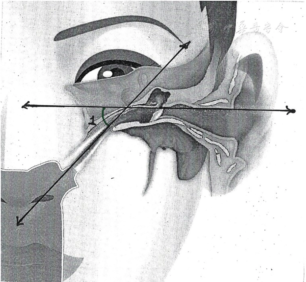
The ETA with respect to Reid’s plane was significantly acute on the COM side compared to the healthy side (28.13±1.35 vs. 27.97±1.86, p=0.00). Cohen’s d = 0.31 indicated a small-to-medium effect size ( Figure 1).
5. Pretympanic Diameter (PTD)
The PTD was significantly smaller on the COM side compared to the healthy side (3.30 ± 0.77 vs. 3.91 ± 0.56, p < 0.001). Cohen’s d = 1.01 reflected a large effect size ( Figure 2).
6. Tubotympanic Angle (TTA)
The TTA was significantly wider on the COM side (148.86 ± 3.57 vs. 144.76 ± 2.15, p < 0.001). Cohen’s d = 0.96, indicating a large effect size ( Figure 3).
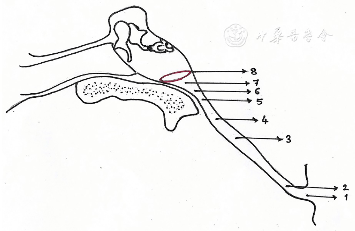
Labels: 1-Pharyngeal orifice, 2-Pharyngeal portion, 3-Mid-portion, 4-Pre-isthmus, 5-Isthmus, 6-Post-isthmus, 7-Pretympanic, 8-Tympanic orifice.9
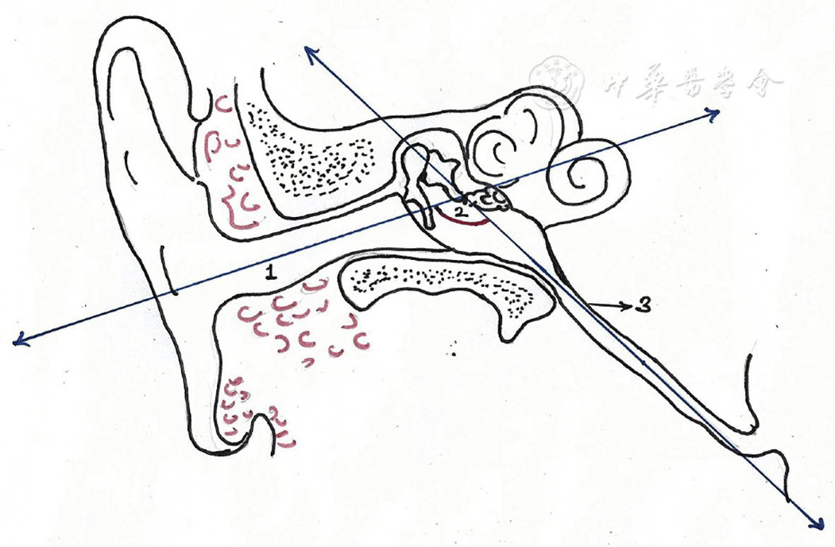
1-External auditory canal, 2-Tubotympanic angle, 3-Eustachian tube.9
Radiological investigations, particularly HRCT, are used as an adjunct to clinical and audiological assessments in the diagnosis of middle ear diseases and planning surgeries.10 As COM is a multifactorial disease, otologists need to know the anatomical variants to detect the risk factors and plan treatment accordingly.11,12 By studying unilateral COM, an underexplored manifestation of the disease, this study offers insights into these anatomical structures playing roles in the pathogenesis of COM.
Among anatomical variations in the nose and paranasal sinuses, DNS, particularly with a greater angle, and concha bullosa were associated with increased COM risk. These nasal pathologies can impede nasal airflow to the ET, thus affecting mastoid cell aeration.13,14 This finding corroborates the findings of Damar et al. and Atila et al., who observed that anatomical alterations in the nose, such as DNS, concha bullosa, and inferior turbinate hypertrophy disrupt nasal airflow, ET pressure, and mastoid aeration, and thus increase the incidence of COM.6,15 These nasal pathologies may affect the ipsilateral ear, helping explain why unilateral COM is associated with nasal or sinus abnormalities on the same side. However, contrary to the findings of Atila et al. and Khaksari et al., no association of COM was observed with hypertrophy of turbinates or sinusitis.6,14
The mastoid air cells were less pneumatized and predominantly sclerotic on the COM side, while 24% (n=12) of the contralateral normal temporal bones were also sclerotic. According to Jadia et al., the contralateral temporal bones in unilateral COM exhibit poor pneumatization, ranging from 17.8% in mucosal diseases to 55.9% in squamous diseases.16 This supports the theory that unilateral COM represents a continuous process with an increased risk of disease development in the contralateral ear.4,16
KS is a bony partition extending from the petrosquamosal fissure to the anterolateral surface of the mastoid process, and is postulated to reduce mastoid aeration. Toros et al. found no significant relationship between KS and temporal bone aeration, whereas Elibol et al. found KS more frequently in patients with COM.3,17 By studying purely unilateral COM cases, the authors found no association between COM and KS presence or thickness.
The genetic theory claims that pneumatization of the temporal bone is genetically determined, rendering poorly pneumatized bones susceptible to COM. ET orientation and angulation can affect mastoid pneumatization.18 The two commonly studied ET angles are the ETA and TTA. The ETA is defined as the angle between the right plane and the tympanic pharyngeal orifice. The angle between the longitudinal axes of the ET and the bony external auditory canal is called the TTA.19 Nemade et al. detected ETA 27.56 ± 3.62 in healthy ears and 25.41 ± 2.57 in diseased ears. They found the TTA to be 148.12 ± 3.43 °in diseased ears and 145.14° ± 4.34° in healthy ears.5 In a nutshell, they found significantly less ETA and significantly less TTA obtuse in patients with COM. In patients with unilateral COM, the authors found a significantly acute ETA, wider TTA, and narrower PTD on the COM side of the skull than on the healthy temporal bones. A narrower ETA may facilitate nasopharyngeal reflux, thereby increasing the risk of infection and inflammation in the middle ear. Additionally, a wider TTA can enhance pathogen entry into the middle ear, contributing to chronic inflammation and infection. A narrower PTD can impede airflow and ventilation of the mastoid. These results highlight the impact of ET anatomical variations on COM pathogenesis, further supporting the theory that specific anatomical features predispose individuals to middle ear infections.
Despite these significant findings, this study has several limitations. The sample was drawn from a single center. Furthermore, detailed assessments of subtypes were not included, which could have offered a more comprehensive understanding of the COM subtypes and their anatomical correlates. Nevertheless, the authors’ institution is a major referral center that enhances the representativeness of the study sample. Future multicenter studies should explore a broader population for these anatomical variations and correlate these with COM subtypes.
Significant anatomical differences were observed between the diseased and healthy sides of the skull of patients with unilateral COM. Among the rhinologic variants, ipsilateral DNS with a greater angle and concha bullosa were associated with COM. Poor mastoid pneumatization, acute ETA, narrow PTD, and wide TTA were significantly associated with COM. These anatomical factors should be considered during the diagnostic evaluation and treatment planning for COM.
Preregistered data analysis: This research was not preregistered.
The experimental protocols were approved by the ethical standards of the Institutional Review Board of the Khyber Teaching Hospital. The study was conducted in accordance with the ethical standards of the Institutional Review Board of Khyber Teaching Hospital, Peshawar, Pakistan (registration no. 495/DME/KMC issued on 18/08/2023). Written informed consent was obtained from all participants included in this study.
The abstract of this manuscript was accepted for a poster presentation at the American Otological Society AOS/COSM Spring Meeting, 2025.
Despite these significant findings, this study had some limitations. The sample was drawn from a single center. Furthermore, detailed assessments of subtypes were not included, which could have offered a more comprehensive understanding of the COM subtypes and their anatomical correlates.
| Views | Downloads | |
|---|---|---|
| F1000Research | - | - |
|
PubMed Central
Data from PMC are received and updated monthly.
|
- | - |
Provide sufficient details of any financial or non-financial competing interests to enable users to assess whether your comments might lead a reasonable person to question your impartiality. Consider the following examples, but note that this is not an exhaustive list:
Sign up for content alerts and receive a weekly or monthly email with all newly published articles
Already registered? Sign in
The email address should be the one you originally registered with F1000.
You registered with F1000 via Google, so we cannot reset your password.
To sign in, please click here.
If you still need help with your Google account password, please click here.
You registered with F1000 via Facebook, so we cannot reset your password.
To sign in, please click here.
If you still need help with your Facebook account password, please click here.
If your email address is registered with us, we will email you instructions to reset your password.
If you think you should have received this email but it has not arrived, please check your spam filters and/or contact for further assistance.
Comments on this article Comments (0)