Keywords
Endoscopy lithotripsy, Impacted bile duct stone, Obstructive jaundice
Endoscopy lithotripsy, Impacted bile duct stone, Obstructive jaundice
Bile duct stone is a common diagnosis made by digestive surgeons in routine practice. Although usually asymptomatic, bile duct stone can obstruct the biliary tract, leading to symptoms such as jaundice, right-upper quadrant abdominal pain, and changes in urine and stool color. Most cases of biliary tract obstruction occur in the common bile duct (CBD).1 Giant bile duct stone are a rare presentation in cases of gallbladder stones and are defined as stones larger than 5 cm or 50 mm. In cases of impacted giant bile duct stone, standard non-invasive procedures like Endoscopic Retrograde Cholangiopancreatography (ERCP) are not feasible, thus management involves percutaneous approaches or open surgery.2,3 Lithotripsy, a device that emits sound waves, is commonly used to break up kidney or ureter stones to facilitate their spontaneous expulsion. This device also holds potential for breaking up giant bile duct stone, easing the removal of these stones. This case report aims to document the successful management of an impacted giant bile duct stone using endoscopy-guided ureteroscopy lithotripsy for the visualization and fragmentation of larged impacted stones located higher in the biliary tract.
A female in her sixties presented with intermittent right upper quadrant abdominal pain over several weeks. Initially, the patient experienced intermittent pain in the upper right abdomen for two months. Jaundiced developed several weeks before admission, symptoms included yellowing of the eyes, dark urine resembling strong tea, and pale clay-colored stools similar to putty, with no occurrence of black stools. Although these symptoms subsided on their own, abdominal pain recurred week ago, accompanied by increased jaundice. The patient reported nausea and vomiting but no fever and began to feel itchiness all over the body. No otrher relevant previous and familial history.
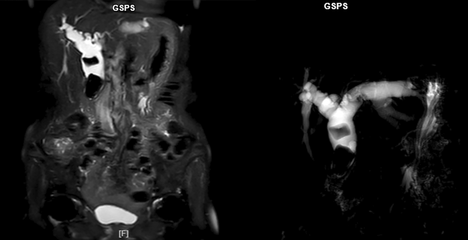
Upon physical examination, the patient was fully conscious with stable hemodynamic. The sclera was icteric, and the conjunctiva was not anaemic. The abdomen was soft and tender in the upper right quadrant, with a positive Murphy’s sign and no guarding. Laboratory tests showed a slight decrease in haemoglobin and haematocrit levels, normal leukocyte count, normal prothrombin time and activated partial thromboplastin time. There was an increase in bilirubin levels, with total bilirubin above 9.0 mg/dL and increased in total direct bilirubin above 7.0 mg/dL. Magnetic resonance cholangiopancreatography (MRCP) revealed dilatation of the common bile duct (CBD) and intrahepatic bile ducts on both sides, along with multiple giant stones in the CBD and common hepatic duct (CHD) ( Figure 1). The diagnosis was obstructive jaundice due to multiple giant stones in the common hepatic duct and common bile duct with distal CBD stenosis.
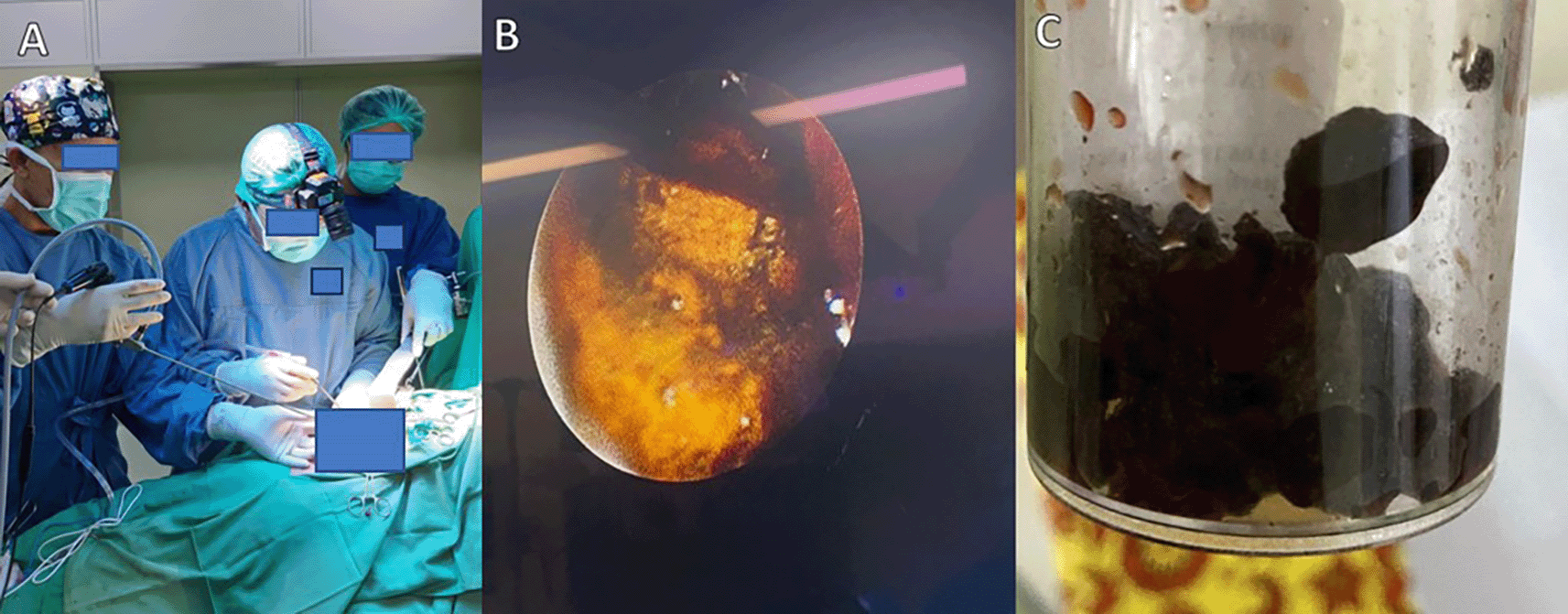
The surgical intervention consisted of CBD exploration and choledochotomy followed by a side-to-side choledochoduodenostomy and cholecystectomy. Intraoperatively, after cholecystectomy, CBD exploration showed dilatation extending to the right and left hepatic ducts. Choledochotomy was performed, revealing multiple stones in the CBD and a large, impacted stone in the CHD. Stone removal was facilitated using guided URS and lithotripsy, breaking the stone into fragments for evacuation (Figure 2A and 2B). The size of the stone was approximately 4 cm (Figure 2C). Proximal probing showed no stones, and distal probing indicated stenosis of the CBD with a probe size of 3 Fr. Proximal bile flow was smooth. A decision was made to perform a side-to-side bypass from the CBD to the duodenum (choledochoduodenostomy). The patient recovered well and was discharged within a week. At follow-up, she remained symptom-free.
This case report focuses on the use of endoscopy-lithotripsy for impacted giant common hepatic duct and common bile duct stones. The patient presented with typical symptoms of obstructive jaundice, including yellowing of the eyes, dark urine, and pale, clay-colored stools. As the disease progressed, the patient experienced abdominal pain accompanied by increased jaundice. These symptoms are common in cases of obstructive jaundice. It is important to determine whether the jaundice in this patient is prehepatic, intrahepatic, or posthepatic. To differentiate these, bilirubin levels were measured, showing increased total, conjugated, and unconjugated bilirubin levels. An increase in direct bilirubin above 1.0 mg/dL is noted as an anomaly when total bilirubin increases.4 Elevated conjugated bilirubin levels indicate obstructive jaundice primarily caused by cholestasis and hepatocellular dysfunction.5 The increase in unconjugated bilirubin in this patient might be due to the high total bilirubin already accumulated in the body, thus also raising the unconjugated bilirubin, although this increase is not as significant as that of conjugated bilirubin.
MRCP for this patient revealed multiple giant stones in the CBD and CHD. The occurrence of giant bile duct stone, defined as stones larger than 5 cm, is rare and associated with higher complication rates and technical difficulties in surgical management.2 The main management modality for bile duct obstruction due to bile duct stone is Endoscopic Retrograde Cholangiopancreatography (ERCP). However, the ERCP technique is known to have a low success rate for stones larger than 10 mm and is therefore not feasible for giant bile duct stone cases.3 The standard management for these bile duct stone is open surgery with CBD exploration, although there are some case reports of laparoscopic procedures for giant bile duct stone.6,7 In this case, the patient was decided to undergo open surgery with common bile duct exploration, choledocostomy, and cholecystectomy due to the presence of multiple bile duct stone involving many bile ducts. Additionally, open surgery was chosen due to the location of the impacted giant bile duct stone being higher than the bile duct which was in the CHD.
A highlight in this case report is the use of lithotripsy, which can break down giant bile duct stone with the aid of endoscopy ureteroscopy in patients undergoing open surgery. Lithotripsy is a fragmentation technique using sound waves typically used to break up kidney and ureter stones. The use of lithotripsy for breaking bile duct stone has been reported by this case report and other previous reports. Wang, et al. reported the use of Fluoroscopy-guided percutaneous lithotripsy using a FREDDY laser in patients with giant bile duct stone and reported a 100% success rate for bile duct stone clearance with a residual stone rate of 18.8% of cases.8 Endoscopic lithotripsy has also been reported by Khalil, et al. in patients undergoing percutaneous cholecystectomy. In this case report, Khalil, et al. used a pediatric video gastroscope for direct visualization of the gallbladder before applying electrohydraulic lithotripsy. The management was deemed successful in breaking the bile duct stone into small fragments that were drained using a catheter.9 In this case report, lithotripsy was performed using ureteroscopy. There are previous case reports that also used a ureteroscope to break giant bile duct stone with lithotripsy. Loffeld, et al. used ureteroscopy lithotripsy to visualize and break bile duct stone using a percutaneous approach.10
Based on literature review, previous case reports tended to use a percutaneous approach in performing lithotripsy because in those cases, the giant bile duct stone occurred at only one location. This case report is the first to apply ureteroscopy lithotripsy in patients undergoing open CBD exploration surgery where open surgery was chosen because the impacted bile duct occurred above the biliary tract. In normal cases, a higher incision is required to reach the location of the impacted giant bile duct stone, but in this case, by using endoscopic ureteroscopy visualization and lithotripsy to break the bile duct stone, the incision made in the patient could be reduced to only the CBD. The patient in this case report underwent overall stone removal management with good follow-up results and no complaints up to 14 days after surgery.
This case underscores the benefits of adapting endoscopy lithotripsy for biliary system use, particularly for challenging cases involving impacted giant stones in less accessible locations of the biliary tract. This technique reduced the necessity for large surgical incisions to reach the giant impacted bile duct stone in the common hepatic duct by fragmenting the stone. This case provides a new direction for future cases involving impacted bile duct stone in less accessible place.
| Views | Downloads | |
|---|---|---|
| F1000Research | - | - |
|
PubMed Central
Data from PMC are received and updated monthly.
|
- | - |
Provide sufficient details of any financial or non-financial competing interests to enable users to assess whether your comments might lead a reasonable person to question your impartiality. Consider the following examples, but note that this is not an exhaustive list:
Sign up for content alerts and receive a weekly or monthly email with all newly published articles
Already registered? Sign in
The email address should be the one you originally registered with F1000.
You registered with F1000 via Google, so we cannot reset your password.
To sign in, please click here.
If you still need help with your Google account password, please click here.
You registered with F1000 via Facebook, so we cannot reset your password.
To sign in, please click here.
If you still need help with your Facebook account password, please click here.
If your email address is registered with us, we will email you instructions to reset your password.
If you think you should have received this email but it has not arrived, please check your spam filters and/or contact for further assistance.
Comments on this article Comments (0)