Keywords
Spigelian hernia, strangulation, surgical repair, mesh.
Although Spigelian hernia is rare, it should not be underestimated because of its significant risk of strangulation. The scarcity of studies and the absence of large, comprehensive case series on this pathology often makes clinical decision-making, despite appearing straightforward, both challenging and ambiguous.
This is a retrospective, monocentric study, conducted between January 2013 and December 2022, involving 14 patients who underwent surgery for a Spigelian hernia in the General Surgery Department of Charles Nicole Hospital in Tunis. Various epidemiological, clinical, radiological, and therapeutic factors were analysed using univariate analysis and logistic regression analyses.
Fourteen patients with Spigelian hernia were recruited. The mean patient age was 62 years. All patients had predisposing factors for increased intra-abdominal pressure, and 50% had a history of abdominal surgery. The most common symptom was lateralized abdominal pain. Diagnosis was supported by imaging in 85.7% of cases, with CT scans being particularly helpful in emergency settings. Surgical repair was performed in all patients, predominantly via an open approach (92.8%). Primary suture repair was used in 64.2% of cases, while mesh repair was performed in 35.7%. The postoperative course was uneventful in 85.7% of patients. Morbidity was 14.2%, with no mortality. One recurrence was observed during follow-up.
Despite its rarity, Spigelian hernia should be considered in the differential diagnosis of localized abdominal pain. Imaging, particularly CT, plays a key role in diagnosis. Surgical repair, whether by suture or mesh, offers favorable outcomes with low morbidity and recurrence rates.
Spigelian hernia, strangulation, surgical repair, mesh.
Spigelian hernia is a defect of the anterolateral abdominal wall. It is a rare entity, representing only 1 to 2% of all abdominal wall hernias. 1 It can lead to the protrusion of bowel loops or other organs, with a significant risk of hernia strangulation estimated at 30%. Owing to this risk, surgical intervention is necessary as soon as the diagnosis is confirmed. 2 The clinical diagnosis of Spigelian hernia is often challenging, therefore radiologic exploration is required. Abdominal tomography is the key examination that confirms the diagnosis, determines the hernia contents, and identifies potential complications.3 Therapeutic modalities are not yet consensual. Although surgery is the treatment of choice, the nature of the repair—suturing versus synthetic mesh use—as well as the choice of approach—laparotomy versus laparoscopy—remain to be further validated.
Through this work, we conducted a study on a retrospective series of 14 cases aiming to highlight the diagnostic challenges and various therapeutic modalities of this pathology.
This was a retrospective, monocentric study including 14 patients managed in the General Surgery Department of Charles Nicolle Hospital, Tunis, for Spigelian hernia between January 2013 and December 2022.
We included all patients younger than 18 years of age who underwent surgical repair for Spigelian hernia. We excluded patients with:
Epidemiological, clinical, radiological, and therapeutic data were collected using a standardized form. The following parameters were analyzed:
- Epidemiological variables: age, sex, medical and surgical history, and risk factors for increased intra-abdominal pressure (e.g., obesity, chronic cough, multiparity).
- Clinical variables: reason for consultation (pain, swelling, incidental finding), duration of symptoms, physical examination findings (size of swelling, hernia neck, reducibility, tenderness).
- Radiological variables: type of imaging, diagnostic accuracy, and detailed imaging findings.
- Intraoperative variables: hernia sac and neck size (measured with sterile surgical calipers), contents of the sac (omentum, bowel, empty sac), viability of the bowel, surgical approach, repair technique, mesh characteristics, fixation method, operative time, and intraoperative complications.
- Postoperative variables: early complications within 30 days (surgical, medical, specific, or nonspecific), length of hospital stay, late outcomes (recurrence, rehospitalization).
Ultrasound examinations were performed using a Philips Affiniti 70 (Philips Healthcare, Netherlands) with a 7.5 MHz linear transducer. Computed tomography (CT) scans were acquired using a Siemens SOMATOM Definition AS (Siemens Healthineers, Germany), with 5-mm axial and coronal slices and intravenous contrast administration (Iopamidol 370 mg/mL, Bracco, Italy). Imaging was interpreted independently by two senior radiologists. Discrepancies were resolved by consensus.
All procedures were performed under general anesthesia by senior surgeons specialized in abdominal wall repair. Prophylactic intravenous antibiotics (cefazolin 2 g) were administered at induction.
- Open repair: A horizontal or slightly oblique incision (average length 5–6 cm) was made over the hernia site. The sac was dissected, contents reduced or resected if necessary, and the sac either closed or inverted. Herniorrhaphy was performed with 2-0 slowly absorbable monofilament sutures (PDS®, Ethicon, USA) in cases with a narrow hernia neck (<2 cm). For mesh hernioplasty, a polypropylene mesh (Prolene®, Ethicon, USA or Biomesh®), usually 10 × 10 cm, was tailored to the defect size and fixed using interrupted cardinal sutures of 2-0 monofilament polypropylene in the preperitoneal space or between the internal and external oblique muscles.
- Laparoscopic repair: In selected cases, a three-trocar approach (two 10 mm and one 5 mm) was used. Mesh was placed in the preperitoneal space and fixed with absorbable tacks.
When small bowel resection was required for strangulation, an ileo-ileal anastomosis was performed using a two-layer hand-sewn technique with 3-0 absorbable sutures (Vicryl®, Ethicon, USA) for the inner layer and 3-0 silk sutures for the outer layer. A closed-suction Redon drain (14 Fr) was used in selected cases with large dissection planes.
A total of fourteen patients diagnosed with spigelian hernia were managed in our department between January 2013 and December 2022. The average incidence was 1.4 new repairs per year. Of these, 13 (92.9%) were female and 1 (7.1%) was a male, with a sex ratio of 0.7.
The mean age of the patients was 62 years.
Figure 1 summarizes the age distribution of patients included in the study.

Legend: Bar graph showing the age distribution of patients diagnosed with Spigelian hernia in the cohort. The average age was 62 years.
Eleven patients in our cohort, accounting for 78.5% of cases, had comorbidities of cardiovascular, metabolic, or other chronic conditions.
Hernia risk factors leading to increased intra-abdominal pressure were observed in all of our patients as illustrated in Figure 2.
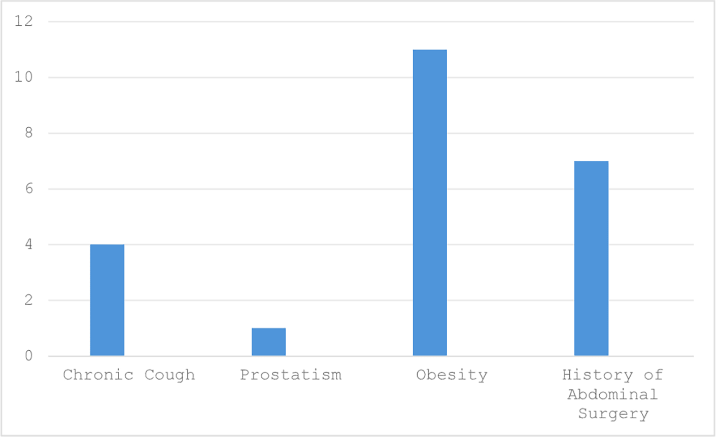
Legend: Diagram illustrating the distribution of risk factors associated with increased intra-abdominal pressure among the patients (e.g., obesity, chronic cough, multiparity).
The most common reason prompting consultation was lateralized abdominal pain, experienced by 92.8% of patients (n=13).
Table 1 summarize the different clinical symptoms experienced by patients with Spigelian hernia during consultation, covering pain features, signs of obstruction, and incidental discoveries.
The duration between the onset of symptoms and consultation was 12 months, with a range from 4 to 72 months.
On physical examination, abdominal pain was noted in 9 patients (64.2%). An abdominal mass was palpated in two patients (14.2%), and abdominal distension was observed in one patient (7.1%).
The swelling was located along the left para-rectal line above the level of the umbilicus in both cases. The average diameter of the swelling measured approximately 4 cm. Clinically, the hernias were found to be reducible, painless, and exhibited a positive cough impulse, consistent with the characteristics of uncomplicated abdominal wall hernias.
Twelve out of the fourteen patients in our cohort, accounting for 85.71%, underwent radiological evaluation.
Table 2 presents the imaging techniques utilized and evaluates their success in confirming Spigelian hernia diagnoses, with CT scans showing the greatest diagnostic accuracy.
| Radiological examination | Number of patients (Percentage) | Positive diagnosis rate |
|---|---|---|
| Abdominal CT scan | 4 (28.75%) | 100% |
| Abdominal ultrasound | 8 (57.14%) | 75% |
| No radiological exploration | 2 (14.28%) | - |
Figure 3 demonstrate the radiological findings of Spigelian hernia.
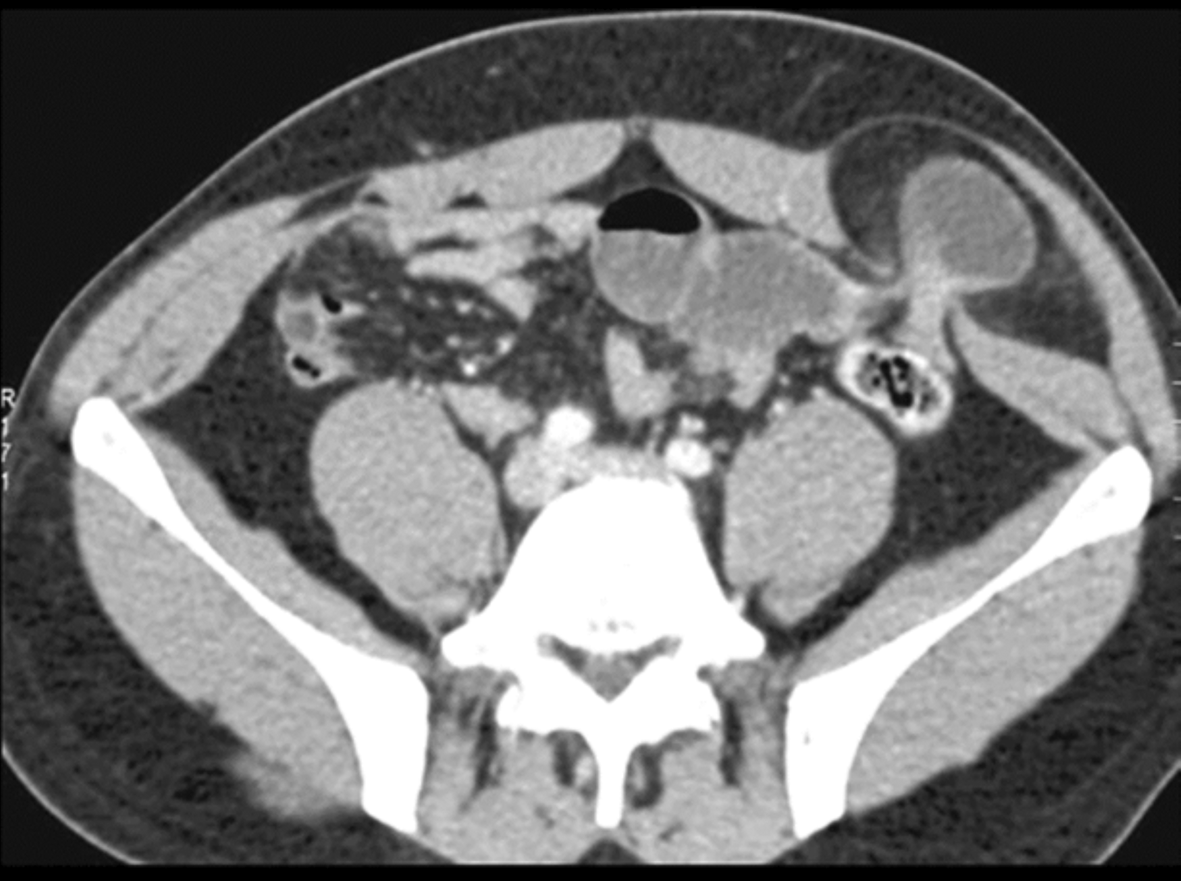
Legend: Axial contrast-enhanced CT image showing a parietal defect along the Spigelian line, with hernial sac containing a loop of small intestine on the left paraumbilical region.
All patients in our cohort underwent surgical intervention under general anesthesia. Prophylactic antibiotics were administered at the time of anesthetic induction in all cases. The procedures were performed by a senior surgeon. Regarding the surgical approach, the majority of patients (13 out of 14, or 92.8%) underwent conventional open surgery. In these cases, the hernia sac was accessed directly through a horizontal or slightly oblique incision, with an average length of 5.3 cm. Only one patient (7.1%) was managed using a laparoscopic approach, in which three trocars were inserted: two measuring 10 mm and one measuring 5 mm. Figure 4 below summarizes the anatomical distribution of parietal defects among the study cohort.
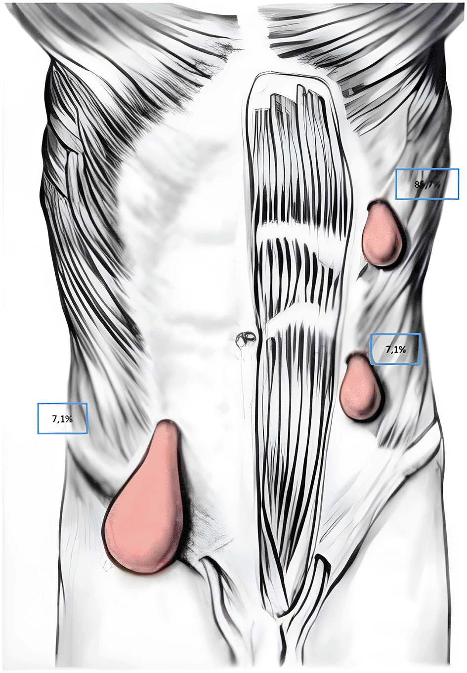
The defect was located along the left para-rectal line, above and beside the umbilicus in 12 patients (85.7%), below and beside the left umbilicus in one patient (7.1%), and below and beside the right umbilicus in one patient (7.1%).
In our sample, the hernial sac had an average of 4.5 cm ± 1.5. The average size of the neck was evaluated at 2 cm ± 1.3.
The content of the hernial sac was empty in nine patients, representing 64.2% of the cases. An intraluminal omental fringe was observed in 3 of our patients (2.14%). Small bowel was incarcerated in the hernial sac in 2 cases (14.2%). The small bowel was viable in one patient and gangrenous in the other.
Table 3 details the surgical methods used in the treatment of Spigelian hernias, specifying the proportion of patients who underwent primary herniorrhaphy versus those treated with mesh hernioplasty.
| Nature of repair | Number of patients | Percentage |
|---|---|---|
| Herniorrhaphy | 9 | 64.2% |
| Hernioplasty | 5 | 35.7% |
In all cases, the material used for hernia repair was polypropylene mesh (Prolene®, Biomesh®). Among the patients who underwent prosthetic hernioplasty, pre-cut polypropylene meshes measuring 10 × 10 cm were used in five cases. These meshes were subsequently tailored intraoperatively to fit the size of the parietal defect, specifically the hernial neck. Regarding the placement and fixation of the mesh, the prosthetic material was deployed in the preperitoneal space in one patient. In the remaining four cases, the mesh was positioned between the internal and external oblique muscles. Fixation was performed using cardinal sutures with slowly absorbable monofilament 2/0 sutures.
The hernial sac was resected in 78.5% of cases (11 patients) and subsequently closed with a continuous suture using 2/0 absorbable thread. In the remaining 3 patients (21.5%), the sac was reduced into the intraperitoneal cavity without resection.
A small bowel resection with ileo-ileal anastomosis was performed in one patient who underwent emergency surgery for a strangulated Spigelian hernia containing necrotic small bowel. A Redon drain was placed adjacent to the mesh in only one patient (7.1%), in whom the hernial sac measured 6 cm.
No intraoperative complications, such as vascular or digestive injuries, were observed in our cohort. The mean duration of the surgical procedure was 112 minutes, with a range of 60 to 240 minutes.
Postoperatively, the average hospital stay was 1.5 days, varying between 1 and 3 days. The immediate postoperative outcomes were favorable in the majority of cases, with 85.7% of patients (n=12) experiencing an uncomplicated recovery. No mortality was recorded. The overall morbidity rate was 14.2% (n=2), with no general or non-specific complications observed. Figure 5 details the type and frequency of specific postoperative complications observed in the study cohort.

Legend: Bar graph showing specific postoperative complications encountered, including ileus, wound infection, seroma, and postoperative pain.
In our study, the median follow-up duration was 3 months, with a range from 1 to 24 months. Only one case of recurrence was reported after one year of follow-up.
Spigelian hernia is the protrusion of preperitoneal fat, a peritoneal sac, or abdominal organs through a defect in the Spigelian aponeurosis—a fibrous layer situated between the rectus abdominis muscle and the semilunar line. This condition typically manifests in the Spigelian zone, a region of structural vulnerability where the aponeuroses of the transversus abdominis and internal oblique muscles become thinner. 1, 2 It is an uncommon type of ventral abdominal wall hernia, accounting for 1 to 2% of all abdominal wall hernias.3,4
This finding aligns with the results of our study, which reported an average incidence of 1.4 Spigelian hernia repairs per year. According to our data, Spigelian hernias are exceptionally rare, accounting for only 0.28% of all operated abdominal wall hernias in our department.
Spigelian hernia is a rare condition with no clear sex predominance in the literature. In a French series by Guivarch et al., involving 51 patients, the sex ratio was reported as 1.3 Spangen’s series noted a slight female predominance.4 In contrast, our Tunisian cohort demonstrated a marked female predominance (sex ratio 0.07), likely attributable to the limited sample size and regional risk factors such as obesity and multiparity—pregnancy being an exclusive female herniogenic factor.
The mean age of onset in our study aligns with Swedish data (~40 years),3 differing from French reports (71 years), 1 possibly reflecting demographic variations.
Chronic cough, a poorly documented risk factor, emerged as significant in our series (14 cases).
Several predisposing factors have been proposed in the literature.
83.3% of the female patients in our cohort were multiparous (≥2 pregnancies), corroborating findings from Moroccan and Tunisian studies that have established a significant association between multiparity and the development of Spigelian hernias.5,6
Our study, with a relatively larger series of 14 cases, also supports a strong association between chronic cough—particularly in the context of chronic obstructive pulmonary disease (COPD)—and the development of Spigelian hernia.
Concurrent abdominal wall defects or a history of prior hernia surgery were documented in 28.6% of cases, reflecting findings from previous studies that report frequent associations between Spigelian hernias and other hernia types.7,8
Spigelian hernias pose significant diagnostic difficulties due to their unique anatomical location. When small, the hernia remains tightly constrained, making it difficult to palpate. Larger defects or those occurring in a thin abdominal wall may become visible in a standing position.
Clinical symptoms vary, ranging from mild discomfort to sharp pain.
According to studies published by Bouali5 and Neto6 (2021), Spigelian hernias frequently present at an advanced, complicated stage, with strangulation being a common finding.
These observations align with our findings: the majority of patients (92.8%) reported paroxysmal abdominal pain, while 50% described subocclusive episodes and 28.5% were admitted on an emergency basis for continuous abdominal pain associated with vomiting.
The reliability of physical examination alone remains limited. A palpable mass with a cough impulse is rare and is more likely to be detected when the hernia sac is large and/or the patient is thin.9 In two studies published in 20023 and 2006,10 Larson et al. found that clinical diagnosis of Spigelian hernia was established in 64% of cases,3 rising to 71% in the series by Malazgirt.10
These findings are consistent with those of our cohort. Physical examination led to diagnosis in only two patients (14.2%), in whom a mass was palpated along the pararectal line.
Given the limitations of clinical assessment, imaging plays a crucial role in diagnosing Spigelian hernias. While ultrasound is useful for initial evaluation, abdominal computed tomography (CT) remains the gold standard due to its superior sensitivity and ability to assess hernia contents.
Ultrasound enables differentiation between a Spigelian fascial defect and a solid mass with high sensitivity, reported to range between 83% and 100%. 2
In our study, the diagnostic sensitivity was slightly lower than reported in the literature, reaching approximately 64.2%. Ultrasound enabled diagnosis in 9 of the 14 patients: it identified the parietal defect in 8 cases and visualized intra-sac digestive content in 1 case.
Due to the frequent misdiagnosis of Spigelian hernias as lipomas or other abdominal wall pathologies—and the fact that small hernias may not traverse all muscle layers—CT is the preferred diagnostic tool.11 It clearly visualizes the aponeurotic defect at the lateral border of the rectus abdominis muscle, the hernia sac, and—in certain cases—its intra-sac contents. CT is also critical for differential diagnosis in cases of suspected strangulation, helping to exclude tumors, diverticulitis, or acute appendicitis. In our cohort, CT was performed only in emergency case.
Due to the risks of complication, it is recommended a surgical repair for Spigelian hernias. The choice between open and laparoscopic approaches in the surgical management of Spigelian hernias remains a subject of ongoing debate, largely due to the absence of high-level evidence guiding clinical practice.
Several studies have examined the role of laparoscopy in managing lateral ventral hernias, including one of the earliest by Moreno-Egea et al. in 2002. They demonstrated a statistically significant difference between the two approaches, with laparoscopy being associated with reduced postoperative morbidity and shorter hospital stays.4 However, these findings were challenged by Clark et al.,12 who argued that the disadvantages observed with the open approach were largely attributable to anesthetic agents rather than the surgical technique itself.
Although laparoscopy facilitates better visualization of the hernia defect and allows the detection of occult or multiple hernial orifices, its utility may be limited in cases involving large defects. In such scenarios, open repair—particularly with preperitoneal mesh placement—remains a viable and sometimes preferable option.
In our cohort, the majority of patients (13 out of 14, or 92.8%) were treated via an open approach. Only one patient underwent laparoscopic repair, and outcomes were comparable between the two approaches.
In a 2024 study of 43 patients with Spigelian hernia, Clark12 reported postoperative complications in 36% of cases treated via the open approach, compared to 13% for the laparoscopic group. Moreno-Egea et al. also reported higher long-term recurrence rates with the open approach.4
Numerous surgical approaches and repair techniques are available, including open herniorrhaphy or mesh repair (either preperitoneal or preaponeurotic) and, via laparoscopy, simple suture or prosthetic repair using intraperitoneal (IPOM), transabdominal preperitoneal (TAPP), or totally extraperitoneal (TEP) methods.
Although associated with higher recurrence rates, herniorrhaphy remains an option in Spigelian hernia repair, particularly when the hernia neck is narrow (<2 cm).13,14 Some surgeons still opt for simple sutures in emergency settings. Recent studies evaluating the infection risk of prosthetic repair in strangulated hernias suggest that this risk is not significantly higher than in elective surgery.12 In our study, nine patients (64.2%) underwent herniorrhaphy. These included the four patients operated on in emergency settings
In contrast to simple suture repair, prosthetic hernioplasty is associated with virtually no recurrences. In the Mayo Clinic series, three recurrences occurred among 75 patients treated with suture repair, while no recurrences were observed in the group that received prosthetic repair—corresponding to recurrence rates of 4% and 0%, respectively.3 Similarly, in a 2002 paper, Moreno-Egea et al. reported no recurrences in 22 patients treated with mesh repair.4
With the efficacy of mesh repair firmly established, particularly in terms of recurrence, the current debate focuses on mesh placement. Open repair may involve preperitoneal or preaponeurotic positioning, while laparoscopic repair can use IPOM, TAPP, or TEP techniques. At present, there are no strong recommendations favoring one approach over the others, as each has its advantages and limitations.
In our series, all patients treated via the open approach in non-emergency settings (9 out of 14, or 64.2%) underwent mesh repair. In one case, the mesh was placed in the preperitoneal space; in four others, it was positioned between the internal and external oblique muscles.
Recently, case reports have described successful robotic-assisted repair of Spigelian hernias, with promising preliminary outcomes.15,16
Table 4 provides a comparative overview of major studies addressing Spigelian hernia repair, focusing on surgical techniques, recurrence rates, and associated complications.
| Year | Design | Author | Patients | Open | MIS | LOS, days | Morbidity, n (%) | Recurrence, n (%) | Follow-up, months |
|---|---|---|---|---|---|---|---|---|---|
| 2002 | RCT | Moreno-Egea et al. | 22 | 11 | 11 (8*; 3**) | Open: 3, MIS: 1*; 1.4** | Open: 4 (18%), MIS: 0 | 0 | 40 |
| 2002 | Retrospective | Larson et al. | 76 | 75 | 1** | N/A | 8 (11%) | Open: 3 (4%), MIS: 0 | 96 |
| 2006 | Prospective | Palanivelu et al. | 8 | 0 | 8*** | 1.2 | 0 | 0 | 41 |
| 2006 | Retrospective | Malazgirt et al. | 6 | 0 | 6*** | 4.1 | 1 (18%) | 0 | 30 |
| 2007 | Retrospective | Nirmal et al. | 3 | 0 | 6*** | 1.2 | 1 (17%) | 0 | 6 |
| 2012 | Retrospective | Perrakis et al. | 15 | 15 | 1** | Open: 3.5, MIS: 1** | 2 (13%) | 0 | 98 |
| 2012 | Prospective | Zuvela et al. | 8 | 0 | 8 | Δ | 0 | 0 | 23.5 |
| 2014 | RCT | Moreno-Egea et al. | 16 | 0 | 16 (7*; 9**) | 0 | 0 | 0 | 48 |
| 2017 | Retrospective | Webber et al. | 101 | 68 | 33 | N/A | N/A | N/A | N/A |
| 2018 | Retrospective | Rankin et al. | 33 | 27 | 6*** | Elective: 1.6, Emergency: 5.6 | 7 (21%) | 0 | 32 |
| 2020 | Retrospective | Ruiz de la Hermosa et al. | 39 | 30 | 9** | Elective: 2.6, Emergency: 4 | 2 (5%) | 2 (5%) | N/A |
Immediate postoperative morbidity following abdominal wall surgery is not negligible. It is mainly due to local wound complications including seroma formation (5-15%), hematomas (3-8%), surgical site infections (2-10%), skin necrosis (1-3%), prosthetic infection (1-5%), enterocutaneous fistula formation (0.5-2%), and postoperative ileus (5-12%). The few randomized studies on Spigelian hernia, including those by the Mayo Clinic and Moreno-Egea, compared open and laparoscopic mesh repair. Both showed a consistent trend favoring the laparoscopic approach, with lower rates of superficial wound complications (1.5% vs. 10.1%) and shorter hospital stays. These differences failed to reach statistical significance in pooled analyses. 1, 2
Long-term outcomes following abdominal wall hernia repair are evaluated through recurrence rates, chronic postoperative pain, and quality of life measures. The few available studies on Spigelian hernia repairs showed that mesh repairs are associated with significantly fewer recurrences compared to simple sutures. In the Mayo Clinic series, only 3 out of 75 sutures led to recurrence (4%), whereas none of the 6 patients who received mesh experienced recurrence. Similarly, in the Moreno-Egea study, no recurrence was reported among 22 mesh repairs. 1, 2
Meta-analytic data from general abdominal wall repairs corroborate these findings, demonstrating equivalent long-term efficacy between open and laparoscopic mesh techniques (3.6% vs 3.4% recurrence, respectively) in a pooled analysis of 526 patients.
Despite these favorable outcomes, some concerns remain about the intraperitoneal placement of mesh due to potential risks such as fistulas, adhesions, or intestinal obstruction. However, a cohort study by Hawn found no significant increase in such complications, even though 5% of meshes had to be partially or completely removed. In our own study, we observed similar results, with a low recurrence rate and no significant rise in complications after intraperitoneal mesh placement. Nonetheless, careful technique selection tailored to the hernia characteristics and surgeon’s expertise remains essential.
Spigelian hernia, though rare, poses a significant risk of strangulation, which can threaten patient prognosis. Accurate diagnosis relies on a combination of clinical and radiological data, with abdominal tomography often necessary for confirmation and surgical planning. Our study highlights the importance of an appropriate surgical approach, although the optimal technique and surgical access route require further investigation. The results suggest that with timely and tailored surgical intervention, Spigelian hernias can be managed effectively, leading to satisfactory outcomes, low morbidity, and a limited recurrence rate.
For this study, no conflicts of interest were reported by the authors. We committed to conducting this study in accordance with medical ethical standards. This study was approved by the Surgery Department of Charles Nicolle Hospital, Tunis, Tunisia. As the study involved a retrospective review of anonymized data, no formal Institutional Review Board (IRB) reference number was issued. Access to the archives was authorized by the head of the General Surgery Department “A” at Charles Nicolle Hospital in Tunis. Data collection and the preparation of iconographic materials were carried out on-site within the department.
Written informed consent was obtained from all participants (or their legal guardians) prior to their inclusion in the study. For the retrospective review of anonymized data, the requirement for additional consent was waived by the Surgery Department of Charles Nicolle Hospital.
All data underlying the results are available in Zenodo under the following URL: https://zenodo.org/records/15838533 (Mabrouk A, Fatnassi O, Boukhchim M, Jedidi L, Ben Lahouel S, Karmous N, Ben Moussa M. Dataset for: Spigelian Hernia: Diagnostic Challenges and Therapeutic Approaches: Report of Fourteen Clinical Cases. Zenodo. 2025. doi: 10.5281/zenodo.15838533).
The dataset includes raw numerical values used to generate all figures and tables, including means, standard deviations, ROC curve values, and statistical parameters. Data are available under a CC-BY 4.0 license.
The authors extend their gratitude to the medical and paramedical staff of the General Surgery Department at Charles Nicolle Hospital for their invaluable assistance in patient management and data collection.
| Views | Downloads | |
|---|---|---|
| F1000Research | - | - |
|
PubMed Central
Data from PMC are received and updated monthly.
|
- | - |
Is the background of the case’s history and progression described in sufficient detail?
No
Is the work clearly and accurately presented and does it cite the current literature?
No
If applicable, is the statistical analysis and its interpretation appropriate?
No
Are all the source data underlying the results available to ensure full reproducibility?
No
Are the conclusions drawn adequately supported by the results?
No
Is the case presented with sufficient detail to be useful for teaching or other practitioners?
No
Competing Interests: No competing interests were disclosed.
Reviewer Expertise: hernia, education
Is the background of the case’s history and progression described in sufficient detail?
No
Is the work clearly and accurately presented and does it cite the current literature?
No
If applicable, is the statistical analysis and its interpretation appropriate?
No
Are all the source data underlying the results available to ensure full reproducibility?
Partly
Are the conclusions drawn adequately supported by the results?
No
Is the case presented with sufficient detail to be useful for teaching or other practitioners?
No
Competing Interests: No competing interests were disclosed.
Reviewer Expertise: Pediatric Surgery, Statistics
Alongside their report, reviewers assign a status to the article:
| Invited Reviewers | ||
|---|---|---|
| 1 | 2 | |
|
Version 1 19 Sep 25 |
read | read |
Provide sufficient details of any financial or non-financial competing interests to enable users to assess whether your comments might lead a reasonable person to question your impartiality. Consider the following examples, but note that this is not an exhaustive list:
Sign up for content alerts and receive a weekly or monthly email with all newly published articles
Already registered? Sign in
The email address should be the one you originally registered with F1000.
You registered with F1000 via Google, so we cannot reset your password.
To sign in, please click here.
If you still need help with your Google account password, please click here.
You registered with F1000 via Facebook, so we cannot reset your password.
To sign in, please click here.
If you still need help with your Facebook account password, please click here.
If your email address is registered with us, we will email you instructions to reset your password.
If you think you should have received this email but it has not arrived, please check your spam filters and/or contact for further assistance.
Comments on this article Comments (0)