Keywords
Dental implants, All-on-Four, Stackable surgical guide, Immediate loading

Dental implants, All-on-Four, Stackable surgical guide, Immediate loading
Dental implantation is considered one of the most effective solutions for managing complete edentulism.1 All-on-4 concept has emerged as an innovative option to compensate for all teeth of one jaw with a fixed prosthesis using only four implants per jaw.2 This protocol is based on placing the implants in anterior area with higher bone density, with the posterior implants tilted at an angle ranging between 30° and 45°.3 The purpose of this tilting is to avoid injury to important anatomical structures such as the maxillary sinus in the upper jaw or the inferior alveolar nerve canal in the lower jaw.4 This technique allows for achieving excellent primary stability and reducing the need for bone grafting. This leads to shortening the treatment period and significantly reducing material costs and traumatic procedures.5
In this context, the stackable surgical guide has recently appeared, which differs from previous guides in that it is multi-layered, often five layers, each with its own specific role, based on a foundation guide, while previous guides relied on only one basic layer.6 This guide plays a fundamental role in improving the accuracy of the procedure and reducing the risks associated with deviations in implant guidance.7 Its design is based on three-dimensional computed tomography (CBCT) and facial scanning techniques via facial scan and computer-aided design/computer-aided manufacturing (CAD/CAM) technologies, which allows for virtual planning of the implant path before surgery.8 It also ensures that the implants are placed in ideal positions, while maintaining a correct and functional fit of the temporary prosthesis with the implants, and an ideal occlusal relationship of the prosthesis with the opposing jaw.9
The use of the stackable guide achieves multiple benefits in addition to what the traditional guide achieves in reducing surgery time and reducing unnecessary surgical interventions, as it secures the application of abutments and the temporary prosthesis through it. In addition to the possibility of performing pre-planned bone trimming through this guide if necessary.10 This contributes to reducing the rate of complications and improving the patient’s experience. Moreover, this technique allows the surgeon full control over the procedure, which enhances the integration between the surgical and prosthetic aspects.11
This report presents a case that used an integration of the All-on-4 concept with stackable surgical guide, that may provide an integrated solution characterized by accuracy, efficiency, and durability, which may make it a gold standard for treating cases of complete edentulism.
A 53-year-old nonsmoker female patient presented to the outer clinics of the faculty of dental medicine in Damascus University seeking functional and aesthetic teeth. Left lower lateral incisor and lower canine were considered untreatable due to advanced periodontal disease. The patient’s medical history showed that, she was free of systemic diseases or any contraindications for dental implant surgery, the patient asked for a fixed dental prosthesis for her missing lower teeth.
A comprehensive evaluation revealed that the oral cavity was free of pathological lesions or structural abnormalities. The dental examination showed significant periodontal attachment loss affecting the mandibular left lateral incisor and canine. In contrast, the mandibular left second molar demonstrated good stability and was not indicated for extraction. Additionally, an adequate width of attached gingiva was observed in the mandibular arch. ( Figure 1).
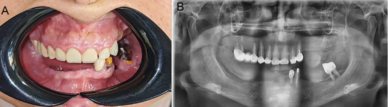
A: intraoral view of maxillary and mandibular. B: The orthopantomography image.
Radiographic Analysis: The CBCT (Ez3D Plus, Vatech, Korea) showed sufficient volume and density (D3 bone density) of the remaining bone to perform implantation without any need for bone grafting in the anterior region. While, the bone height behind the mental foramen on both sides was low and did not allow for the placement of implants with appropriate lengths, which indicated the need for implantation according to the All-on-Four concept.
Clinical Steps: Dental impressions were taken in a traditional way using alginate, and then cast and the plaster models were scanned (UP3D, Shanghai UP 3D Technology Co., China) to have a digitally model. A facial scan of the patient was also performed using a facial scan (RAYFace, RAY Co., Ltd, Gyeonggi-do, South Korea) to consider facial features during prosthesis design.
The CBCT and facial scan files were integrated with digital scans of the diagnostic cast models to create an accurate and comprehensive virtual treatment plan. AnyOne-type implants (AnyOne, Megagen, Korea) were used in this case. The anterior implants measured 13 mm in length with a diameter of 3.5 mm, while the posterior implants measured 13 mm in length with a diameter of 4.0 mm. The anterior implants were positioned perpendicular to the alveolar bone, whereas the posterior implants were angulated at 29.5° on the left side and 29° on the right side ( Figure 2). The temporary prosthesis was digitally designed to optimize both functional performance and facial esthetics.
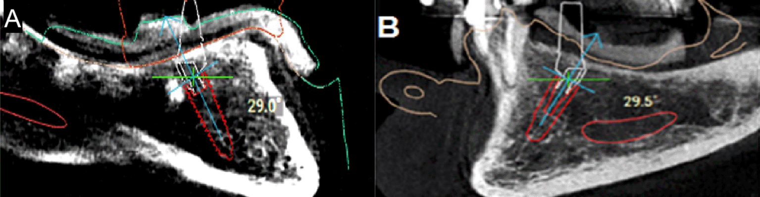
A: 29°Angled dental implant on the right second premolar side. Figure 2. B: 29.5°Angled dental implant on the left second premolar side.
The “All-on-Four” concept was chosen to maximize the use of the available bone and support the prosthesis. The parts of the stackable surgical guide were made of resin (designed by R2Gate software (R2Gate®, Megagen, Korea) and Exocad software (EXO; exocad GmbH, Darmstadt, Germany) and printed by 3D printer (Phrozen Technology, Hsinchu City, Taiwan)). The main components included ( Figure 3):
• Foundation Guide: a screw-retained guide with bone supporting, which used as a primary guide that carries the rest of the elements of the stackable guide, in our case three screws were used to fix the foundation guide to the bone to prevent any movement of this guide during the rest of the procedures.
• Guide for inserting the foundation bone guide: It is a component that rests on the teeth as a reference guide to fix the foundation guide in its correct place. It is connected to it and then removed after it is fixed.
• Implant placement guide: This stackable component is printed with holes guide the drills, that navigate the path and the angle of bone drilling according to the CBCT image that was studied and the implant locations were determined on it. It is fully guided, meaning that the bone preparation is done completely and the implants are inserted through it.
• Abutment installation guide: It is installed on the foundation guide to help determine the final position of the abutments placed on the implants, in our case multi-unit abutments with temporary cylinders were used.
• Temporary prosthesis guide: a guide that rests on the foundation guide carrying the temporary prosthesis, which ensures its placement in the correct planned location.
• Temporary Prosthesis: It is made of polymethyl methacrylate (PMMA) and is used during the healing period to provide aesthetics, maintain the vertical dimension and jaw relationship, and increase patient acceptance. The virtual diagnostic wax patterns planned along with position of implants for temporary prosthesis. This prosthesis is placed for four months post-surgery ( Figure 4).
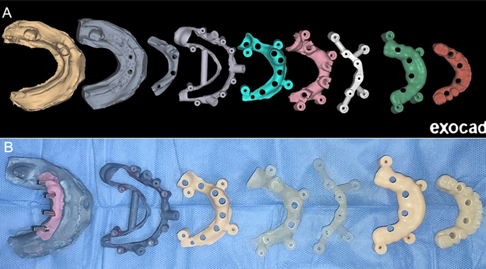
A: Designing the stackable guide components. B: 3D printed stackable guide.
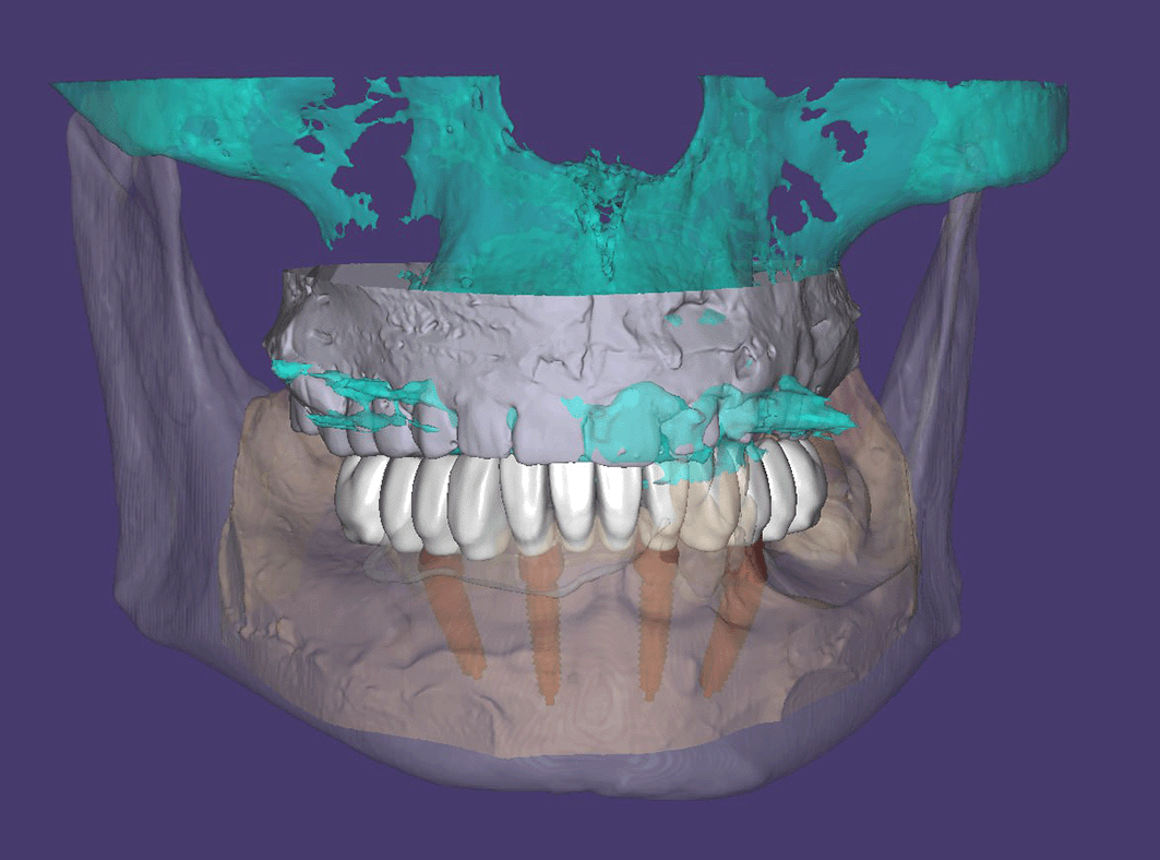
The surgical guide was sterilized by placing it in glutaraldehyde for ten hours before the surgery.
After intraoral disinfection using povidone-iodine, as well as wiping the face with povidone. Sterile drapes were spread around the surgical work area, then a local anesthesia of the mental foramen was performed on both sides using 2% lidocaine with adrenaline 1/105, and infiltration anesthesia was used under the mucous membrane on the lingual side.
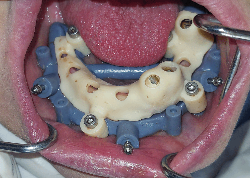
1. The foundation bone guide was installed after adjusting its position by means of its designated guide, which was supported by the teeth, and then the designated guide was removed after fixing the foundation guide ( Figure 5).
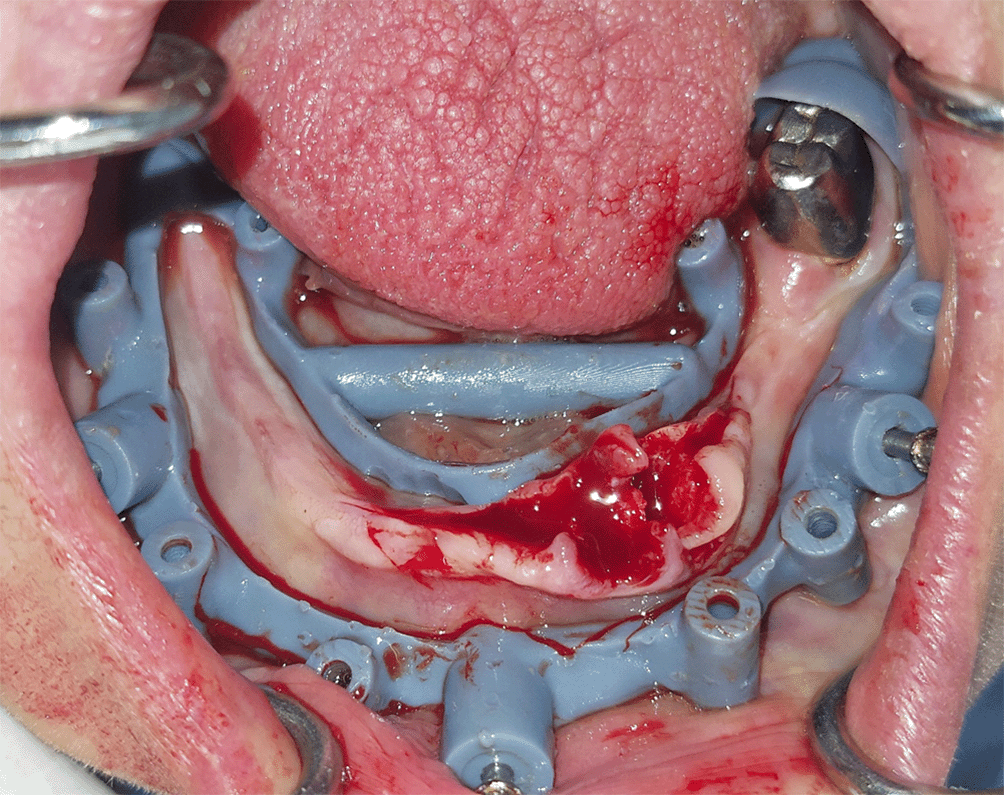
2. The mandibular left lateral incisor and canine were extracted and the molar was preserved, in addition to debriding the alveolar socket in extraction areas ( Figure 6).
3. A full-thickness flap was raised to expose the bone edges, and the size of the flap was determined based on the size of the guide.
4. Bone trimming using a large spherical bur was performed in the places that needed for cutting the sharp edges.
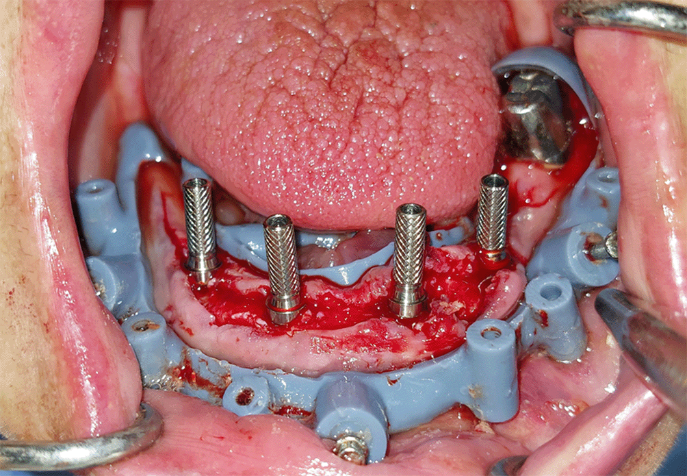
5. The surgical guide for implant placement was installed on the foundation bone guide ( Figure 7), then the fully guided bone preparation was performed to place the implants in their positions, then this guide was removed. The insertion torque for each implant took a value greater than 35 N-Cm and the implant stability quotient (MEGA ISQ II, Megagen, Korea) took values between 69 and 83.
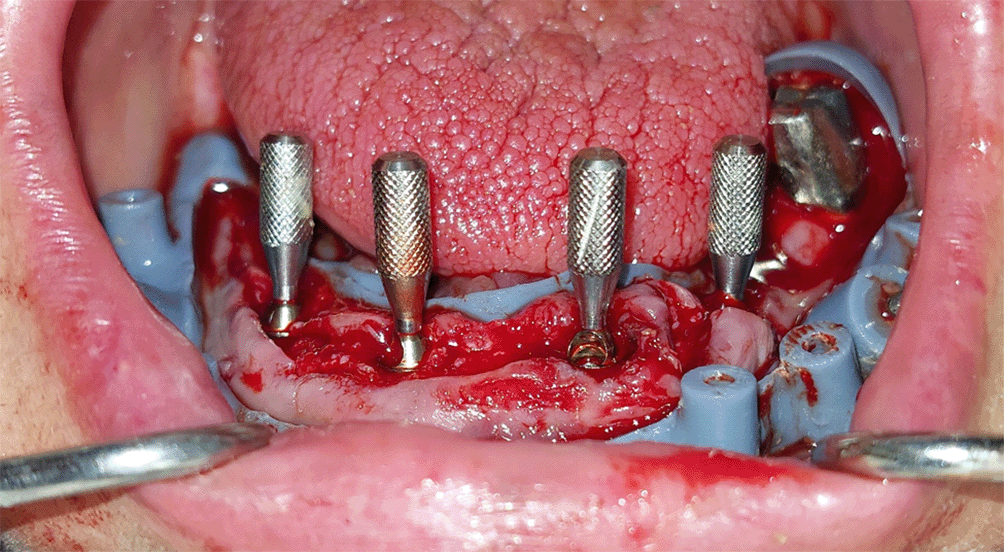
6. The abutment guide was installed on the foundation guide, which allowed for ideal guidance and placement of the multi-unit abutments on the implants ( Figure 8), then these abutments were tightened on the implants according to the manufacturer’s instructions and this guide was removed.
7. The temporary cylinders were installed on the multi-unit abutments and tightened with finger pressure and the flap was sutured with 4–0 silk sutures ( Figure 9).
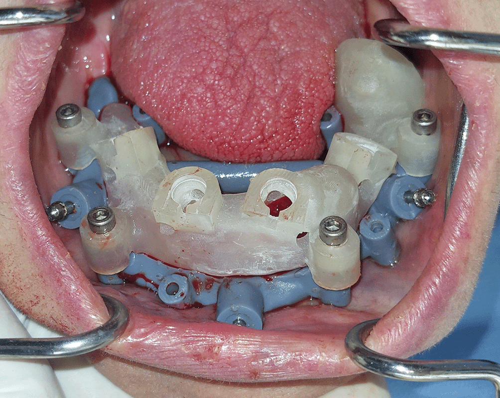
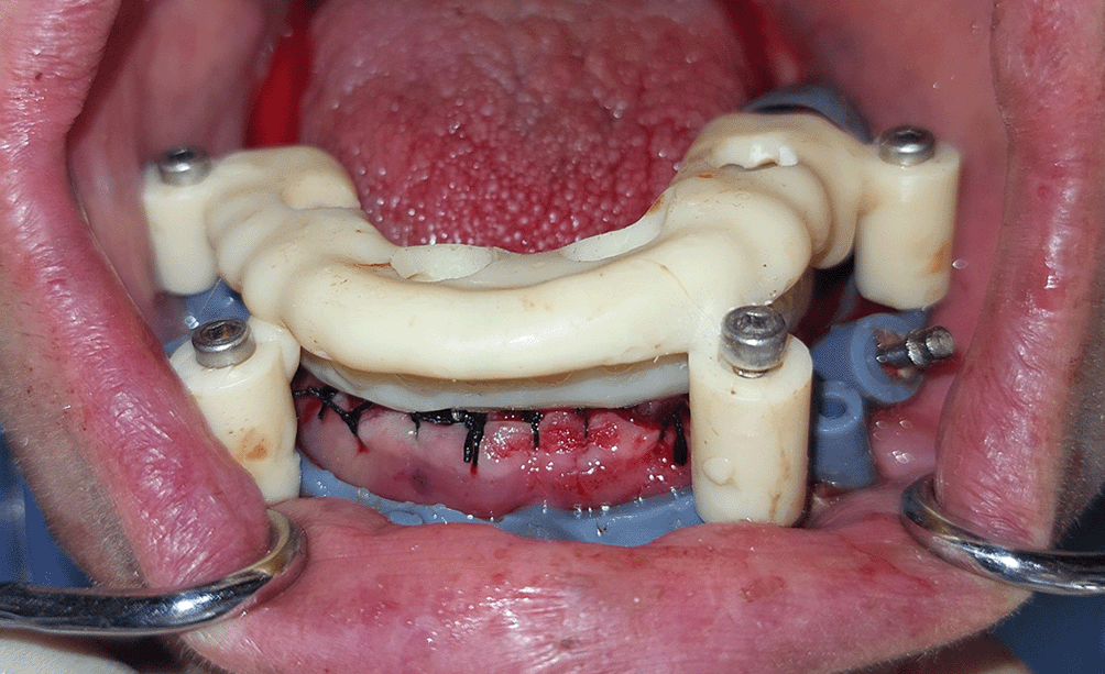
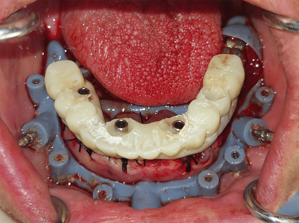
8. The temporary prosthesis guide was installed on the foundation guide, which carries the temporary prosthesis ( Figure 10), and the temporary prosthesis was fixed to the temporary abutments using flow-able composite (Tetric PowerFlow, Ivoclar Vivadent, Schaan, Liechtenstein), and then this guide was removed ( Figure 11).
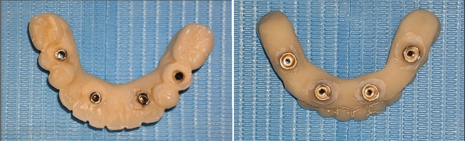
9. The temporary prosthesis was removed from the patient’s mouth, and the excess composite was removed and polished ( Figure 12), then it was re-fixed in the patient’s mouth, and the screw holes were closed with teflon and light-cure flow-able composite. An orthopantomography image was taken after temporary prosthesis fixed ( Figure 13).
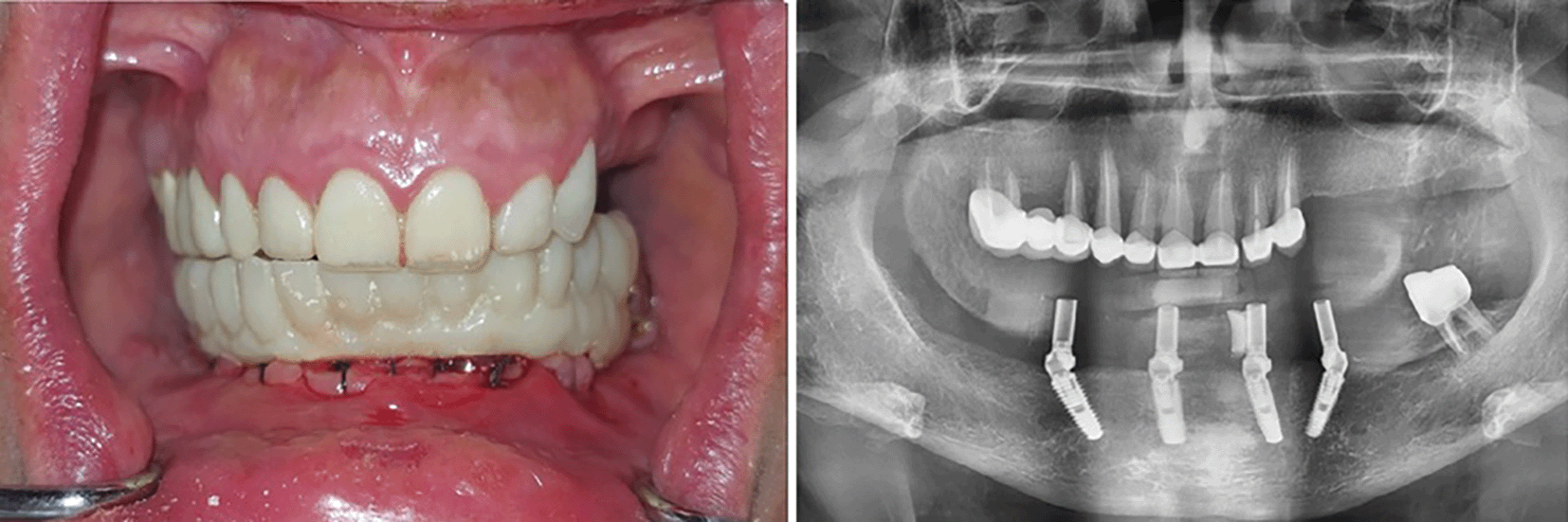
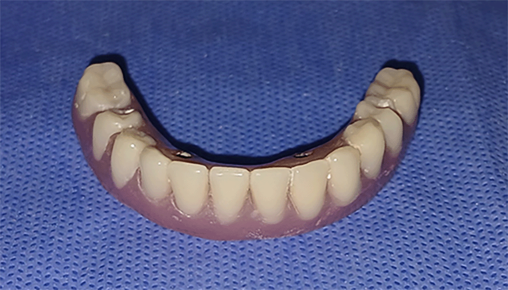
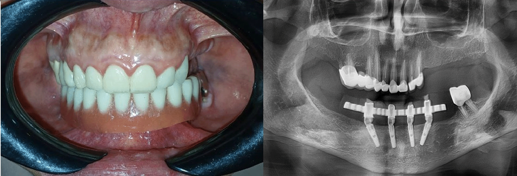
The patient was followed up at one day, one week, one month, four months, and one year after the surgical procedure. A CBCT radiographic image was obtained on the first day postoperatively and at one year to evaluate marginal bone loss around the implants. On the first postoperative day, the patient reported mild pain, which was effectively managed with paracetamol 500 mg (Unadol, Unipharma, Syria), administered four times daily. Additionally, the patient was prescribed amoxicillin with clavulanic acid tablets 1000 mg (Augmentin, Maatouk Pharma, Syria), taken twice daily, to reduce the risk of postoperative infection. Postoperative instructions included avoiding strenuous activities on the day of surgery and initiating warm saline mouth rinses starting the day after surgery. During the follow-up period, only minor and expected postoperative complications were observed, which gradually resolved during the healing phase. After four months, the patient was recalled for replacement of the provisional. The provisional prosthesis appointment included replacing the temporary cylindrical abutments with permanent abutments, and then replacing the PMMA prosthesis with a permanent prosthesis, including a metal bar and acrylic resin prosthetic teeth. The temporary prosthesis is relied upon to reach the final prosthesis, which copy the vertical dimension and the relationship between the jaws ( Figure 14) ( Figure 15).
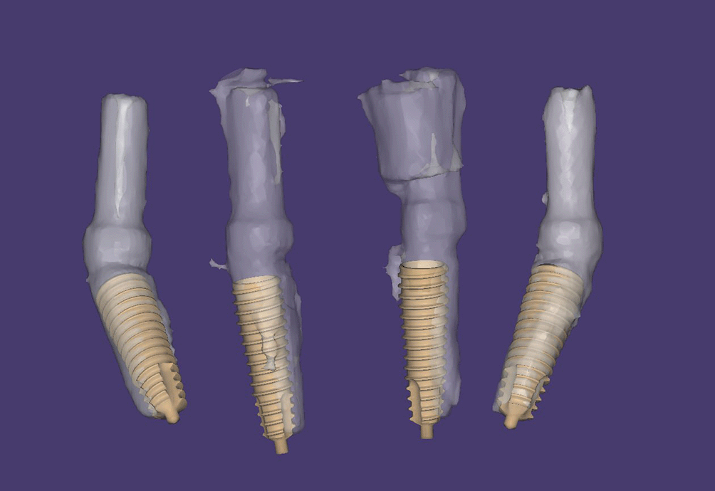
At the 12-months follow-up, we noticed a good stability of the bone levels around the implants in the CBCT (the average bone loss 0.6 ± 0.11 mm) with good patient satisfaction. Comparative radiographic analysis (Geomagic Studio 2012 software, 3D Systems; Mor risville, NC) for confirm the accuracy of implant placement between what was planned and what was surgically achieved ( Figure 16) was presented in the ( Table 1).
The stackable surgical guide represents a significant advancement in computer-guided implant surgery. By incorporating multiple layers, each with specific functions, this guide provides superior precision compared to traditional single-layer guides. The integration of CBCT imaging, facial scanning, and CAD/CAM technology enables comprehensive three-dimensional planning before surgery, allowing for optimal implant positioning while avoiding critical anatomical structures.8,12,13
The type of the placement of the surgical guide is important for the accuracy of the implant guidance, and the best option is based on the remaining teeth in the jaw. Therefore, transferring details of the remaining teeth inside the mouth to the laboratory is of utmost importance.14,15 In our case, the attention was taken to obtaining a high-quality impression using individual stamps and then matching it with the patient’s cone beam computed tomography.
All-on-Four concept, when combined with the stackable surgical guide, offers a comprehensive solution for complete edentulism management. This combination reduces surgical time, minimizes bone resorption, and provides immediate functional and aesthetic rehabilitation.12 The use of temporary prostheses allows for immediate loading while maintaining proper vertical dimension and occlusal relationships during the osseointegration phase.16
The accuracy achieved with this technique, as confirmed by radiographic comparison between planned and actual implant positions, demonstrates the reliability of computer-guided surgery. The minimal postoperative complications observed in this case reflect the reduced surgical risk in complex treatments associated with guided surgery compared to conventional freehand techniques.17
Regarding the implant stability measurements, the digital surgical guides did not provide explicit ISQ values, but the guided implants increase the confidence between the planning and the actual results which help in choosing the best implant site for both the size and the density resulting a good ISQ records.18,19 Regarding marginal bone loss, comparisons show that bone stability was mostly acceptable, Yang and colleagues indicated small marginal bone loss in one year followed by All-on-4, reflecting weak changes in bone levels when using this protocol.20 In our case, the marginal bone level changes ranged between 0.46–0.74 mm a year post-implant, which is similar to those reports. Therefore, the current data does not show radical differences in bone resorption rates between using implant guides (including stackable guides) and other guided All-on-4 techniques, and remain within the expected ranges for the immediate All-on-4 concept.
Digital planning and 3D printing provide important clinical advantages in complex tooth compensation. It has become possible to combine CBCT images, oral scanning, and facial scanning to prepare a three-dimensional virtual model of the patient, which enables the development of a plan based on the shape and functional needs of each patient. Before surgery, the compatibility of the prosthesis and the position of the implants can be examined practically on the digital model, which reduces the need for additional adjustments during surgery.1,21
To reinforce this, this study used digitally designed surgical guides and printed layers (SLA) with biocompatible materials. Manufacturing tools in this way allowed for precise control of their shape and fixation characteristics (such as cylindrical cavities and magnetic attachments). It was noted that cumulative guides divide the process into stages (fixation base, drilling branch, then prosthesis base), which reduces cumulative deviation errors and total surgery time.9,10,22 For example, facial digital planning contributes to placing implants in aesthetically and functionally optimal locations, while the printed design of the guide ensures ideal fit for each tool.
In general, these techniques are vital for completing a synchronous implant-centered procedure (surgery and prosthetics). They reduce laboratory time in the clinic and reduce patient discomfort, and also allow for pre-determining the minimum amount of bone to be removed or modified. Thus, this approach combines safety and efficiency and enhances integration between diagnostic, surgical, and prosthetic phases, which supports the stability of functional and aesthetic results in the short term.21,23
We found an apical deviation between 0.8–1.4 mm and a coronal deviation between 1.3–1.8 mm, with an angular deviation 2.4–3.9 degrees. These records are slightly above the range identified by Abad-Coronel et al. in their review of ten studies on surgical guide accuracy.24 In our case, unilateral tooth-supported guide was used, while the contralateral support was mucosal. Shi et al. found relatively lower accuracy with a surgical guide tooth-supported on one side compared to a surgical guide supported bilaterally.19
All published studies to date (case reports or case series) are limited in number and there are no prospective designs or randomized trials comparing the technique to its alternatives. It was previously mentioned in recent systematic reviews that the lack of methodological quality and small sample sizes and short follow-up currently may limit confidence in conclusions. Therefore, it is important that the results of this case (and similar studies) be taken as preliminary indicators and not as conclusive evidence of the superiority of the described approach.
This clinical case highlights the high effectiveness of using the stackable surgical guide in achieving precise and successful dental implantation according to the All-on-Four concept with immediate loading. Clinical and radiographic results showed good agreement between pre-planning and surgical execution, with acceptable bone resorption rates and without significant complications. These data support the adoption of the stackable surgical guide as a reliable and modern option to improve dental implant results, with emphasis on the importance of future studies to support these results on a broader scale.
Written informed consent was obtained from the patient for publication of this case report and accompanying images. A copy of the written consent is available for review by corresponding author on request.
This study was approved by the Research Ethics Committee at the Faculty of Dentistry, Damascus University. The ethical approval was granted on 14 July 2024 under the reference number DN-140724-270. All procedures were conducted in accordance with the ethical standards of the institutional and national research committee and with the 1964 Helsinki declaration and its later amendments.
| Views | Downloads | |
|---|---|---|
| F1000Research | - | - |
|
PubMed Central
Data from PMC are received and updated monthly.
|
- | - |
Provide sufficient details of any financial or non-financial competing interests to enable users to assess whether your comments might lead a reasonable person to question your impartiality. Consider the following examples, but note that this is not an exhaustive list:
Sign up for content alerts and receive a weekly or monthly email with all newly published articles
Already registered? Sign in
The email address should be the one you originally registered with F1000.
You registered with F1000 via Google, so we cannot reset your password.
To sign in, please click here.
If you still need help with your Google account password, please click here.
You registered with F1000 via Facebook, so we cannot reset your password.
To sign in, please click here.
If you still need help with your Facebook account password, please click here.
If your email address is registered with us, we will email you instructions to reset your password.
If you think you should have received this email but it has not arrived, please check your spam filters and/or contact for further assistance.

Comments on this article Comments (0)