Keywords
Autism Spectrum Disorder, Emotion Detection, EEG, Muse 2, Physiological Signals, Youth with Autism, Behavioural Monitoring, Early Warning
This article is included in the Fallujah Multidisciplinary Science and Innovation gateway.
Autism Spectrum Disorder, Emotion Detection, EEG, Muse 2, Physiological Signals, Youth with Autism, Behavioural Monitoring, Early Warning
AI has recently had a major impact on human medical research and applications that call for sophisticated representation, computationally demanding calculations, and decision-making procedures.1–4 The primary goal of this work is to identify and detect human emotional responses in individuals with Autism Spectrum Disorder (ASD), which encompasses a wide range of neurodevelopmental conditions. People with ASD frequently display repetitive behavioural patterns, and their emotional responses are strongly associated with difficulties in communication and social interaction.5,6 Autistic people frequently exhibit irregular emotional behaviour, which can be challenging to understand or identify using standard observational techniques. Self-harm, hand flapping, rocking, and prolonged staring are common behaviours associated with autism. Rather of being misbehaviour, such conduct usually reflects stress, overburden, or unfulfilled needs. Giving physicians and caregivers a novel physiological approach to deduce the internal states of individuals with ASD is one of the goals of our work.1–4 We investigate an option that uses a simple EEG montage and wearable neurophysiology. Numerous researchers have used EEG signals extensively to analyse human-specific behaviour.7,8
By developing a distinct differentiator between ASD behavioural response and otherwise normal reactions, this work seeks to build a reference base for autistic behaviour response. The present study creates a testbed for further research in which we assess the EEG signals of people with and without ASD. Keep in mind that the objective is to find tiny, comprehensible signal features that accurately identify symptoms of ASD rather than to construct a model for brain activity in general. In order to facilitate early screening and emotion-aware intervention, such identification is needed.
This method expands on earlier research on emotion recognition utilizing viable, small signal sets, where the researchers looked at fifteen distinct physiological markers. EEG, skin conductivity, blood pressure, heart rate variability (HRV), and other factors were assessed. The importance of EEG signals, which frequently independently disclose respondents' inner emotional status, was one of the study's main conclusions.1–4
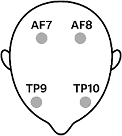
In order to determine if a small collection of EEG characteristics may distinguish ASD from neurotypical controls, this study offers a proof-of-concept analysis using a four-sensor headband. Over a period of ten to fifteen minutes, the EEG bands (alpha, beta, delta, gamma, and theta) are measured. Four distinct sensors are positioned at the AF7/AF8 (frontal) and TP9/TP10 (temporo-parietal) to measure each band.9
Five neurotypical controls and seven people with autism spectrum disorder (ASD) are involved in the pilot experiment. The findings set the stage for a longer follow-up with more participants and recordings that are controlled for artifacts.
In summary, the objective of this article aligns with several sustainable development goals (SDGs), such as: SDG 3: Good Health and Well-Being, by helping to identify the emotions of individuals with ASD, which may help clinicians and parents understand their needs. By empowering the educational process for people with ASD through the use of emotion detection tools, it also advances SDG 4: Quality Education. It also aligns with SDG 10: Reduced Inequalities, which is exemplified by reducing educational and social barriers. Additionally, our endeavour advances SDG 9: Industry, Innovation, and Infrastructure by introducing a novel health data-driven innovation.
The authors of this study examined the EEGs of twelve patients, including five neurotypical controls (n = 5) and seven individuals with ASD (n = 7). Alpha, beta, delta, gamma, and theta bands are the five band-isolated time series that each participant's deployed sensors monitor and report. The data taken by the patients using a written ethical approval received by the ‘Arab Village for Special Challenges’ under approval number (ز/1810). Two sensors were positioned in front of the subject's head for this arrangement, specifically the AF7 sensor on the left and the AF8 sensor on the right, or above the eyes. However, as seen in Figure 1, two additional sensors, TP9 and TP10, were placed toward the rear, or close to the ears (TP9 on the left mastoid region and TP10 on the right mastoid region). The sensor data is reported and stored as channels 1, 2, 3, and 4 using Muse 2 technology. Before statistical testing and modeling, the sessions that some participants completed were combined inside the subject.
The most challenging aspect of this study was gathering data from participants with ASD; the authors intend to include at least 30 people with ASD (ages 10–20) from nearby clinics and centers in their next investigation. Participants who can tolerate wearing a head-mounted sensor and have a confirmed diagnosis of ASD will be included. People with other neurological conditions, including epilepsy, that could interfere with EEG recordings will not be included by the authors.10,11
To put it succinctly, all study methods will be carried out with Institutional Review Board (IRB) permission, and prior to participation, participant assent and parental agreement will be sought.
The Muse 2 headband is used in this study to take measurements.12,13 Although Muse 2 may record many metrics, the pilot study solely looks at EEG data. The authors of a prior study4 employed a NeXus Q32 system with 14 physiological indicators, including EEG, to identify human emotions. EEG signals (alpha, beta, delta, gamma, and theta) may be adequate to identify the emotion without the need to measure additional signals, according to earlier thorough research. Actually, the primary problem with depending solely on EEG is that it is very challenging, if not impossible, to use EEG signals in a non-invasive way.
The measuring sessions take place in a pleasant environment with video observation, and they last only ten to fifteen minutes. Every session involving subjects with ASD was carried out under the guidance and assistance of a therapist, who assisted in keeping the sessions organized and ended them when needed. As a result, this illustrates the challenges the writers encountered when gathering information for their paper.
When using this type of data, where participation is voluntary and withdrawal is possible at any time, confidentiality is obviously essential. An example of the data is shown in Table 1. With an average duration of 15 minutes, each data capture yielded over 250,000 readings, or a reading every 3.6 milliseconds for one normal/healthy (X) and one ASD (Y).
The power per band per electrode (Pband,e) is computed as the mean of squares of the time series. For each subject and band:
To reduce inter-subject scale differences, the relative power at each electrode is given by
This study uses a number of additional statistics to give a thorough comparison of EEG properties between the Autism and Healthy groups. To get a statistically sound conclusion, the authors used the relevant statistical analysis in addition to the relative power band displayed above. When data are independent and group variances are roughly similar (pooled-variance assumption), the independent-samples t-test, which assesses whether the mean value varies between the groups, is appropriate. Welch's t-test (unequal variances) is recommended if variances are obviously different.14,15
Hedges' g is also used to measure the standardized effect size, or the practical magnitude of the difference. Hedges' g is a standardized mean difference that, after applying a small-sample bias correction, indicates the distance between two group means in units of their pooled standard deviation. Hedge’s g is computed as:
x1, x2 are group means and Sp is the standard deviation
“g” is expressed in units of pooled standard deviations and corrected for small-sample bias. In fact, g values are considered to be as follows; small as |g| ≈ 0.2, medium as |g| ≈ 0.5 and large as |g| ≈ 0.8. The power value and Hedges’ g are helping to understand the statistical significance as well as the magnitude and potential practical relevance of the effect.16,17
For multiple tests (ADS and Control with 5 different bands each), we use the false discovery rate (FDR) using Benjamini et al. method.18,19
We employ the Area Under the ROC Curve (AUC), where ROC stands for Receiver Operating Characteristic, to account for discrimination performance. The AUC calculates the likelihood that a randomly selected positive rate will be ranked higher than a randomly selected negative rate using the Mann–Whitney formulation.20–22 AUC (area under the curve) describes discrimination (1.0 = perfect, 0.5 = chance); ROC, according to Mann-Whitney, is a curve that displays True Positive Rate (sensitivity) vs. False Positive Rate (1 − specificity) across all decision thresholds. The following section goes into more detail on the application of the AUC and ROC. Future studies will employ a bigger sample size to examine the existing results utilizing the AUC approach in more detail.
We employed leave-one-subject-out (LOSO) cross-validation to estimate performance on unseen individuals and avoid leaking from repeated sessions. In each test, the model was trained on the remaining recordings while all of the recordings from a single person were kept out for testing. After that, we assessed three lightweight models and presented three ROC curves:
1. Two-feature model using relative beta at TP9 and relative gamma at TP10 (features selected a priori from univariate screening).
2. Log-band model using the log10 of mean power in alpha, beta, gamma, delta, and theta.
3. The Combined model included the five log-band features plus the two relative features (7 total). All analyses were run using CSV files exported from the Muse 2 Application.
No further artifact rejection was applied beyond the preprocessed data.
Figures 2A–2E show the difference in average power alpha, beta, delta, gamma and theta bands; each figure shows the overall power average for the four sensors/channels (AF7/AF8 and TP9/TP10). The blue bars in the figure represent the normal control subjects, while the yellow bars represent the ADS) subjects. The average power of a given band, e.g., alpha, is computed as the average across all participants (e.g. with ADS) over a given sensor, for example TP9. Then we take the average across all ADS participants. Since the sample is small, we use an error range by computing the deviation of the average from the standard deviation. Figures 2A–2E shows the summary of the results, with the yellow bar indicating the relative power average for the ADS sample, while the blue bar represents the normal control sample. The thin black line represents the error bar for each result. For example the Alpha@TP9 autism mean 661.4 with SE (error) ≈ 495.8. In other words, the Alpha@TP9 range is (661.4-495.8) to (661.4+495.8) or minimum of 165.6 to maximum of 1157.2.
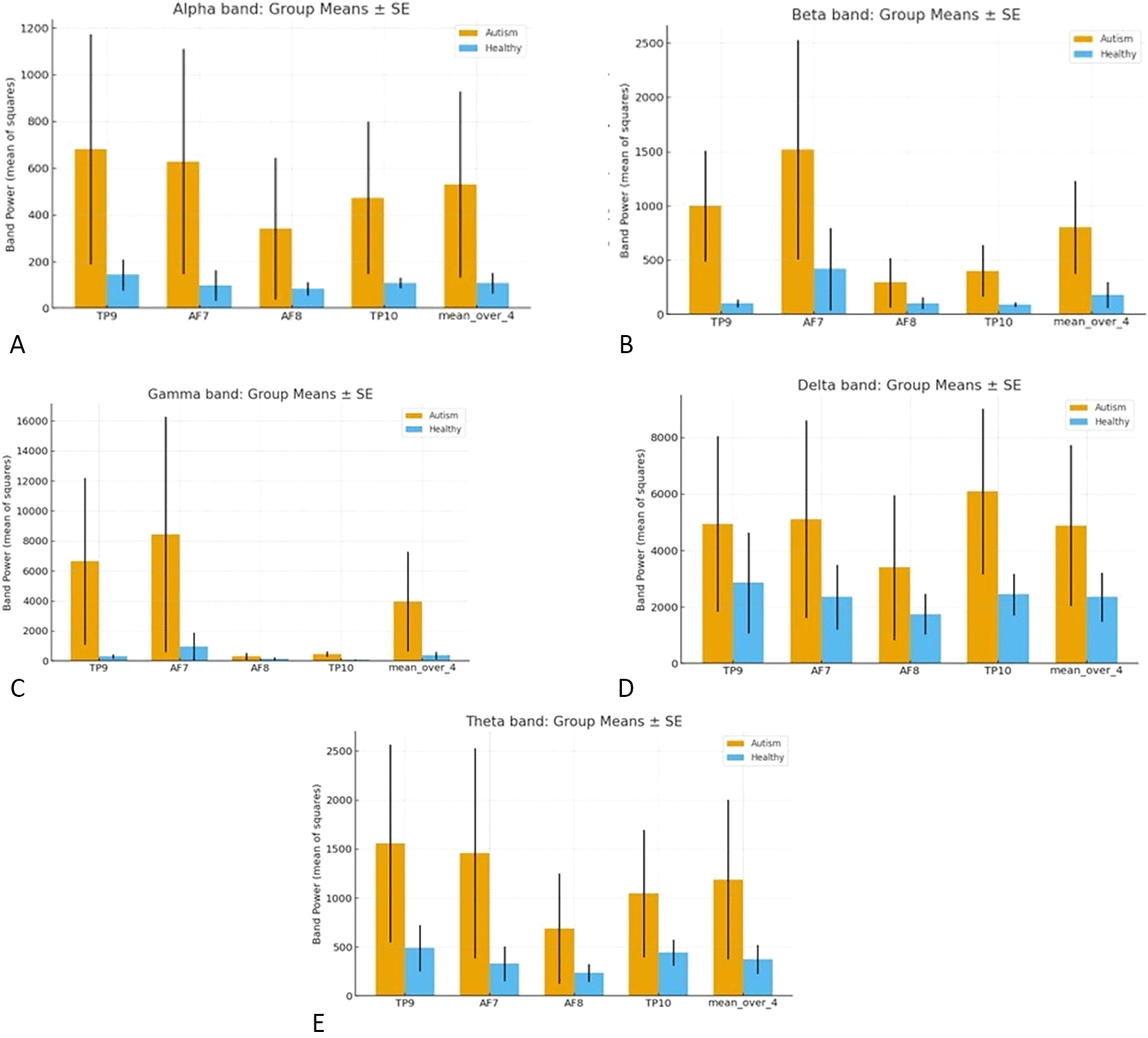
Across all the Figures 2A–2E, the Autism group’s exhibit higher power than the Healthy group, indicating higher absolute power on average. However, the relative β at TP9 and relative γ at TP10, are the clearest discriminators of ASD vs. controls.
The absolute-power differences observed for gamma at TP10 (right temporal) with a Hedges’ g ≈ 0.94 (Autism > Healthy) indicating large effect. For beta at TP9 (left temporal) with g ≈ 0.79 (Autism > Healthy), the difference index is moderate to large.
Using the single–band discrimination based on log10 overall band power (per subject), the gamma band ranked highest (AUC ≈ 0.71), with beta at AUC ≈ 0.69 next highest. Alpha showed modest overall power at (AUC ≈ 0.63), and theta/delta at (AUC ≈ 0.57/0.54).
Figure 3 summarizes the results for the Area under ROC curve, where we utilized the leave-one-subject-out (LOSO) principle. The figure illustrates the Receiver Operating Characteristic (ROC) for the three models (two-feature, log band, and combined 7 features)
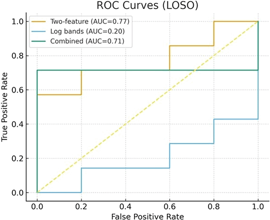
The two-feature model (Beta and Gamma bands) with AUC = 0.77, reveals a focused classifier using the beta band at TP9 and gamma band at TP10. Note that the entire 2-factor curve sets above the diagonal line, which represents the medium chance (FPR=TPR). The two feature model exhibited the best discrimination between ADS subjects and normal control subjects. Hence, the two features model may be sufficient to do most of the work in distinguishing between the groups. This findings, however, needs to be further investigated with more experiments and subjects, especially in light of other research results, which emphasize the alpha band.23
The log-bands baseline (AUC = 0.20) is a broader feature set using log-transformed band power across all sensors (alpha, beta, gamma, theta, delta). LOSO. The entire ROC curve falls below the diagonal, which represents the 0.5 chance (FPR=TPR). This indicates weak or inconsistent signal in these generic summaries for this dataset.
The combined model with AUC = 0.71 is a representation of both models, the 2 feature model and the log band model. The combined model outperformed the log-bands baseline. However, it trailed behind the two-feature model. This behavior indicates that adding weak or collinear features may weaken the signal and reduce generalization.
Even, at low false-positive rates, performance remains meaningful despite the small sample. For example, at FPR ≈ 0.20 (1/5 healthy misclassified), the two-feature model detects ≈57% of Autism cases. The combined model (at FPR = 0.2) detects ≈71%. At FPR ≈ 0.40, the same pattern holds (two-feature ≈57%, combined ≈71%).
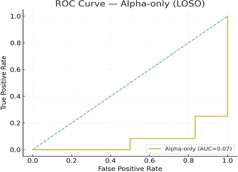
The alpha effects in our dataset are modest ( Figure 4), although, several studies detect alpha abnormalities in Autism. Figure 4 shows that only at >0.8 FPR, the Alpha band begins to detect autism cases. This does not necessarily contradict the alpha-centric literature, were these studies frequently rely on other sensors such as eyes-closed reactivity, individual alpha peak frequency, or connectivity measures. Our experiments continue to reliably detect autism to a good degree without the use of these extra parameters. Within these constraints, the temporal beta/gamma measures provide strong enough and reliable discrimination between the two sets.
The ROC overlay demonstrates that a minimal, two-feature sensors, namely the relative beta at TP9 and the relative gamma at TP10, deliver the highest cross-validated AUC and favorable sensitivity. This avoids the need to rely on a larger sensor array or complex preprocessing. This simple design is practical for real-world screening. The two temporal sensors are fast to place, and relative features aren’t easily thrown off by changes in overall amplitude. Table 2 provides a summary of key performance/results.
Even with ROC curves and LOSO cross-validation, models can appear overly optimistic especially with relatively low sample. However, in our study this risk is acknowledged, and remedied by aggregating repeated sessions within subject, used leave-one-subject-out (LOSO) to hold out entire participants, and controlled multiple testing via FDR, steps intended to reduce overfitting and selection bias. We also emphasized effect sizes (Hedges’ g) alongside ROC to avoid relying on a single metric and noted that findings are preliminary pending larger, multi-site validation with stricter artifact control and broader coverage, which we outline as future work.
Because the study involved minors, informed consent was obtained from the participants’ legal guardians. The study received ethical approval from ‘Arab Village for Special Challenges’ under approval number (ز/1810), and all procedures followed the Declaration of Helsinki guidelines. The consent was verbal, as approved by the ethics committee at Arab Village for Special Challenges, due to cultural considerations, and logistical reasons. The approval of the research was granted after review of the research protocol to ensure compliance with ethical standards for research involving human participants. Generally, all disabilities centers in Jordan are following the rules under ‘The Higher Council for the Rights of Persons with Disabilities’, and Arab Village for Special Challenges is listed under this council and follow its rules. The centre has as ethics review process that reviews research proposals prior to approval that aligned with the Higher Council for the Rights of Persons with Disabilities specifications. All legislation including (laws, instructions and international agreements) are available in Higher Council for the Rights of Persons with Disabilities website.
| Views | Downloads | |
|---|---|---|
| F1000Research | - | - |
|
PubMed Central
Data from PMC are received and updated monthly.
|
- | - |
Provide sufficient details of any financial or non-financial competing interests to enable users to assess whether your comments might lead a reasonable person to question your impartiality. Consider the following examples, but note that this is not an exhaustive list:
Sign up for content alerts and receive a weekly or monthly email with all newly published articles
Already registered? Sign in
The email address should be the one you originally registered with F1000.
You registered with F1000 via Google, so we cannot reset your password.
To sign in, please click here.
If you still need help with your Google account password, please click here.
You registered with F1000 via Facebook, so we cannot reset your password.
To sign in, please click here.
If you still need help with your Facebook account password, please click here.
If your email address is registered with us, we will email you instructions to reset your password.
If you think you should have received this email but it has not arrived, please check your spam filters and/or contact for further assistance.
This is a timely and clinically motivated proof-of-concept study exploring whether a minimal, wearable EEG setup can distinguish individuals with ASD from neurotypical controls. The use of a ... Continue reading Overall Assessment
This is a timely and clinically motivated proof-of-concept study exploring whether a minimal, wearable EEG setup can distinguish individuals with ASD from neurotypical controls. The use of a low-cost, accessible consumer-grade device is a genuine practical strength, and the research direction is valuable. With some clarifications and adjustments, this work has good potential to contribute meaningfully to the field.
Major Points for Consideration
1. Title and Framing
The title would benefit from a small revision - the current subtitle reads as a fragment and could be made more complete. A suggestion might be something like "EEG-Based ASD Screening Using Temporal Beta and Gamma Features: A Pilot Study." Additionally, the phrase "Emotion Detection" may set slightly different expectations than what the study delivers - the paper focuses on classifying ASD versus neurotypical status rather than detecting specific emotional states like happiness or fear. Aligning the title and framing with the actual classification goal would help readers immediately understand the contribution and avoid any confusion.
2. Clarifying the Research Question
Related to the above, the introduction occasionally moves between "emotion detection" and "ASD classification" as if they are interchangeable. Since these are genuinely different tasks, a brief sentence early in the paper explicitly defining the target variable - group discrimination between ASD and neurotypical participants - would significantly improve the manuscript's clarity and scientific coherence.
3. Artifact Control
The authors helpfully acknowledge that no artifact rejection was applied beyond the device's onboard preprocessing. It would strengthen the paper considerably to discuss this more explicitly in the limitations section, particularly since gamma-band power - the top discriminating feature - is known to be susceptible to EMG contamination from muscle activity. The authors need not have solved this problem in a pilot study, but a candid discussion of how this might affect the interpretation of the gamma finding would reassure readers and reviewers alike.
4. Reporting p-values
Table 2 notes that p-values for band-wise group tests were not reported. Including these, along with their FDR-adjusted counterparts, would make the statistical analysis complete and allow readers to evaluate the significance of the findings more fully. This is a relatively straightforward addition that would meaningfully improve the manuscript.
5. Tempering the Conclusions
The conclusion's suggestion that these findings could support rapid clinical screening is an exciting possibility but given the sample size of 12 participants and the single-site design, it may be worth softening this claim slightly - framing it as a promising direction warranting further investigation rather than a near-term clinical tool. This would make the contribution feel more credible, not less.
Minor Suggestions
Individual data points. With n=12, showing individual data points overlaid on the group bar charts (e.g., as dots or a strip plot) would give readers a clearer sense of variability and group separation. This is a simple addition that adds a lot of transparency.
Error bar labeling. The error bars in Figure 2 are labeled as SE in the figure but described in the text in a way that suggests SD. A quick check and correction of this labeling would avoid ambiguity.
ASD vs. ADS. There are a few instances where "ADS" appears in place of "ASD" in the figures, tables, and body text. A careful find-and-replace pass would clean this up.
Literature engagement. The paper would benefit from engaging with a slightly broader set of EEG-ASD studies, particularly work on connectivity biomarkers, mu-rhythm suppression, or prior consumer-EEG ASD classification efforts. This would situate the contribution more firmly in the existing literature and reduce the appearance of over-reliance on the authors' own prior work.
Results/Discussion separation. Separating or clearly sub-sectioning the Results and Discussion would improve readability and help readers follow the flow from findings to interpretation.
Strengths Worth Highlighting
The two-feature model using relative beta at TP9 and relative gamma at TP10 is an elegant and practical finding - it achieves the highest cross-validated AUC (0.77) using just two sensors, which is promising for low-cost, real-world deployment. The LOSO cross-validation strategy is well-chosen for a small sample, and the use of Hedges' g alongside AUC to avoid over-reliance on a single metric reflects good statistical thinking. The effect sizes observed (g ≈ 0.94 for gamma at TP10) are genuinely noteworthy and provide strong motivation for a larger follow-up study.
Recommendation
Minor-to-moderate revision. The core idea is promising, and the methodology is reasonable for a pilot study. Addressing the framing, reporting the p-values, acknowledging the artifact concern in the limitations, and making the smaller corrections above would prepare this manuscript well for a revised submission.