Keywords
Posterior shoulder dislocation, Reverse Hillsach lesion, Modified McLaughlin Procedure, Deltopectoral approach, Lesser Tuberosity, Subscapularis muscle
This article is included in the Fallujah Multidisciplinary Science and Innovation gateway.
Posterior shoulder dislocation, Reverse Hillsach lesion, Modified McLaughlin Procedure, Deltopectoral approach, Lesser Tuberosity, Subscapularis muscle
Shoulder dislocation is a common and frequent presentation in the emergency department. However, posterior shoulder dislocation is rare, accounting for <2% of all cases of shoulder dislocation.1
The mechanisms of posterior shoulder dislocation are indirect forces that produce marked internal rotation and adduction (occurring during a fit or convulsion, or with an electric shock) or falling on an outstretched hand or on the flexed adducted arm.2 A direct blow to the front of the shoulder may also be considered as a direct mechanism of posterior shoulder dislocation.1
Posterior shoulder dislocation is difficult to diagnose initially and is frequently missed because the dependence is mainly on a single A-P view, which is similar to normal as well as the forgetfulness of thinking about this condition during examination of the patients.3
Radiologically, the anteroposterior (AP) view may show an electrical bulb sign and empty glenoid fossa.1 A lateral and axillary view is essential (although it is painful for patients), which may show posterior subluxation or dislocation and sometimes a deep indentation on the anterior aspect of the humeral head (Reverse Hillsach lesion).4 A CT scan is also required to confirm the diagnosis.1,2
Posterior shoulder dislocation may be associated with fractures of the lesser tuberosity, posterior part of the glenoid, or the surgical neck.4 The treatment is closed reduction; if failure occurs, then surgical or open reduction.1,2 Posterior shoulder dislocation may be complicated by osteoarthritis, stiffness, instability, or aortic Necrosis.5
This clinical case series aimed to highlight a rare condition that can be easily missed in acute presentation in the emergency room due to the lack of clinical features and inadequate radiological assessment. Three cases of chronic posterior shoulder dislocation, missed in diagnosis, will be discussed, with an explanation of the methods used to reach the diagnosis and treatment.
A 25 years old male patient presented to a private clinic in Fallujah city on 11/10/2024 with left shoulder pain and limitation of movement for 2 months. The forearm was clasped to his chest with inability to perform external rotation of the forearm. The patient had a history of electrical shock injury prior to his compliant.
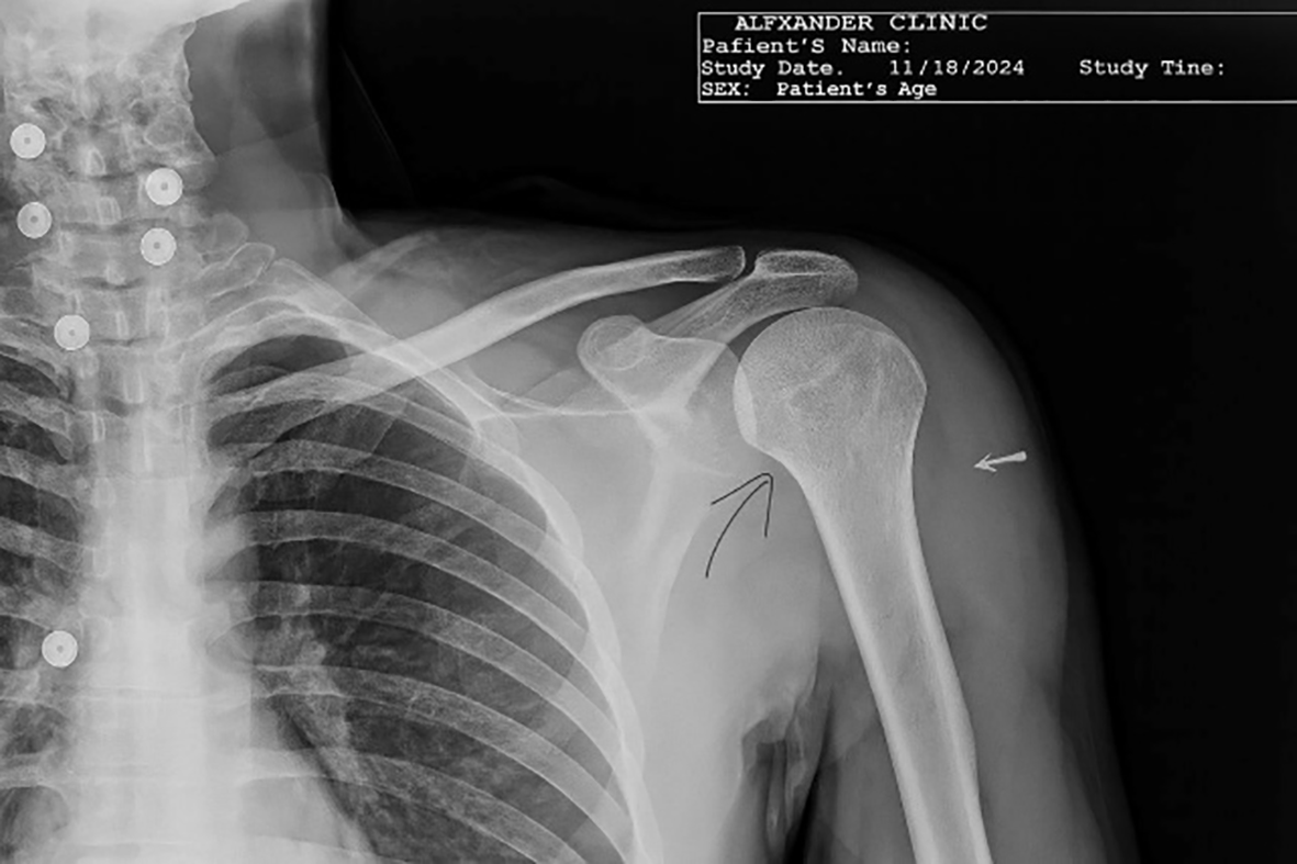
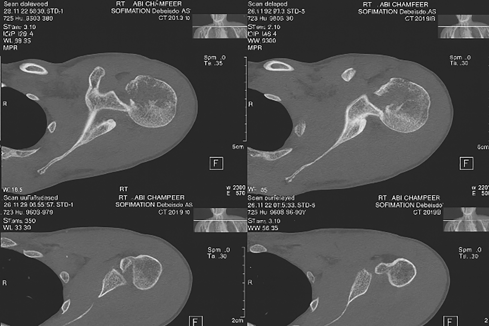
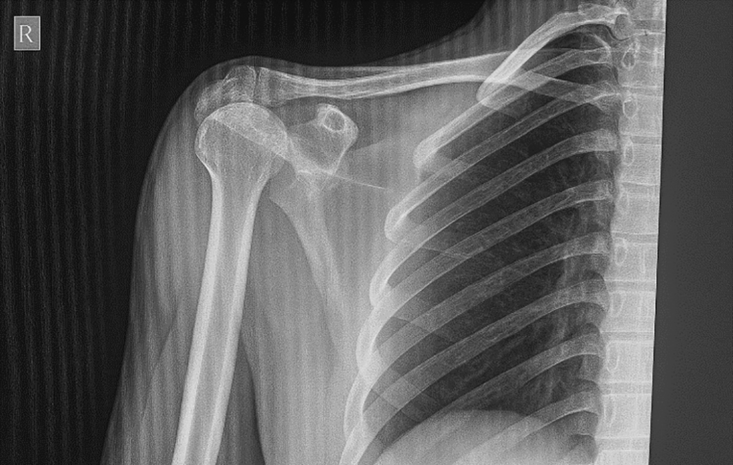
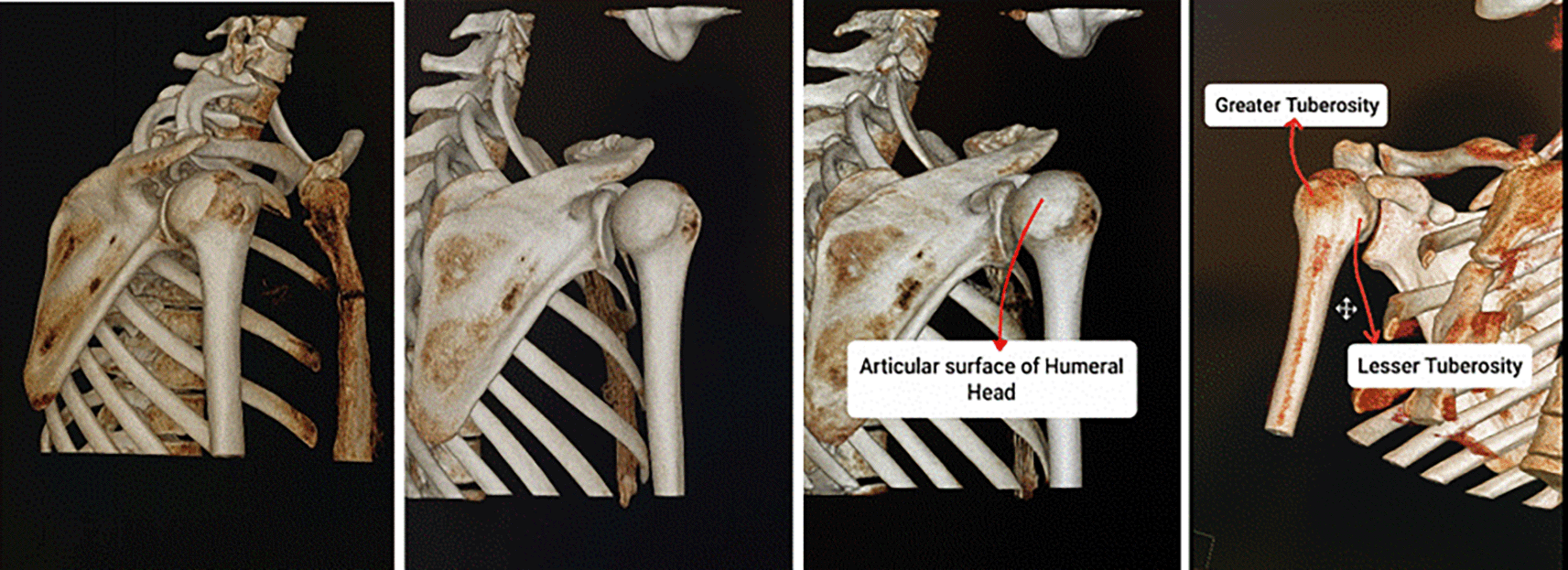
On examination, there was loss of shoulder contour with limitation of movement with a positive posterior drawer test. Radiographs showed a light-bulb appearance ( Figure 1). The CT scan showed Reverse Hillsach lesion ( Figure 2).
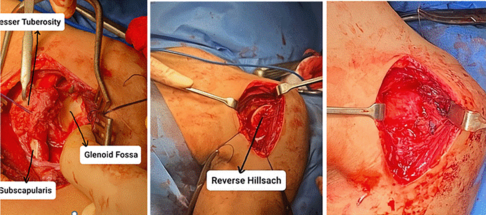
The management plan involved surgical reduction of this dislocation using the Modified McLaughlin Procedure. The patient was admitted to the operative room in Fallujah Teaching Hospital to undergo the surgical procedure using the deltopectoral approach to reduce the dislocation by the open method with transfer of the subscapularis with its lesser tuberosity to the defect (Reverse Hillsach) on the anterior part of the Humeral Head ( Figure 3).
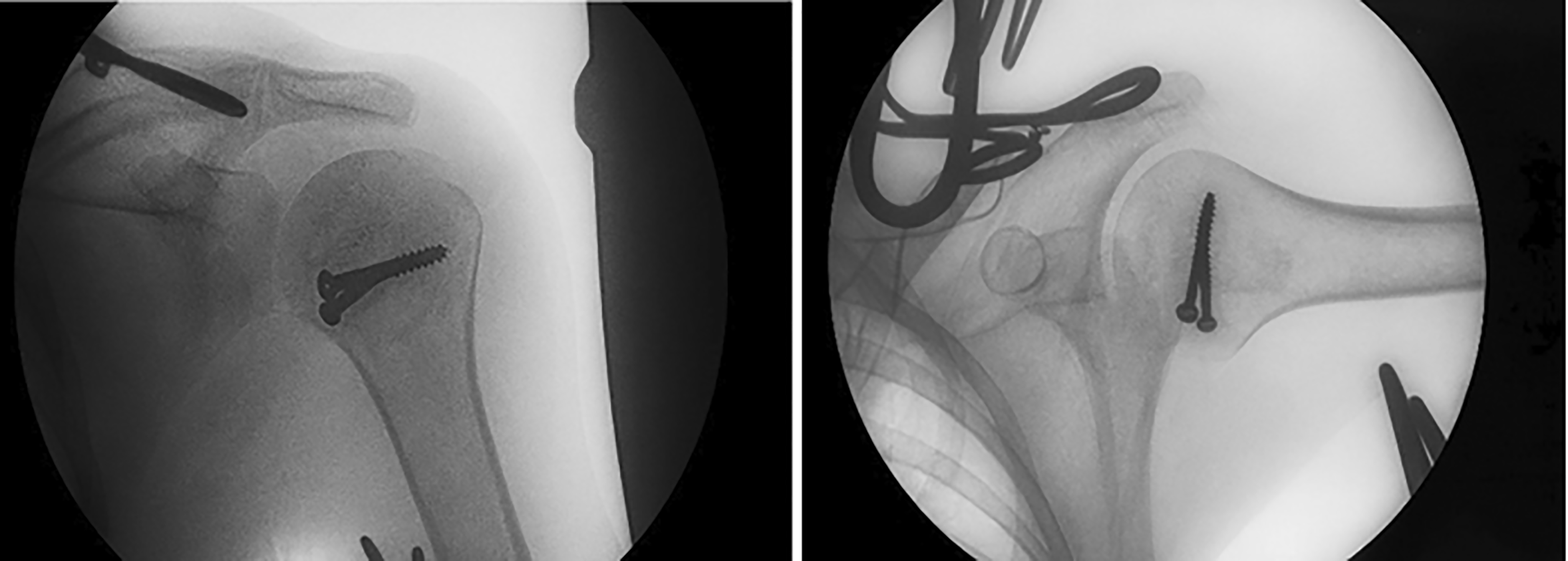
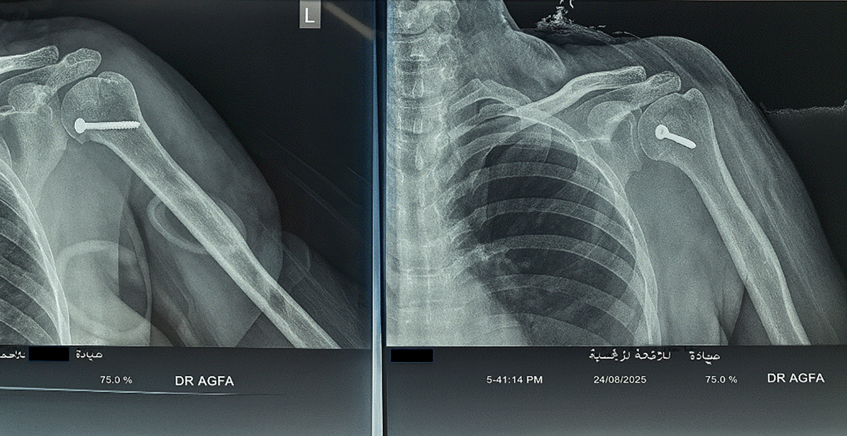
Postoperatively, the patient wore a Gunslinger sling for 4 weeks, followed by exercise. The postoperative radiograph is shown in ( Figure 4).
The patient expressed significant satisfaction with the outcome, noting that he could finally return to his daily activities without the constant pain and severe limitation he had experienced for two months following the injury.
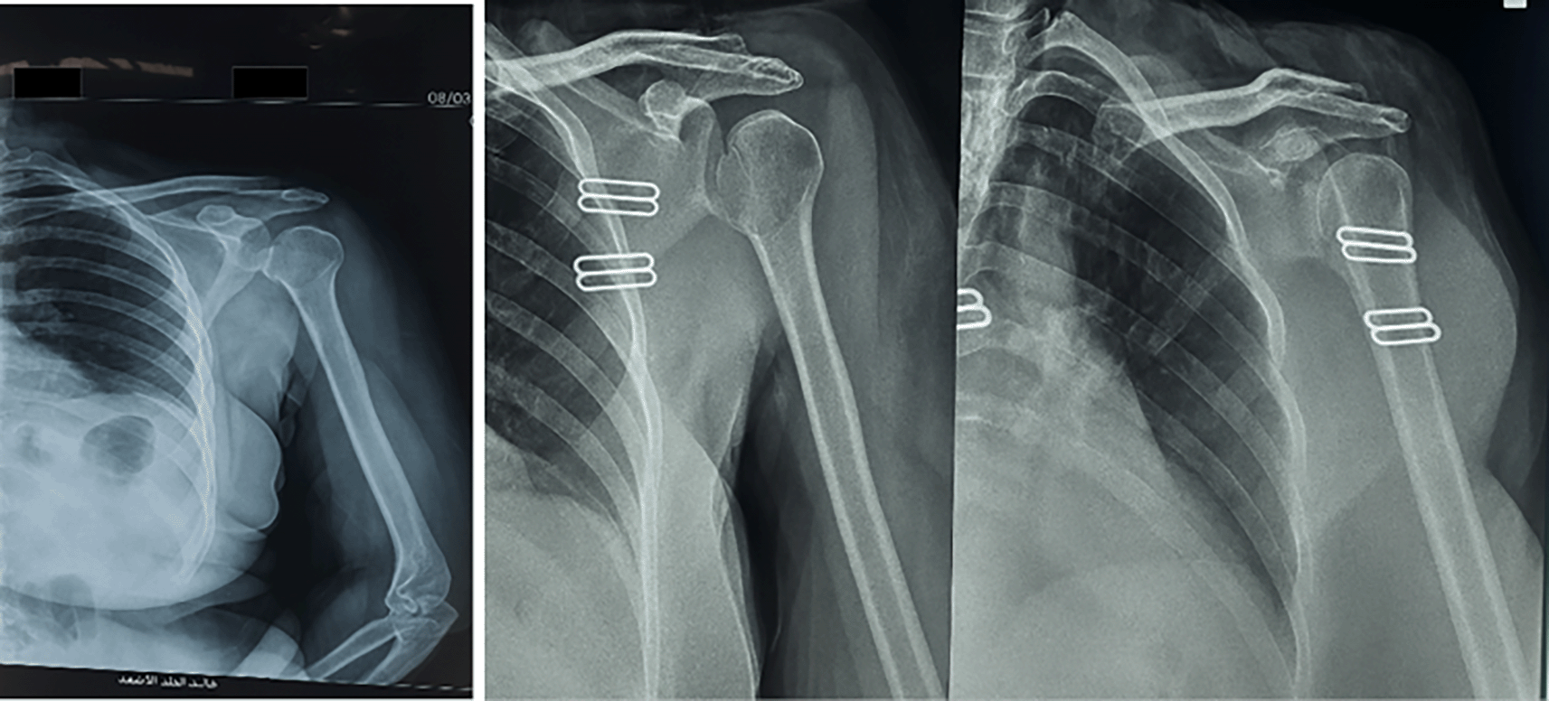
A 52 years old female patient presented to a private clinic in Fallujah city on 8/3/2025, with left shoulder pain and limitation of movement for 3 months. Her symptoms occurred after an episode of convulsions.
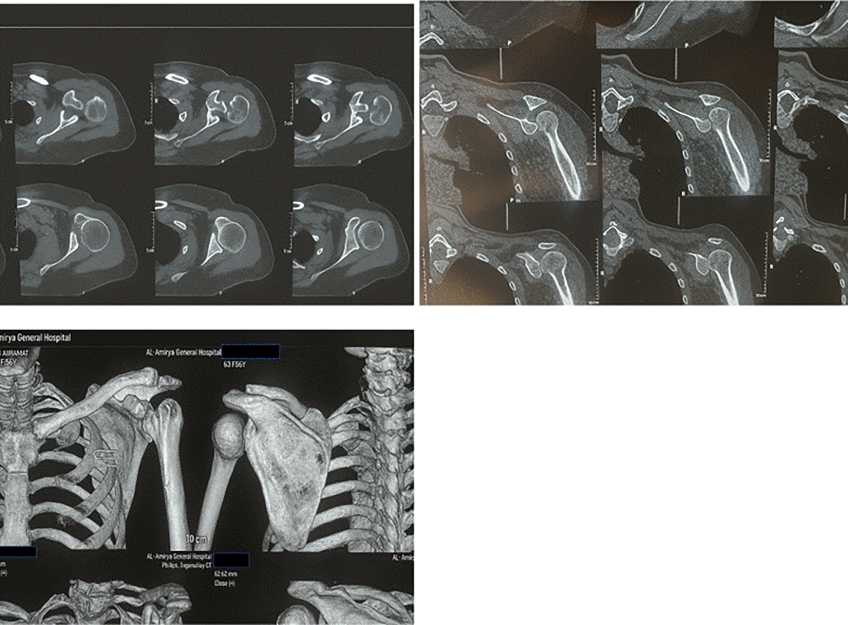
On examination, there was loss of external rotation movement of the shoulder. The X-rays showed a light-bulb appearance ( Figure 5). The CT scan: shows Reverse Hillsach lesion ( Figure 6).
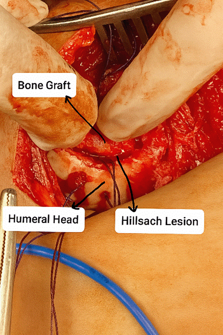
The management plan involved surgical reduction of this dislocation using the Modified McLaughlin procedure. The patient was admitted to the operative room at Fallujah Teaching Hospital to undergo the surgical procedure using the deltopectoral approach to reduce the dislocation by the open method with transfer of the subscapularis with its lesser tuberosity to the defect (Reverse Hillsach) on the anterior part of the Humeral Head with Bone Graft ( Figure 7).
Postoperatively, the patient wore a Gunslinger sling for one month followed by exercise. The postoperative radiograph is shown in ( Figure 8).
The patient reported a high level of satisfaction after the surgery, specifically highlighting the restoration of her shoulder’s external rotation, which allowed her to regain independence in her personal care.
An 18 years old male patient presented to a private clinic in Fallujah city on 12/7/2025, with right shoulder pain and limitation of movement for 2 months. He had a history of falling, was admitted to the emergency department, and was told that he had a shoulder dislocation (there was no radiological document for his first dislocation). The patient underwent closed reduction (as he said), and later on, the patient still complained of limitation of movement of the right shoulder with severe pain and was admitted to a private clinic seeking relief.
The patient was sent for X-rays that showed a light-bulb appearance ( Figure 9). The CT scan showed the impaction of the Humeral Head in the glenoid fossa with an internally rotated Humeral Head that locks the head in situ (inside the glenoid fossa) without exiting the humeral head outside the glenoid socket ( Figure 10).
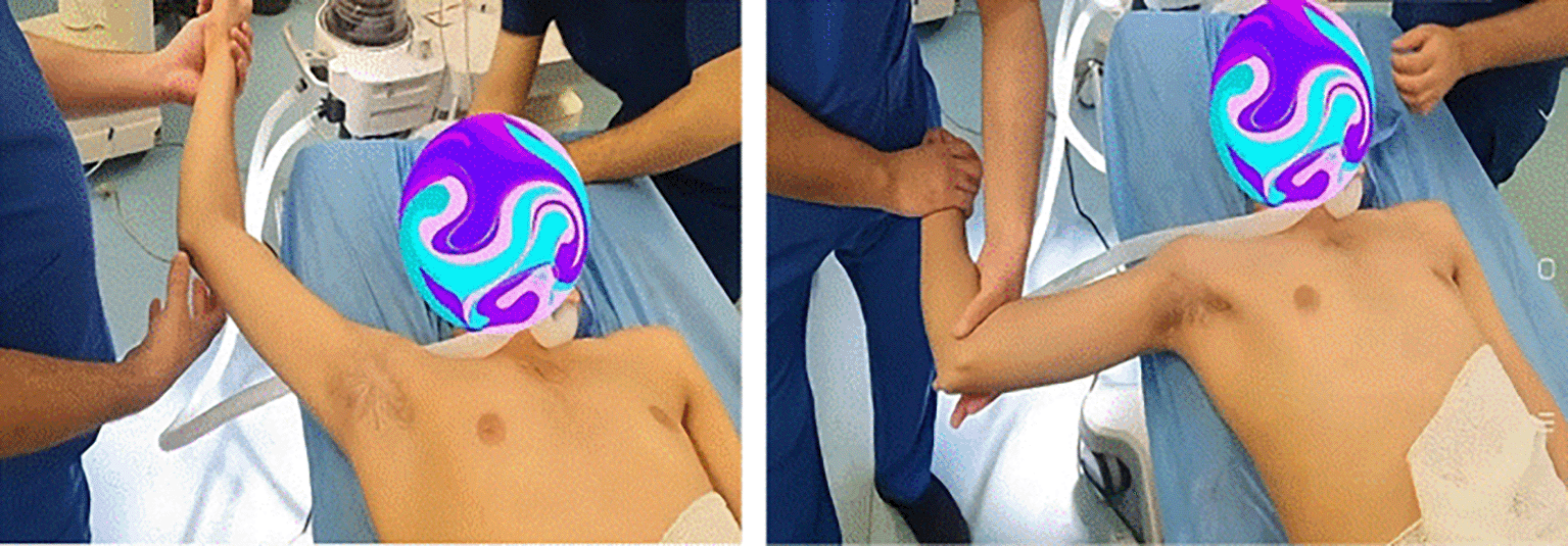
The management plan was discussed with the patient and his family, which was to try closed reduction under general anesthesia (although the rate of success is very low because of the chronicity of the case). If closed reduction fails, we proceed to open reduction.
Under General Anesthesia, closed reduction successfully reduced the head with external rotation of the Humeral Head with a snap. Examination of shoulder joint movement under anesthesia revealed a full range of movements ( Figure 11).
Subsequently, the patient wore an arm sling for two weeks, followed by exercise. MRI was performed to exclude ligament injuries.
Posterior Shoulder dislocation includes acute (time less than 6 weeks), chronic (>6 weeks), and recurrent dislocations. Its etiology is either direct (high-energy trauma) or indirect (epilepsy or electrical injury).
The usual presentation is that the arm claspes to the chest, posterior bulge of the shoulder, hollow anterior shoulder with a projecting coracoid, limitation of movement, and inability to perform external rotation of the shoulder.
Radiology included AP, lateral, and axillary views. Computed tomography (CT) scans are required in doubtful cases. Radiological signs included a light bulb appearance, empty glenoid sign (anteroposterior, AP view), or indentation of the anterior part of the humeral head (in Lateral or Axillary view).
Treatment is a trial of closed reduction under anesthesia; if it fails, open reduction is required, and the stability of the joint is performed after reduction.
Posterior shoulder dislocation is difficult to diagnose because of the rarity of cases, shortage of clinical signs, and inadequate radiological assessment. Therefore, clinical suspicion aids in the diagnosis.
Early diagnosis leads to early management, which decreases the risk of complications. Converting the condition to chronic dislocation worsens complications and leads to osteochondral lesions and joint destruction.
| Views | Downloads | |
|---|---|---|
| F1000Research | - | - |
|
PubMed Central
Data from PMC are received and updated monthly.
|
- | - |
Provide sufficient details of any financial or non-financial competing interests to enable users to assess whether your comments might lead a reasonable person to question your impartiality. Consider the following examples, but note that this is not an exhaustive list:
Sign up for content alerts and receive a weekly or monthly email with all newly published articles
Already registered? Sign in
The email address should be the one you originally registered with F1000.
You registered with F1000 via Google, so we cannot reset your password.
To sign in, please click here.
If you still need help with your Google account password, please click here.
You registered with F1000 via Facebook, so we cannot reset your password.
To sign in, please click here.
If you still need help with your Facebook account password, please click here.
If your email address is registered with us, we will email you instructions to reset your password.
If you think you should have received this email but it has not arrived, please check your spam filters and/or contact for further assistance.
Comments on this article Comments (0)