Keywords
Congenital epulis, Congenital granular cell tumor, Neumann's tumor, Hydroxyprogesterone, Case report
Congenital epulis, Congenital granular cell tumor, Neumann's tumor, Hydroxyprogesterone, Case report
Congenital epulis (CE), also known as a congenital granular cell tumor (CGCT) or Neumann's tumor, is a rare benign oral soft tissue tumor that is present at birth.1 The condition was first described by Neumann in 1871.2 Its incidence is very low, with fewer than 250 reported cases in the literature.16 It has a dramatic and well-documented female predilection, with a female-to-male ratio reported between 8:1 and 10:1.17 To our knowledge, this is the first case reported from Yemen in the available literature.
Congenital epulis typically presents as a solitary, smooth-surfaced, pedunculated, or sessile mass, which can vary in size from a few millimeters to several centimeters.4,12 It most commonly arises from the anterior maxillary alveolar ridge, with mandibular occurrences being less frequent.3,15 The exact cause of congenital epulis is unclear. The higher incidence of tumors in females hints at a potential hormonal influence.5 However, this hypothesis has been difficult to confirm, as studies have consistently failed to identify estrogen or progesterone receptors within the tumor cells.12,13
While smaller lesions may be asymptomatic and can even regress spontaneously,6 a larger tumor can cause significant feeding difficulties and, rarely, airway obstruction.9 Surgical excision is usually recommended as it can interfere with the newborn's feeding and respiration, with a marked psychological impact on parents.7 The prognosis following simple excision is excellent, with no reported cases of malignant transformation or recurrence, even after incomplete removal.8 To our knowledge, this report presents the first documented case of a large congenital epulis from Yemen, distinguished by its anterior alveolar origin and a maternal history of hydroxyprogesterone therapy during pregnancy.
A 5-hour-old female neonate was referred from the maternity department to our emergency department for the evaluation of a large mass protruding from her mouth. The baby was born at 40 weeks of gestation via spontaneous vaginal delivery with episiotomy to a 30-year-old primiparous mother. The birth weight was 2.5 kg, and APGAR scores were 8 and 9 at 1 and 5 min, respectively.
The mother’s antenatal history was significant for a threatened abortion diagnosed in the early second trimester, which was managed with complete bed rest and weekly intramuscular injections of hydroxyprogesterone caproate at a dose of 250 mg, which was continued until 37 weeks of gestation. The mother attended only two specific antenatal care visits during the entire pregnancy, a factor that likely contributed to the lack of prenatal diagnosis. Consequently, routine prenatal ultrasound screening was limited, and the oral anomaly was not detected in utero, leading to a surprising diagnosis at delivery. The patient had no family history of congenital or hereditary anomalies.
On presentation, the neonate was afebrile and showed signs of mild dehydration, with no signs of respiratory distress. The random blood sugar level was 46 mg/dL due to an inability to breastfeed, which was managed immediately with intravenous dextrose correction, and a nasogastric (NG) tube was inserted to ensure nutritional support while awaiting definitive treatment.
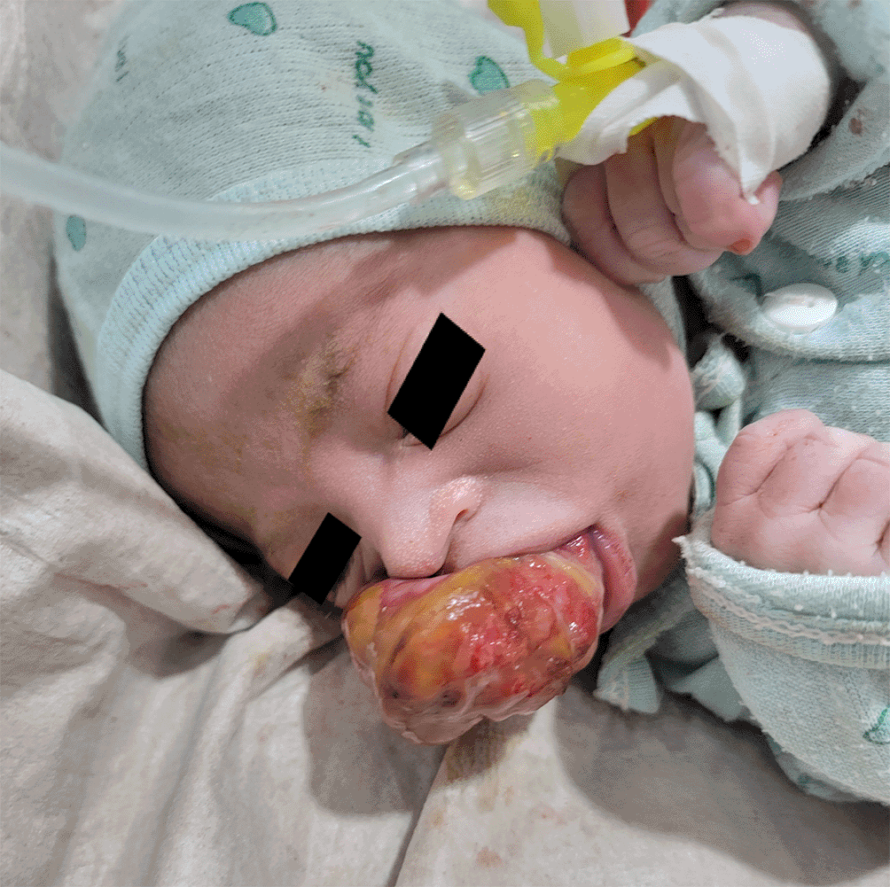
On examination, there was a large pedunculated fleshy mass approximately 4×3 cm in size in the anterior aspect of the upper alveolar ridge (maxillary gingiva) protruding extra orally, preventing complete mouth closure and separating the upper lips from the alveolar ridge ( Figure 1).
Based on clinical presentation, a provisional diagnosis of congenital epulis was made. The differential diagnoses included teratoma, hemangioma, and rhabdomyosarcoma.
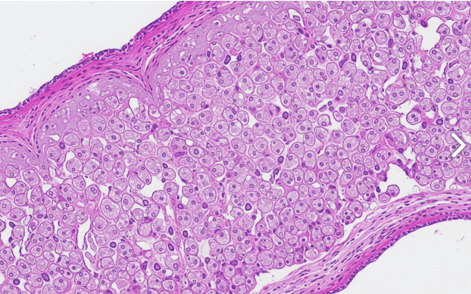
As the mass was large and interfered with breast feeding, the patient's parents were counseled regarding surgical excision of the lesion. On the 3rd day, the patient was referred to a pediatric maxillofacial surgeon, and the mass was excised at the base using electrocautery under local anesthesia. The excised mass was subjected to histopathological examination. The report described the presence of sheets of large polygonal cells with abundant eosinophilic granular cytoplasm and eccentrically placed small round nuclei ( Figure 2). These findings are pathognomonic for congenital granular cell tumors (congenital epulis).
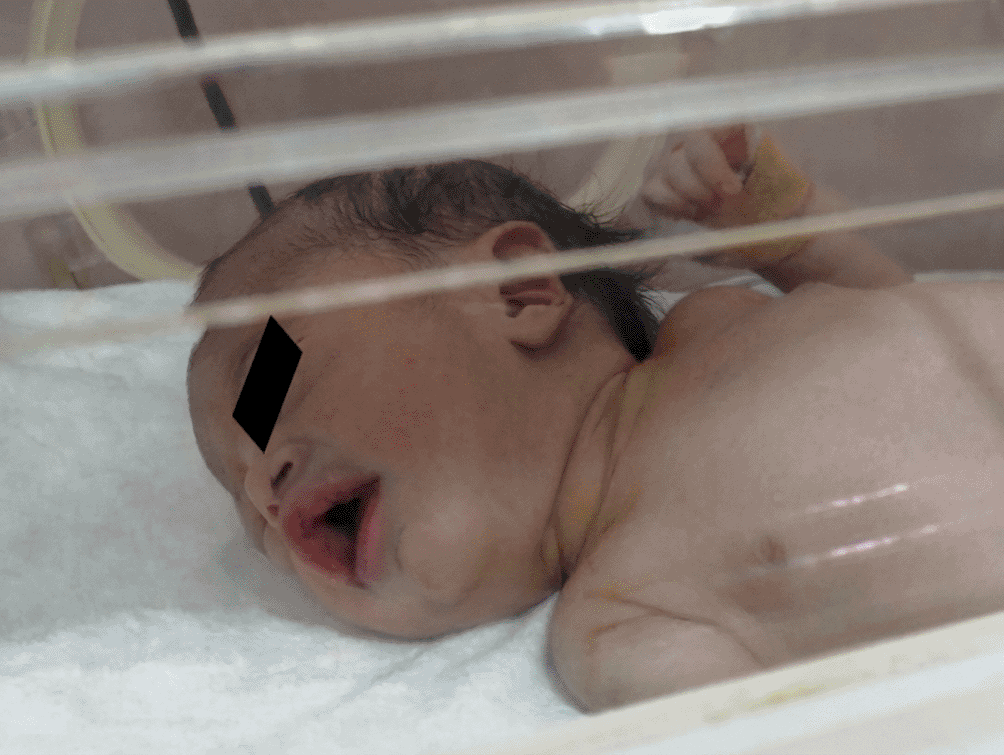
The postoperative course was uneventful. The nasogastric tube was removed, and breastfeeding was successfully started on the second postoperative day to resolve primary morbidity ( Figure 3). The neonate was discharged on the fourth postoperative day. Regular Follow-up examinations at 2 weeks, 3 months, and 6 months showed excellent healing, with no evidence of recurrence, and normal development.
Congenital epulis is a rare, benign neoplasm of the newborn oral cavity.1 A targeted literature search was conducted using PubMed, Google Scholar, and ResearchGate for reports published between 1871 and November 2025 using the keywords ‘congenital epulis’, ‘congenital granular cell tumor’, and ‘Neumann’s tumor’. After screening relevant publications, 32 cases were included in the analysis. ( Table 2)
A hallmark epidemiological feature of congenital epulis is its profound predilection for female newborns, consistently reported at a 9:1 or 10:1 ratio.18 Congenital epulis should also be differentiated from other neonatal oral masses, including teratoma, hemangioma, and rhabdomyosarcoma, based on clinical presentation and histopathological examination.19 Our review of 32 cases provides a robust validation of this finding. Of the 31 cases in which sex was specified, 28 were female (including this case) and two were male, yielding a ratio of 9.3:1.
A unique feature of this case was the maternal history of weekly hydroxyprogesterone injections. The dramatic female predilection of CGCT has long suggested a hormonal etiology.9,10 However, immunohistochemical studies have consistently demonstrated negative estrogen and progesterone receptor expression in congenital epulis tumor cells, arguing against a direct receptor-mediated mechanism.13,14 In our case, IHC was not performed, so receptor status could not be verified. Therefore, while the clinical correlation with multiple exogenous hydroxyprogesterone levels is evident, it is possible that the hormonal effect is indirect or that the tumor arises from a hormone-sensitive precursor cell that loses receptor expression as it differentiates into the granular phenotype, suggesting that maternal hormonal status may be coincidental or related to a more complex, as-yet unknown, non-receptor-mediated mechanism.
In addition to sex, congenital epulis demonstrates a strong predilection for the anterior alveolar ridge, with the maxilla being the most common location.4,15 The literature often cites a maxillary-to-mandibular ratio of 3:1.18 The 32-case review (1871–2025) confirms this, showing maxillary involvement in 25 cases and mandibular involvement in 11 cases (with 5 cases showing lesions in both), for a ratio of approximately 2.3:1. Our case represents a common, well-documented maxillary presentation.
Historically, immediate surgical excision was the exclusive treatment choice.16,24,25 While excision remains the gold standard for large lesions causing functional impairment,17,20 as in our patient some reports demonstrate that smaller, non-obstructive lesions can be monitored and may even spontaneously regress.18,21,22 Furthermore, recent cases indicate an increasing reliance on prenatal ultrasound and MRI to diagnose these lesions in utero, allowing for better delivery planning.10,11,23 The synthesized findings from the literature review are summarized in Table 1.
Congenital epulis is a rare benign tumor of the newborn that requires prompt recognition and management, especially when it is large enough to impair feeding. While the etiology remains elusive, this report adds to the clinical data regarding the potential hormonal influence on this tumor. The diagnosis rests on a "gold standard" histopathological examination, which must include a definitive immunohistochemical panel. Management strategies are evolving, and surgical excision remains the standard of care (93.9% of cases) to resolve functional interference with feeding or respiration. Finally, the patient’s prognosis was excellent. This comprehensive review found a 0% recurrence rate after treatment.
| Views | Downloads | |
|---|---|---|
| F1000Research | - | - |
|
PubMed Central
Data from PMC are received and updated monthly.
|
- | - |
Provide sufficient details of any financial or non-financial competing interests to enable users to assess whether your comments might lead a reasonable person to question your impartiality. Consider the following examples, but note that this is not an exhaustive list:
Sign up for content alerts and receive a weekly or monthly email with all newly published articles
Already registered? Sign in
The email address should be the one you originally registered with F1000.
You registered with F1000 via Google, so we cannot reset your password.
To sign in, please click here.
If you still need help with your Google account password, please click here.
You registered with F1000 via Facebook, so we cannot reset your password.
To sign in, please click here.
If you still need help with your Facebook account password, please click here.
If your email address is registered with us, we will email you instructions to reset your password.
If you think you should have received this email but it has not arrived, please check your spam filters and/or contact for further assistance.
Comments on this article Comments (0)