Keywords
Breast cancer, Clinicopathological characteristics, Epidemiology, Survival rate
Breast cancer, Clinicopathological characteristics, Epidemiology, Survival rate
Breast cancer is a condition characterized by the uncontrollable growth of abnormal breast cells to form tumors,1 which may spread throughout the body when left untreated. Due to the malignancy status and high global incidence, breast cancer represents a significant public concern.2–4 Globally, it has surpassed lung cancer as the most frequently diagnosed cases and also ranks among the most ubiquitous varieties of cancer in women.5–7 According to Global Cancer Today, in 2022, the worldwide incidence of breast cancer was estimated at 2.3 million cases with a mortality rate of 666,103 people.7
The World Health Organization, citing data from the International Agency for Research on Cancer, reported that the highest crude incidence rate of breast cancer was recorded in North America (162.5), followed by Europe (144.3), Oceania (130.5), Latin America and the Caribbean (65.1), Asia (43.4), and Africa (28.2).8 The highest mortality rate was reported in Europe (37.4), followed by North America (26.4), Oceania (25.1), Latin America and the Caribbean (17.7), Asia (13.9), and Africa (13.0). About 58% of breast cancer cases occurred in developing countries, accounting for nearly 50% of total cases.8
According to the National Cancer Institute, breast cancer ranks as the second leading cause of cancer deaths in women in the United States, after lung cancer.5 In Asian women, breast cancer is also the second leading cause of cancer-related deaths globally. In Asia, breast cancer has reached an incidence rate of 46 per 10,000 population, making the region the fifth highest in terms of incidence and mortality worldwide.5 The incidence of breast cancer has been steadily rising, with an estimated mortality rate of 15 per 100,000 population in Southeast Asia.9,10 In Indonesia, it is the most diagnosed type of cancer.11,12 According to the Indonesian Ministry of Health, the mortality and incidence rates of breast cancer were 17 per 100,000 population and 42.1 per 100,000 population, respectively. Further, cancer represents the second highest cost to Indonesia’s Universal Health Coverage.11
Survival rate has been shown as the key indicator of a successful treatment.13 In addition, age, race, lifestyle, tumor stage and size, histopathological type, lymph node status, variety of treatment, and hormonal receptor status have been used as variables for predicting the survival rate of breast cancer patients.14,15 However, survival rate varies significantly worldwide. The five-year survival rate in developed countries was >80%, approximately 60% in middle-income countries, and only 40% in low-income countries.16 Coleman et al. (2008) reported a five-year survival rate as high as 99% for breast cancer patients with localized tumors, 84% for those with regional spread, and 23% for those with advanced metastatic cases.16
Age, another critical determinant, shows a significant correlation with survival rate. Females diagnosed at age 46 to 50 years have a ten-year relative survival rate of approximately 70%, while those under 30 years and above 75 years old have 60% and 59% survival rates, respectively.17,18 Various studies in different reports have indicated a statistically significant correlation between breast cancer and clinicopathological characteristics, including tumor size,19 lymph node and blood vessel invasion status, metastatic lymph node,20 lymph vascular,21 neural invasion, perineural invasion,19 invasive breast carcinoma of no special type (NST),22,23 and lobular invasion.24 In this study, we investigated a comprehensive dataset of all clinicopathological characteristics and performed a statistical analysis using the survival analysis method. The aim of this study is to identify the correlation between the survival rate of female breast cancer patients and the clinicopathological data.
This study used a retrospective cross-sectional design and was conducted at EMC Alam Sutera Hospital (formerly known as OMNI Alam Sutera Hospital) in Banten Province, Indonesia. The study population comprised 200 female breast cancer patients who were referred to the hospital between January 2009 and December 2023. After applying the inclusion and exclusion criteria, 173 patients were eligible and included in the final analysis. Patients were included if they were first diagnosed with breast cancer between 2009 and 2023, had complete anatomical-pathological data, and completed the prescribed treatment. Patients were excluded if they had incomplete or unknown clinical or pathological data, discontinued treatment, were lost to follow-up, or received partial or incomplete treatment.
Demographic, clinical, and pathological data were retrieved from medical records, including age, tumor stage, tumor size, lymph vascular invasion, perineural invasion, invasive lobular carcinoma, invasive carcinoma of no special type, axillary lymph node involvement, and metastatic lymph node status. All included patients received one or more treatment modalities, including chemotherapy, hormonal therapy, breast-conserving surgery, or mastectomy, regardless of axillary lymph node surgery status. All treatments were administered during the studied period, from 2009 to 2023. Ethical approval for this study was obtained from the Research Ethics Committee of EMC Healthcare. All participants were adults aged 18 years and above. Written informed consent was obtained from all participants. The research consent was provided as part of the hospital’s standard written informed consent process. Participants were assured that refusal or withdrawal from the study would not affect their clinical treatment or relationship with healthcare providers.
Data were analyzed using Software Package for Statistical Analysis (SPSS), version 23. The Kaplan-Meier analysis was employed to identify survival curves, and validated with a log-rank test. The resulting variables, age (<40 vs ≥40), tumor stage (early [T1, T2] vs advanced [T3, T4]), tumor size (<2 cm vs ≥2 cm), invasive carcinoma of no special type (yes vs no), invasive lobular carcinoma (yes vs no), invasive lymph vascular (yes vs no), invasive perineural carcinoma (yes vs no), lymph node status (lymph node metastasis vs no lymph node metastasis), and axillary lymph node status (≤5 vs >6–≤14 vs >14), were analyzed using the Cox regression gradual approach.
Analysis in this study was conducted in three phases. In the first phase, univariate analysis was performed to describe several variables. It showed figures for Standard Deviation (SD), mean, percentage, and frequency of clinicopathological characteristics. These included age, gender, tumor stage, tumor size, invasive lymph vascular, invasive lobular carcinoma, invasive perineural, invasive carcinoma of no special type, lymph node, axillary lymph node, and survival rate in months. In the second phase, each variable was computed using bivariate analysis with the Kaplan-Meier method to determine the median and cumulative probability. Subsequently, a log-rank test was conducted to correlate the age, tumor stage, tumor size, invasive lobular carcinoma, invasive lymph vascular, invasive perineural, invasive carcinoma of no special type, lymph node, and axillary lymph node to breast cancer survival rate variables.
In the third phase, variables with a P-value of ≤0.05 were examined using a stepwise model as a component of the multivariate analysis. This analysis aimed to investigate the concurrent correlation between the independent variables and the survival of breast cancer patients through the Cox Proportional Hazards Regression model. This model was applied to analyze the survival of breast cancer patients by evaluating risk factors of age, survival status, tumor stage, tumor size, invasive lobular carcinoma, invasive lymph vascular, invasive perineural, invasive carcinoma of no special type, lymph node, and axillary lymph node. The investigation was concluded with a significance level set at 5% and results were considered statistically significant at a P-value of ≤0.05.
A total of 173 out of 200 female breast cancer patients were eligible and included in this study. Twenty-seven patients were excluded, comprising 11 patients with incomplete clinical or pathological data and 16 patients who discontinued treatment or received treatment at another healthcare facility. As shown in Figure 1, patients were categorized into three groups. The first group consisted of 173 patients, including 168 survivors and five deceased patients. These patients had complete clinical and pathological data, completed cancer treatment at EMC Alam Sutera Hospital, and received one or more treatment modalities, including chemotherapy, hormonal therapy, breast-conserving surgery, or mastectomy, regardless of axillary lymph node surgery status. No patient in this group was lost to follow-up.
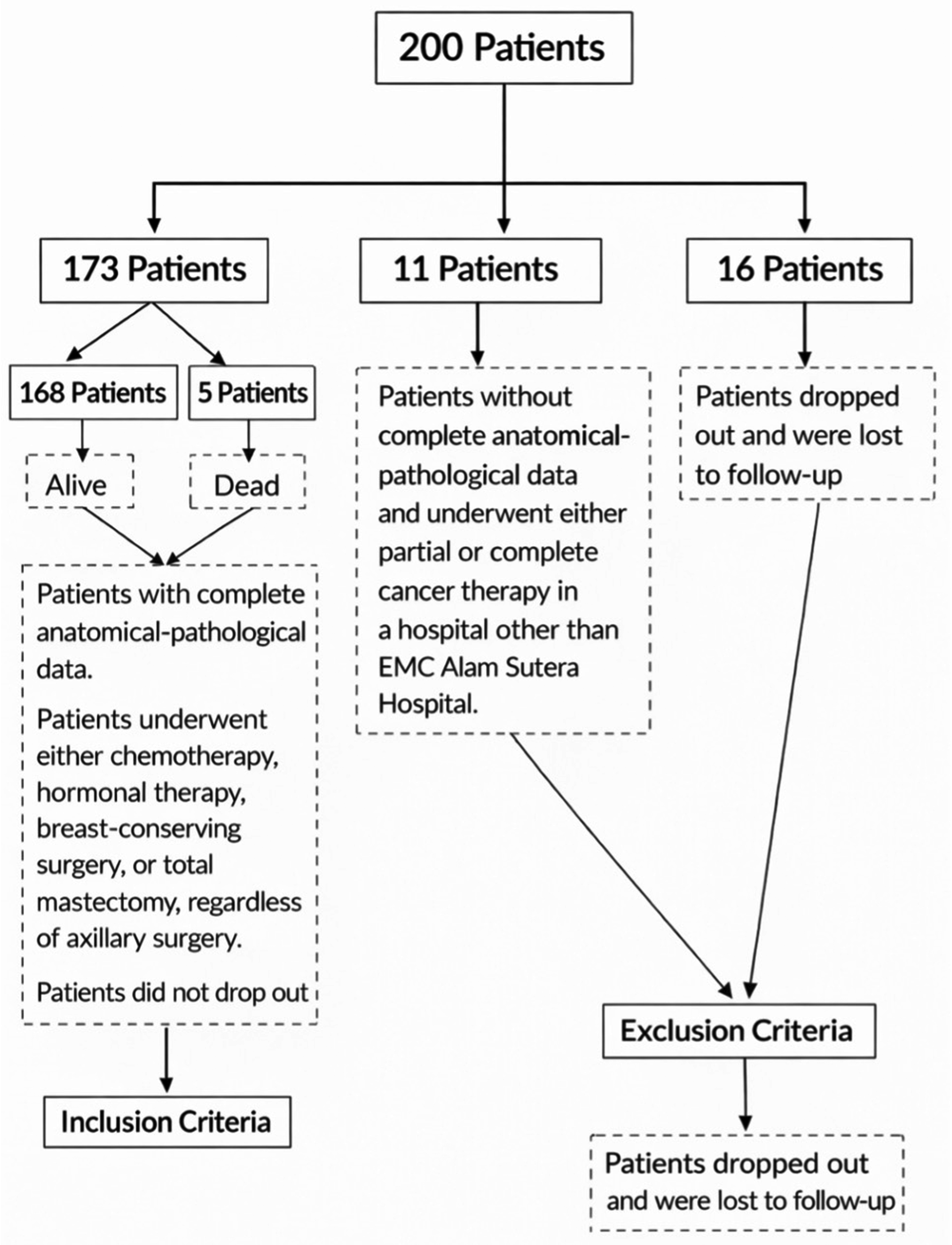
The second group included 11 patients who were excluded due to incomplete cancer treatment, while the third group consisted of 16 patients who received treatment at a healthcare facility other than EMC Alam Sutera Hospital. Accordingly, the final analysis focused exclusively on the first group of 173 patients.
In the study population, 158 patients (91.3%) were aged >40 years, while 15 patients (8.7%) were aged ≤40 years. Clinical tumor stages IIB and IIA were the most prevalent, accounting for 35.8% and 13.9%, respectively. Early tumor stages (T1 and T2) were observed in 70.5% of patients, whereas 29.5% presented with advanced-stage tumors. The majority of tumors measured ≥2 cm in diameter. Invasive carcinoma of no special type was the most common histological subtype, identified in 88 patients (50.9%). In addition, invasive lobular carcinoma, lymph vascular invasion, and perineural invasion were observed in 22.0%, 13.9%, and 3.5% of cases, respectively. Notably, 70.5% of patients had lymph node metastasis. Most patients had five or fewer positive axillary lymph nodes, as shown in Table 1.
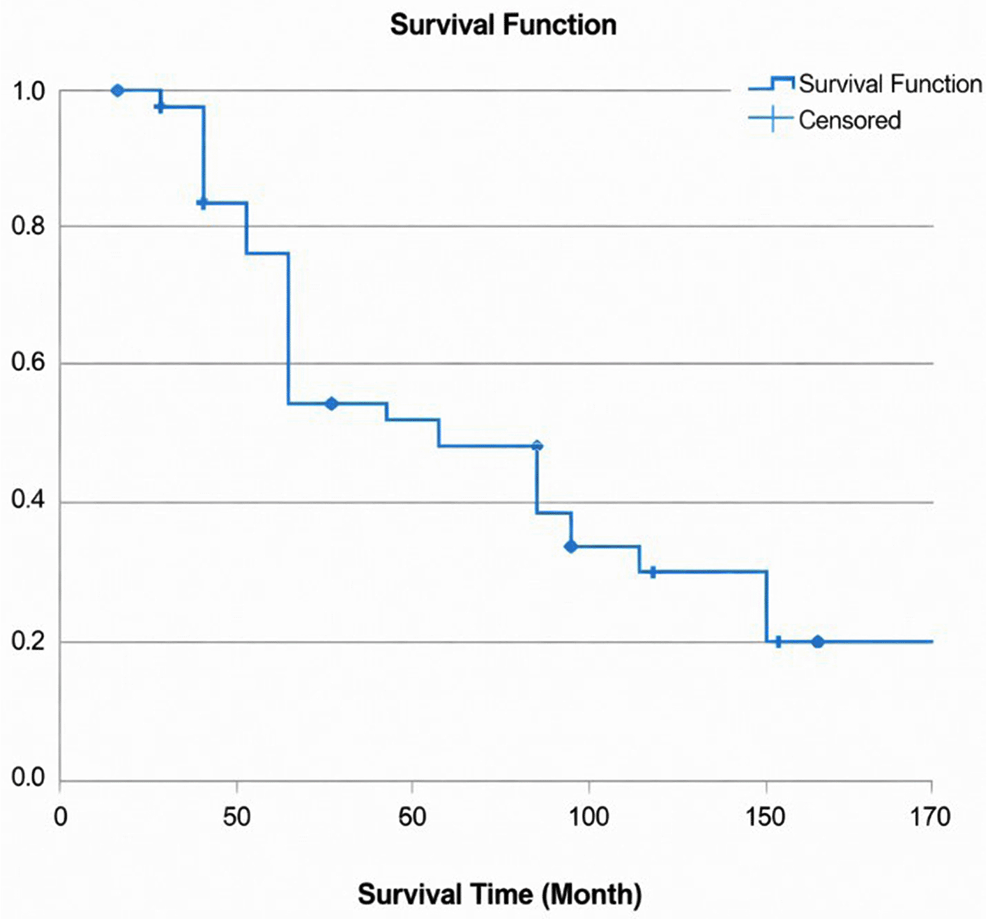
The mean survival time was 68.54 months (SD = 1.97), with a cumulative survival probability of 77.4%, as shown in Figure 2. Five patients were censored, and the first event occurred at 36 months, when one patient died. The probability of breast-cancer-specific survival up to 68 months was 45%, and the median survival time was 60 months, indicating that 50% of patients remained alive.
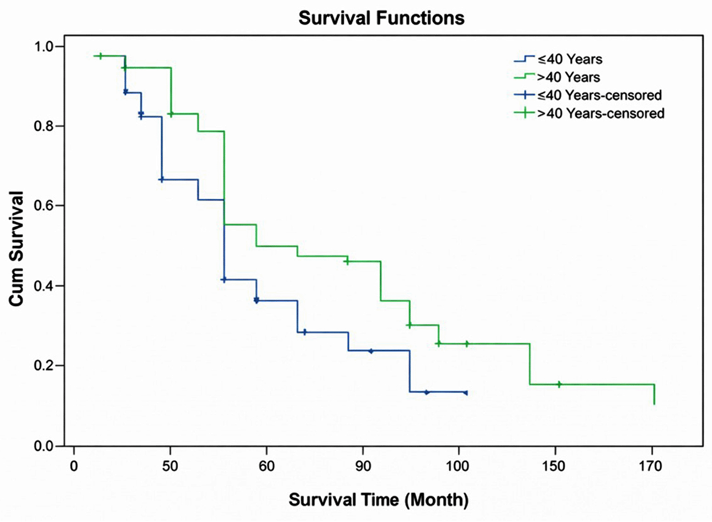
Patients aged >40 years demonstrated better survival outcomes, with a mean survival time of 70.04 months (SD = 2.10) and a cumulative survival probability of 79%, compared to patients aged ≤40 years, with a mean survival time of 52.80 months (SD = 3.47). Survival in the ≤40-year age group was significantly shorter and occurred earlier than in the >40-year group (P = 0.022). During the 144-month follow-up period, all patients aged >40 years experienced the event, whereas in the ≤40-year age group, more than 5% of patients survived beyond 50 months, as shown in Figure 3.
Advanced tumor stage and tumor size ≥2 cm was associated with lower mean survival time and cumulative survival probability compared to early-stage tumor and tumor measuring <2 cm. Patients with invasive carcinoma of no special type, lymph vascular invasion, and perineural invasion also demonstrated reduced survival, with mean survival times of 65.02 months, 59.75 months, and 50.00 months, respectively, compared to patients without these pathological features. Patients with lymph node metastasis had shorter survival than those without metastasis, with mean survival times of 65.94 months and 74.78 months, respectively. Further, patients with five or fewer positive axillary lymph nodes showed better survival outcomes (with mean survival time of 70.05 months), compared to those with six to fourteen or more than fourteen positive axillary lymph nodes, whose mean survival times were 55.63 months and 54.36 months, respectively.
A Kaplan-Meier analysis showed that age, invasive carcinoma of no special type, lymph vascular invasion, perineural invasion, lymph node status, and axillary lymph node status were all statistically significant variables, with P-values of 0.002, 0.049, 0.016, 0.028, 0.045, and 0.029, respectively. However, there was no statistically significant association between breast cancer survival rate and tumor stage, tumor size, or invasive lobular carcinoma, as indicated by P-values of 0.205, 0.328, and 0.438, respectively. The mean survival times for patients with various clinical and clinicopathological characteristics are shown in Table 2. The lack of statistical significance observed for other factors may be attributable to the limited sample size and the relatively high proportion of missing data.
A significant relationship was observed when clinicopathological characteristics were analyzed multivariate analysis in relation to breast cancer survival rate. However, the majority of these clinicopathological factors showed no association with survival outcomes. The results showed that only age was statistically significant. More specifically, this study found that patients aged under 40 years had a 2.14-times higher risk of mortality compared to those aged over 40 years. Further, the risk of death decreased significantly by 21% with each additional year of age (HR = 2.14, 95% CI: 1.228–3.739).
The group with lymph node metastasis showed a 1.2-times increased risk of breast cancer mortality compared to the group without lymph node metastasis, although the difference was not statistically significant. Patients with perineural invasion had a 1.62-times higher non-significant probability of death compared to those without perineural invasion. The non-invasive carcinoma of no special type group had an 88.3% reduced risk of breast cancer mortality, although invasive carcinoma of no special type did not have a statistically significant impact. Similarly, patients with five or fewer positive axillary lymph nodes had an 89.5% lower risk of death, but this factor was not statistically significant ( Tables 3 and 4).
A significant correlation was found between breast cancer survival rate and cancer condition factors (age, lymph vascular invasion, perineural invasion, lymph node status, invasive carcinoma of no special type, and axillary lymph node status) in the calculation performed by bivariate analysis using the log-rank test. In terms of age, a high proportion of breast cancer patients in this study was aged over 40 years (91.3%)—a similar attribute found in studies of the Western and Asian populations.25 A good correlation between age and breast cancer survival rate in this study is consistent with previous studies that older age is associated with longer survival outcomes.26 Our further analysis showed a lower survival rate in the patient group aged younger than 40 years (P = 0.022). This finding was supported by a study conducted in Italy that reported a lower survival rate among females with breast cancer aged less than 40 years. Younger patients have a higher tendency for axillary lymph node-positive disease, high tumor grade, and triple-negative breast cancer. As reported by a study in Germany, younger breast cancer patients typically had more aggressive tumor characteristics than older patients, leading to a lower survival rate.27
Invasive carcinoma of no special type was significantly associated with survival rate in this study’s correlation analysis (P = 0.049). This is consistent with a previous study of 482 female breast cancer patients in Malaysia, which reported a positive association between invasive carcinoma of no special type and survival rate (P = 0.018).7 In two other studies conducted in France and Korea, a positive correlation was identified between survival rate and invasive carcinoma of no special type, lymph vascular invasion (with P-values of 0.01 and 0.001, respectively), and axillary lymph node status (with P-values of 0.001 and 0.008, respectively).23,24 Chamalidou et al. studied 3,245 breast cancer patients in Sweden and found that invasive carcinoma of no special type was significantly correlated with age and metastatic lymph node status.24 Both variables were associated with breast cancer survival rate, each with a P-value of 0.001. Theoretically, a higher degree of invasion increases the risk of cancer spreading to surrounding tissues, thereby reducing the survival rate.28
There was a significant correlation in this study between survival rate and lymph vascular invasion (P = 0.016), which was supported by the observation that all patients with lymph vascular invasion died within 72 months, while over 5% of patients without lymph vascular invasion survived beyond 72 months. This finding is consistent with an earlier study conducted in Turkey, reporting a significant association between lymph vascular invasion and survival rate (P = 0.008) among 697 breast cancer patients.2 In France, Houvenaeghel et al. evaluated 4,205 breast cancer patients and found a significant correlation between lymph vascular invasion and breast cancer survival rate (P = 0.0001).21 In general, patients with lymph vascular invasion have a lower survival rate29 due to the tendency of the cancer cells to spread rapidly after invading the lymphatic system, making it a key prognostic factor in breast cancer.
A markedly association was found in this study between perineural invasion and survival rate (P = 0.028). All patients without perineural invasion from this study died within 144 months, while over 50% of patients with perineural invasion survived beyond 60 months. Similar results were shown by a previous study of female breast cancer patients in Turkey, with a P-value of 0.039.2 Another study in Japan investigated 105 patients with invasive carcinoma of no special type and noted that perineural invasion was significantly correlated with local recurrence and lymph node status, serving as an important prognostic factor.30 Patients with perineural invasion tend to have a lower survival rate, as the infiltration of the cancer cells into the nerves promotes further tumor spread and reflects a more aggressive disease course.31
Lymph node involvement has been shown to serve an important role in survival rate among breast cancer patients. From a correlation analysis of lymph node metastasis status and survival rate in this study, we identified a significant correlation between the two, with a P-value of 0.045, as well as a significant correlation (P = 0.045) between axillary lymph node status and survival rate. Across all 173 patients included in the study, various numbers of lymph nodes impacted were identified. We divided these patients into three categories based on the number of lymph nodes impacted: (1) ≤5 lymph nodes were found in 146 patients; (2) 6–≤14 lymph nodes were found in 16 patients; and (3) >14 lymph nodes were found in 11 patients. In a further statistical analysis, a total 97 (66.4%) of 146 patients from the ≤5 lymph node category showed lymph node metastasis, and two of them died within month 120–144, while the remaining 144 patients survived. A total 49 (33.5%) of these 146 patients showed no lymph node metastasis, with 48 surviving patients and only one deceased patient. In the second category, all 16 patients showed lymph node metastasis, with only one patient deceased and the rest surviving. In the third category, all 11 patients showed lymph node metastasis, with only one patient deceased and the remaining ten patients surviving.
Theoretically, the more lymph nodes impacted the lower the survival rate. On this account, the above ‘inconsistent’ results made us take a deeper look into the molecular-pathological data of the patients to find a conclusive link. The majority of these patients showed a Luminal A molecular pathological pattern (with estrogen and progesterone receptors positive and HER2-positive +2), found in 67 (69%) patients from the first category; in 36 (73.4%) patients from the second category; and in five (45.4%) patients from the third category. Therefore, in this study, the Luminal A molecular pathological pattern was identified as the dominant molecular pathological pattern. However, this finding still has not provided a sufficient explanation for the incongruity.
A cohort observational study in Vietnam of 27 participants showed similar results and reported a significant association between lymph node status and breast cancer survival rate (P = 0.001).3 Research reports from France and China found a significant positive correlation between lymph node status and lymph vascular invasion, with P-values of 0.01 and 0.001, respectively.32 Further, several studies conducted in Malaysia and Japan suggested that lymph node status was significantly correlated with breast cancer recurrence rate and perineural invasion, with P-values of 0.001 and 0.039, respectively.30 This finding is consistent with studies conducted in the United States and Turkey.26 Similar studies conducted in France, Sweden, and Korea reported a significant correlation between axillary lymph node status and invasive breast cancer of no special type, with P-values of 0.001, 0.001, and 0.008, respectively.22,24,33 These data support the premise that lymph nodes serve as a crucial prognostic factor that markedly impacts the survival outcomes of breast cancer patients.34 An increased number of lymph nodes affected by metastasis corresponds to a higher probability of cancer cell dissemination, which in turn reduces the overall survival rate.
No significant correlation was identified by this study between survival rate and tumor stage (P = 0.205), tumor size (P = 0.328), and pathological pattern of invasive lobular carcinoma type (P = 0.438). Similar results were reported by multiple studies in China, Korea, and Iraq,31,35 showing no correlation between tumor stage and breast cancer survival rate, with P-values of 0.632, 0.873, and 0.957, respectively. Another study conducted by De Nonneville et al. of 1,111 patients found no correlation between tumor stage and survival rate, with a P-value of 0.995,26 although some studies in Italy and Malaysia reported a significant correlation between tumor stage and survival rate, with P-values of 0.0001 and 0.001, respectively.32 Moreover, no statistic significant correlation between tumor size and survival rate was found in previous studies conducted in Italy, Sweden, China, and Turkey, with P-values of 0.38, 0.97, 0.967, and 0.223, respectively.31,32 Contrary to this finding, studies undertaken in Malaysia and Italy reported a positive association between tumor size and survival rate, with P-values of 0.005 and 0.001, respectively.7
Notably, early tumor stage (IIB), tumor size ≥2 cm, invasive carcinoma of no special type, and lymph node metastasis were found in higher frequency in this study. Meanwhile, advanced tumor stage, tumor size <2 cm, and lobular, lymph vascular, and perineural invasion appeared in a lower number of cases. The finding of early-stage cancer as the dominant stage in this study (IIA, IIB and T1, T2) is consistent with findings from many other studies conducted in various countries in Asia, Europe, and United States, reporting that early stage cancer was found in a higher proportion compared to advanced stage cancer.27,36 Further, in terms of patient survival, a total of 168 (97.1%) patients survived since their diagnosis of cancer, while the other five were deceased at various times within 3–12 years of their firm diagnosis of the disease.
The above findings further emphasize the immense importance of early detection and diagnosis of breast cancer in women to promote a higher survival rate, particularly among patients aged over 40 years. Considering this, early breast cancer detection programs should be a priority in increasing public awareness of breast cancer and ensuring a good quality of life.37 Further, our study found that around 85% of patients had a tumor size of over 2 cm, with 15% of them having tumor in size between 2–5 cm, while 85% over 5 cm. This finding is supported by a study conducted in Malaysia, reporting that 83.4% of its cases were patients with a tumor size exceeding 2 cm.7
The pathological examination results of this study revealed that invasive carcinoma of no special type accounted for 59.9% of cases, with 6.8% of them involved patients aged over 40 years. This finding is consistent with studies conducted in Iraq, France, and Sweden, where invasive carcinoma of no special type was reported as the predominant histological type, at rates of 78.7%, 62%, and 63.9%, respectively.22 A few of patients aged over 40 years in this study showed invasive lobular carcinoma, invasive lymph vascular carcinoma, and invasive perineural carcinoma, with case percentages of 22%, 13.9%, and 3.5%, respectively. This pathological examination finding is supported by studies in Iraq and Spain, which identified invasive lobular carcinoma as the least frequent histological subtype, with incidences of 6.1% and 4.5%, respectively.36 Studies in France and China reported that invasive lymph vascular carcinoma was found only in 24% and 41.7% of cases, respectively,20 and a study in Japan revealed that invasive perineural carcinoma was the least common histological type, at 45.9%.30
This study found no correlation between invasive lobular carcinoma and survival rate, with a P-value of 0.438. Kumilau et al., in a survival analysis of 482 breast cancer patients, observed that invasive lobular carcinoma was not correlated with recurrence rate (P = 0.139).7 In this study, all patients with invasive lobular carcinoma died within 72 months, whereas, in the non-invasive lobular carcinoma group, more than 5% patients survived beyond 108 months. A previous study in Malaysia reported a significant positive correlation between invasive lobular carcinoma and survival rate (P = 0.018).7 Patients with lobular invasion tend to show better survival outcomes compared to those with no special type or other histological subtypes. Lymph node metastasis is an important parameter that requires close observation. We identified that 70.5% of breast cancer patients had lymph node metastasis and that this percentage was slightly higher than those identified in studies conducted in China, France, and the United States of 66.6%, 40%, and 68.4%, respectively.24 Most cases (84.4%) in this study were presented with five or fewer lymph nodes metastases, consistent with previous studies undertaken in the United States and China that reported rates of 60.5% and 75.8%, respectively, for cases with fewer than five positive axillary lymph nodes.38
However, despite the findings of this study being consistent with multiple studies from other countries, the cumulative survival probability was lower compared to reports from developed countries.7 This discrepancy may have been resulted from the incomplete implementation of the national breast cancer registry system, and the low public awareness of early screening, to which socio-economic factors might have contributed, in Indonesia. Another factor that may have contributed to this discrepancy is the fact that most patients in Indonesia tend to avoid chemotherapy after surgery due to the adverse effects associated with chemotherapy. Yet, surgery alone without neoadjuvant chemotherapy may lead to a reduced survival rate. Patients in developed countries are generally more willing to undergo and complete the full course of chemotherapy, which contribute to better outcomes. Studies in the United States and France have shown that breast cancer patients receiving neoadjuvant chemotherapy followed by surgery and adjuvant chemotherapy had a significantly higher survival rate compared to those who did not.39
The statistical analysis in this study using Cox proportional hazards regression revealed a significant correlation between age and breast cancer survival rate, with a P-value of 0.007. According to numerous studies, the survival rate of breast cancer patients was influenced by several factors, including age.16 This study found that patients aged under 40 years had a 2.14-times higher risk of mortality compared to those aged over 40 years. Further, the risk of death decreased significantly by 21% with each additional year of age (HR = 2.14, 95% CI: 1.228–3.739). In line with past studies, this result indicates that age is a highly significant predictor of survival among breast cancer patients.
Based on the results, Cox regression analysis showed that invasive carcinoma of no special type, lymph vascular invasion, perineural invasion, lymph node status, and axillary node status were not significantly correlated with survival rate (P > 0.05). More specifically, the lymph vascular invasion group was presented with a 1.31-times higher—but statistically non-significant—risk of breast cancer mortality compared to the non-lymph vascular invasion group (HR = 1.31, 95% CI: 0.759–2.292). The non-lymph vascular invasion group had an 86.8% lower risk of death, but the statistical analysis found no significant correlation (P = 0.326). In a similar study conducted in Turkey, a Cox proportional hazards analysis of 697 breast cancer patients found no significant correlation between lymph vascular invasion and breast cancer survival rate (P = 0.118).2 The perineural invasion group suggested a 1.62-times higher risk of death compared to the non-perineural invasion group, although it was not statistically significant (HR = 1.62, 95% CI: 0.631–4.194). In a further Cox regression analysis, patients without perineural invasion had an 83.3% lower risk of breast cancer mortality, but showed no significant correlation with survival rate (P = 0.314). A study conducted by Ozgur et al. in Turkey also proved no significant correlation between perineural invasion and breast cancer survival through a Cox regression analysis, with a P-value of 0.899.2
This study is a comprehensive breast cancer study with evaluation of clinicopathological characteristics, including lymph vascular and perineural invasion, and statistical analysis of their correlation with survival rate.40 Breast cancer survival rate is variably associated with clinicopathological factors, consisting of age, invasive carcinoma of no special type, lymph vascular invasion, perineural invasion, lymph node status, and axillary lymph node status. Age (>40 years) and invasive carcinoma of no special type are important parameters in enabling better survival rate predictions.
Ethical principles were upheld throughout the study. Ethical approval was obtained from the Research Ethics Committee of EMC Healthcare (No. KEP-EMC/001/2025). All data were anonymized prior to analysis, and patient confidentiality was strictly maintained.
In conclusion, age is significantly correlated with survival outcomes and serves as an important predictor of survival among female breast cancer patients. Given the inconclusive findings, particularly from the Cox regression analysis, studies with larger sample sizes and additional parameters are needed to obtain more definitive results.
| Views | Downloads | |
|---|---|---|
| F1000Research | - | - |
|
PubMed Central
Data from PMC are received and updated monthly.
|
- | - |
Provide sufficient details of any financial or non-financial competing interests to enable users to assess whether your comments might lead a reasonable person to question your impartiality. Consider the following examples, but note that this is not an exhaustive list:
Sign up for content alerts and receive a weekly or monthly email with all newly published articles
Already registered? Sign in
The email address should be the one you originally registered with F1000.
You registered with F1000 via Google, so we cannot reset your password.
To sign in, please click here.
If you still need help with your Google account password, please click here.
You registered with F1000 via Facebook, so we cannot reset your password.
To sign in, please click here.
If you still need help with your Facebook account password, please click here.
If your email address is registered with us, we will email you instructions to reset your password.
If you think you should have received this email but it has not arrived, please check your spam filters and/or contact for further assistance.
Comments on this article Comments (0)