Keywords
Faricimab, anti-VEGF, age related macular degeneration, retinal vein occlusion edema, diabetic macular edema.
Faricimab, anti-VEGF, age related macular degeneration, retinal vein occlusion edema, diabetic macular edema.
Among the most common causes of blindness and visual impairment worldwide are retinal and choroidal vascular diseases such as diabetic retinopathy, diabetic macular edema (DME), retinal vein occlusion- related macular edema (RVO), and neovascular age-related macular degeneration (n-AMD).1
The pathophysiological hallmarks of n-AMD, DME, and RVO often intersect, involving oxidative stress, inflammatory cascades, and abnormal angiogenesis. Collectively, these factors converge on vascular endothelial growth factor (VEGF) and angiopoietin 2 (Ang-2) which orchestrates pathological vessel growth and elevates vascular permeability.2,3 Co-expression of Ang-2 and VEGF-A has been linked to accelerated neovascularization.4 VEGF-A facilitates angiogenesis by encouraging the movement, survival, and development of endothelial cells. Ang-2 has been demonstrated to increase proinflammatory signals in endothelial cells and is implicated in vascular leakage and aberrant vessel shape.4
The treatment of RVD has changed over the past 2 decades due to the introduction of anti-vascular endothelial growth factors (anti-VEGFs), which are currently the first-line treatment.5 Anti-VEGF agents have become a powerful and successful tool in the fight against RVO, DR complications, and n-AMD. These agents administered intravitreally block proangiogenic factors' functional activity with varying target selectivity, affinity, and efficacy.6 VEGF inhibitors that are currently used to treat retinal disorders are ranibizumab, aflibercept, brolucizumab, and bevacizumab, the latter is used off-label.7 In recognition of the multifactorial pathophysiology underlying RVD, research efforts are increasingly focused on developing novel therapeutic approaches that extend beyond VEGF inhibition alone.8 Although anti-VEGF therapies have demonstrated efficacy in controlled clinical trials, their effectiveness in real-world practice frequently falls short of expectations. Common obstacles include primary or secondary nonresponse, tachyphylaxis, and recurrence of edema following treatment discontinuation.9
Faricimab is a new bispecific antibody that targets both VEGF-A and Ang-2, was recently developed by Roche/Genentech to treat DME and n-AMD. Faricimab binds and neutralizes Ang-2, a key regulator of vascular stability, as well as VEGF-A, a major contributor to neovascularization and vascular permeability. By inhibiting VEGF-A, Faricimab lessens neovascularization and vascular permeability, preventing the buildup of retinal fluid that causes macular disorders' vision loss. Its attachment to Ang-2 also prevents vascular destabilization, which further prevents the buildup of retinal fluid.10 This means that, in contrast to current VEGF-A inhibitors, the simultaneous blockage of both pathways offers the possibility of comprehensive therapeutic efficacy.1
The study is designed to evaluate the efficacy and safety of intravitreal Faricimab in patients with refractory macular edema secondary to n-AMD, DME, and RVO, who have previously undergone multiple intravitreal anti-VEGF treatments other than Faricimab and exhibited poor or no response.
This study has previously been published as a preprint.11
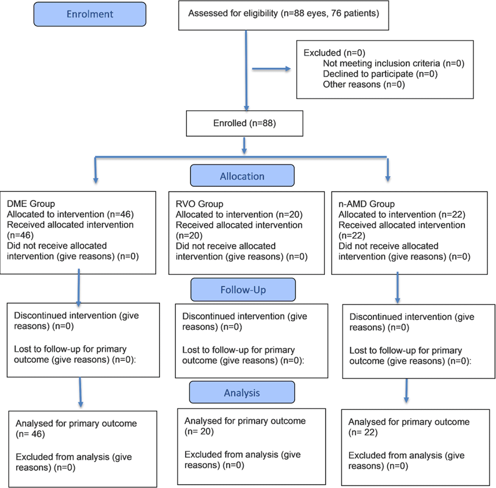
This study was conducted at a specialized Ophthalmic Center in Baghdad, Iraq, between January 2025 and December 2025. The study included 88 eyes of 76 patients diagnosed with primary refractory macular edema secondary to RVO, DME, and n-AMD. Patient recruitment started on March 1 and ended on November 30. The study was conducted and reported in accordance with the CONSORT (Consolidated Standards of Reporting Trials) guidelines.
Ethical and scientific approvals were obtained prospectively from the Research Ethics Committee at the Department of Pharmacology, College of Medicine, University of Baghdad (ethical approval No.03-37 3/1/2025). All participants provided written informed consent after being thoroughly informed about the purpose, risks, procedures, and potential benefits of the research. The research team maintained full compliance with Good Clinical Practice guidelines and adhered to the ethical principles of the Declaration of Helsinki.
Trial name: ClinicalTrials.gov
Trial number: NCT07093385
Registration date: 2025-07-29
RegistrationURL: https://register.clinicaltrials.gov/prs/beta/studies/S000FBYY00000040/results/create
The trial was registered on ClinicalTrials.gov retrospectively due to administrative and institutional requirements. As this trial was conducted as part of a master’s thesis, priority was given to obtaining ethical approval and ensuring compliance with institutional protocols, which led to an unintentional delay in registration. Once the importance of early registration for transparency and compliance was recognized, the process was promptly completed.
2.4.1 Patients inclusion criteria
Patients were considered eligible if they met all the following inclusion criteria:
1. patients aged 18 years old or older diagnosed with refractory DME, RVO, or active n-AMD by clinical evidence, optical coherence tomography OCT and angiography (OCTA).
2. Primary refractory patients that show persistent subretinal or intraretinal fluid with no improvement in BCVA or reduction in central retinal thickness after a minimum of 3 loading doses of Bevacizumab and Aflibercept (CRT>300 μm with BCVA either unchanged or worse compared with baseline).
3. Ability to attend follow-up appointments and comply with treatment protocol.
2.4.2 Patients exclusion criteria
Patients were excluded from the study if any of the following conditions were met:
1. Inconsistent treatment history: missed a dose of anti-VEGF doses, or failure to complete the three loading doses of Faricimab.
2. Ocular comorbidities that could confound outcomes, such as: Visually significant cataract as Grade 2+ or more, corneal opacity, uncontrolled glaucoma (IOP > 25 mmHg on medication) with an increase in cup/disc ratio, Other macular pathologies (e.g., macular hole, epiretinal membrane ERM).
3. Severe baseline vision loss (BCVA < 6/60).
4. Secondary non-responders: patients who initially showed improvement in BCVA and CRT after anti-VEGF therapy, but subsequently experienced deterioration despite continued treatment with bevacizumab and aflibercept.
Baseline assessments and data collection: To be enrolled in the study, eligible patients gave their written informed consent to be included in the treatment regimen. Subsequently, patients' demographic data (age and gender) and past medical history for hypertension and diabetes mellitus were documented in an electronic medical records system, and patients subjected to a combination of tests for complete ophthalmic evaluation. Both eyes' Best-Corrected Visual Acuity (BCVA) was measured using a Snellen chart, which was subsequently transformed into algarithm of the minimum angle of resolution (logMAR) for statistical analysis. Subsequently, topical tropicamide 1% eye drops were used to dilate the patient's pupil. Slit lamp ocular examination was performed for anterior segment examination. A 90 or 78 diopter condensing lens was used for a thorough inspection of the fundus and macula. Goldman Applanation Tonometry was used for intraocular pressure monitoring. Optical Coherence Tomography Angiography OCT-A (Optovue, solix, Japan) was used to diagnose the cases of n-AMD and CNV, also to exclude ischemic maculopathy cases. Spectral Domain Optical Coherence Tomography (Optovue, solix, Japan) was utilized to evaluate intraretinal fluid (IRF), subretinal fluid (SRF) and central retinal thickness (CRT). All visual assessments were performed using the same device and conducted by the same experienced examiner to minimize inter-device and inter-observer variability.
Intervention procedures: After baseline evaluation, all patients receive intravitreal faricimab injection as a loading dose for three consecutive months within two days of examination. Intravitreal administration of faricimab was performed under aseptic conditions following topical anesthesia with tetracaine hydrochloride. The eyelids and eyelashes were disinfected with 10% povidone-iodine solution, and the conjunctival fornices were irrigated with 5% povidone-iodine. Faricimab (6 mg/0.05 mL) was injected into the vitreous via pars plana, approximately 3.5 mm posterior to the limbus in pseudophakic and 4 mm in phakic eye, using a 30-gauge needle. Post-injection, topical tobramycin was prescribed four times daily for 5 days.
Outcome measures: The ophthalmologic assessment was repeated again after one month from the last injection to compare the results and to assess treatment response ( Table 2-4). The response to faricimab was evaluated both functionally and anatomically. The primary outcomes assessed were the mean BCVA, CRT, complete resolution of retinal fluid—defined as the absence of any (SRF) and (IRF) in macular volume patterns on (SD-OCT).
All analyses were conducted using SPSS v.26. Data was represented as tables and figures. For statistical analysis, BCVA measurements were transformed into the logarithm of the minimum angle of resolution (LogMAR). While categorical variables were expressed as percentages, continuous variables were provided as mean ± standard deviation (SD). For numerical variables that were regularly distributed, paired t-tests were used to compare the time points; for nominal distributed variables, McNemar tests were employed. For every analysis, a p-value of less than 0.05 was used to determine statistical significance.
A total of 76 patients were included in the study, with a mean age of 66.14±8.678 years range (46-83) with 43 patients (56.57%) cases ≥ 65 years and 33 (43.42%) case < 65 years. The total number of males was 37 (48.68%) and females were 39 (51.32%) with a ratio of female to male (1.05:1.0). Past medical history (PMH) was positive among 62 (81.58%) patients, with HT among 30 (39.47%) and DM among 47 (61.9%). Some patients 15 (19.73%) have both HT and DM. Table 1 summarizes patient demographic data.
The study enrolled 88 eyes, 44 (50%) right and 44 (50%) left eyes. Previous anti-VEGF injections were Intravitreal aflibercept among 45 (51.14%) eyes, Intravitreal bevacizumab among 32 (36.36%), and mix Intravitreal aflibercept and Intravitreal bevacizumab among 11 (12.50%) eyes; with mean injections of 7.0±1.77 range between (5-12) in the past year. Table 2 summarize eyes baseline characteristic.
Of the total 88 eyes treated with intravitreal faricimab, 46 (52.27%) eyes diagnosed with DME, 20 (22.73%) eyes diagnosed with RVO, and 22 (25.0%) eyes diagnosed with n-AMD. Figure 1 shows ocular diagnosis and Figure 2 shows the flow diagram of the trial.
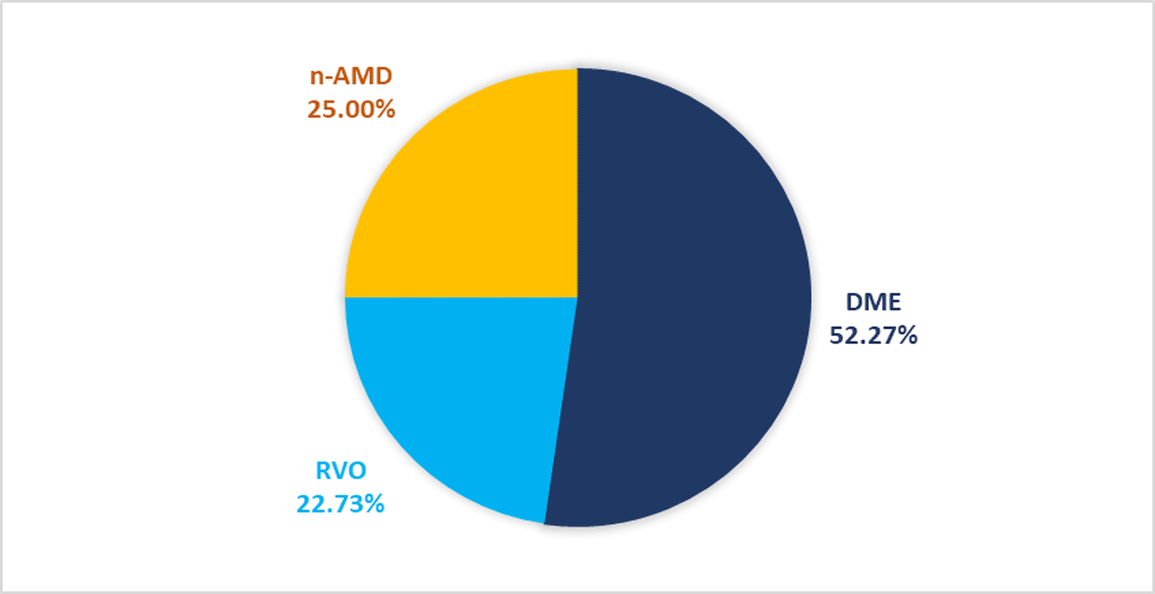
Abbreviation: RVO, retinal vein occlusion edema; DME, diabetic macular edema; n-AMD, neovascular age-related macular degeneration.
Patients diagnosed with DME had a mean age of 64.8 ± 7.9 years range between (54–80) with 17 (44.7%) cases ≥ 65 years and 21 (55.3%) cases < 65 years. All participants 38 (100%) had PMH, including DM in every case 38 (100%) and HT in 10 cases (25.6%), with this subset having comorbid DM. Table 3 summarize demographic data for patients with DME.
The comparison of clinical parameters before and after starting Faricimab was detailed in Table 4. Example on the anatomical outcome showed in Figure 3.
(No=46).
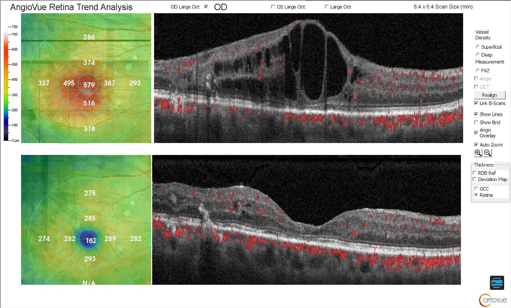
Upper panel: OCT scan at baseline showed persistent cystoid macular edema and intraretinal fluid despite previous aflibercept injections.
Lower panel: OCT Post-treatment demonstrated complete resolution of fluid with improvement in BCVA.
Best corrected visual acuity and central retinal thickness showed a significant improvement (P<0.001). The mean BCVA improved from 0.60 (SD±0.24) to 0.44 (SD±0.24) and mean CRT reduced from 464.74 μm (SD±112.99) to 288.5 μm (SD±85.04), 1 month after the third faricimab injection.
Intraretinal fluid and subretinal fluid show significant reduction (P<0.001). The proportion of eyes with SRF decreased from 15 (32.60%) pretreatment to 2 (4.34%) posttreatment, while the proportion of eyes with IRF decreased from 46 (100%) to 19 (41.30%). Intraocular pressure remained stable over the course of the study (p=0.673).
The study included 19 patients diagnosed with RVO, with a mean age of 63.6±8.6 years range (46-76) with 8 case ≥ 65 years and 11 case < 65 years. PMH was positive in 14 patients (73.7%), including HT in 14 patients (73.7%) and DM in 3 patients (15.9%). From these patients 3 cases (15.9%) had both DM and HT. Table 5 summarize demographic data for patients with RVO.
The comparison of clinical parameters before and after starting faricimab was detailed in Table 6. Example on the anatomical outcome showed in Figure 4.
(No=20).
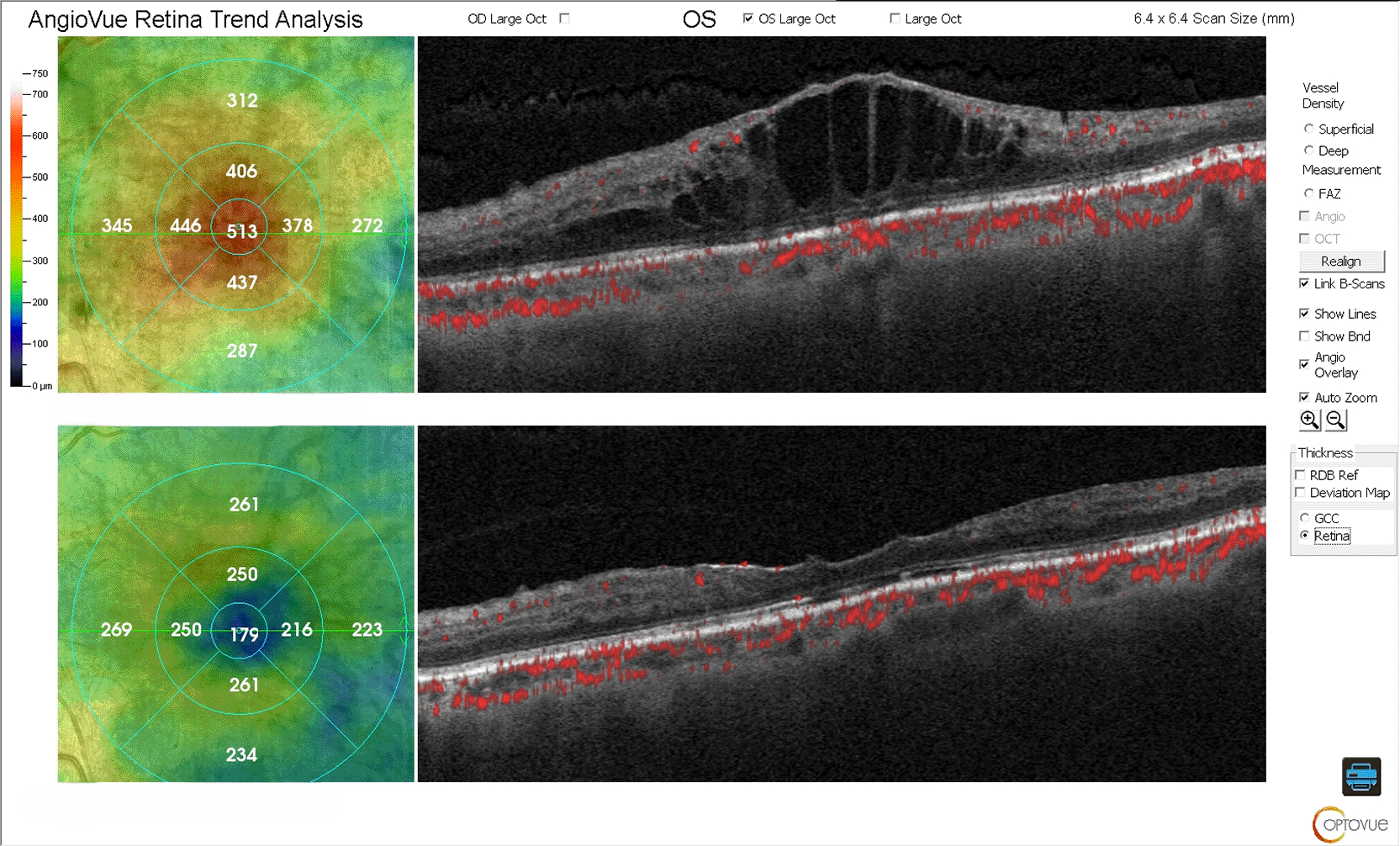
Upper panel: OCT scan at baseline showed significant macular edema and intraretinal fluid with increased central retinal thickness despite previous aflibercept injections
Lower panel: OCT scan after switching to Faricimab showed complete anatomical resolution of intraretinal fluid and retinal thickness reduction.
Abbreviation: CRVO; central retinal vein occlusion edema.
Best corrected visual acuity and central retinal thickness showed a significant improvement (P<0.001). The mean BCVA improved from 0.71 (SD±0.25) to 0.48 (SD±0.27) and mean CRT reduced from 534.3 μm (SD±144.79) to 324.45 μm (SD±88.30), 1 month after the third faricimab injection.
Intraretinal fluid and subretinal fluid show significant reduction (P<0.05). The proportion of eyes with SRF decreased from 10 (50%) pretreatment to 2 (10%) posttreatment, while the proportion of eyes with IRF decreased from 20 (100 %) to 9 (45%). Intraocular pressure remained stable over the course of the study (p=0.408).
The study included 19 patients diagnosed with n-AMD, with a mean age of 74.1±5.4 years range (61-83) with 18 (94.7%) case ≥ 65 years and 1 (5.3%) case < 65 years. PMH was positive in 10 patients (52.6%), including HT in 7 patients (36.8%) and DM in 6 patients (31.6%). From these patients 2 cases (10.5%) had both DM and HT. Table 7 summarize demographic data for patients with n-AMD.
The comparison of clinical parameters before and after starting faricimab was detailed in Table 8. Example on the anatomical outcome showed in Figure 5.
(No=22).
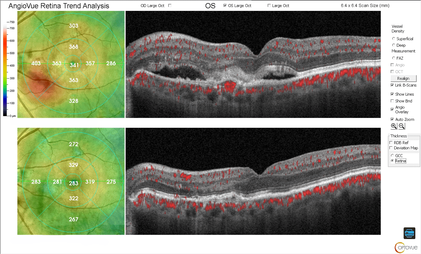
Upper panel: OCT scan showing significant subretinal fluid and macular edema.
lower panel: OCT scan following three loading doses of Faricimab showed subretinal fluid resolution with reduction in central retinal thickness.
Abbreviation: neovascular-AMD, neovascular age-related macular degeneration.
Best corrected visual acuity and central retinal thickness showed a significant improvement (P<0.001). The mean BCVA improved from 0.63 (SD±0.18) to 0.39 (SD±0.28) and mean CRT reduced from 411.23 μm (SD±78.99) to 268.73 μm (SD±58.13), 1 month after the third faricimab injection.
Intraretinal fluid and subretinal fluid show significant reduction (P<0.05). The proportion of eyes with SRF decreased from 22 (100 %) pretreatment to 3 (13.63%) posttreatment, while the proportion of eyes with IRF decreased from 6 (27.27%) to 0 (0%). Intraocular pressure remained stable over the course of the study (p=0.35).
In all three diagnostic groups, the baseline proportion of patients exhibiting retinal fluid (IRF and/or SRF) was 100%. Following treatment, a substantial proportion of patients achieved complete anatomical resolution, approximately 59% in DME, 55% in RVO, and 86.37% in n-AMD. Figure 6 shows retinal fluid resolution.
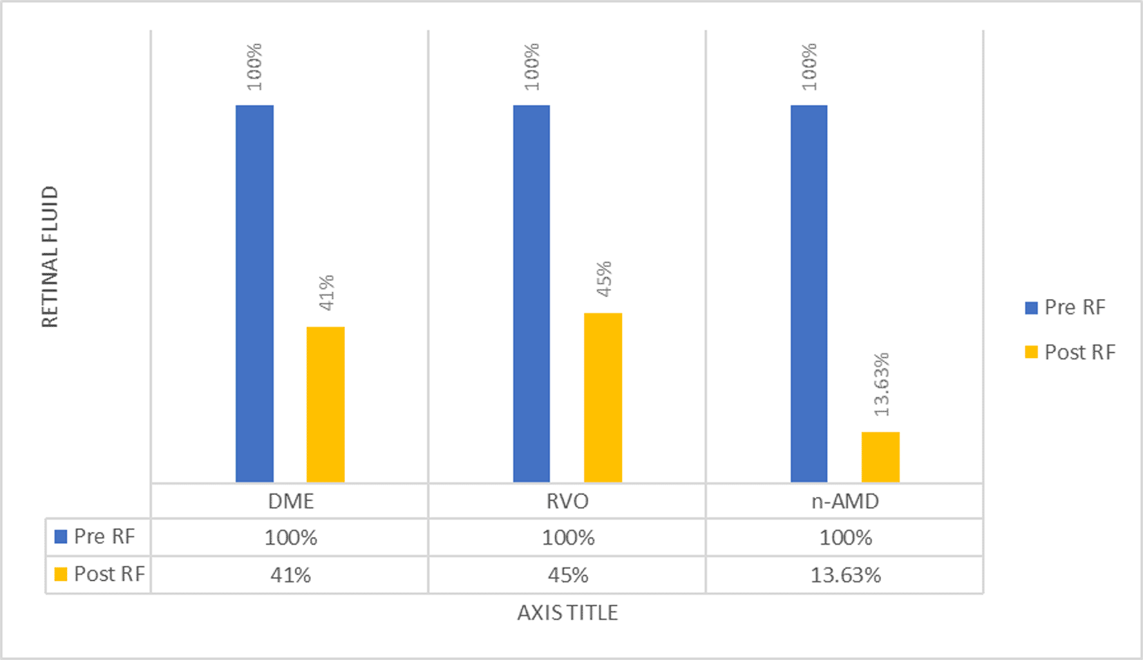
Abbreviation: Pre RF, retinal fluid at baseline; Post RF, retinal fluid after faricimab loading dose; IRF, intraretinal fluid; SRF, subretinal fluid; RVO, retinal vein occlusion edema; DME, diabetic macular edema; n-AMD, neovascular age-related macular degeneration.
This study evaluated 88 eyes from 76 patients, with a mean age of 66.14 ± 8.68 years; 56.57% of patients were ≥65 years, reflecting the strong association between retinal vascular disease (RVD) and aging. These findings are consistent with the study in Nepal which reported a mean age of 69.64 ± 7.31 years and a 52.37% prevalence of retinal abnormalities in individuals aged ≥60 years.12 The female-to-male ratio of 1.05:1 aligns with previous analyses.13
A large proportion of patients (81.6%) had comorbidities, predominantly diabetes mellitus (61.84%) and hypertension (39.47%), with 19.7% presenting both. These conditions are well-known contributors to the development and progression of diabetic retinopathy (DR) and retinal vein occlusion (RVO).14
Among the study eyes, 52.27% had refractory DME, 25.0% had refractory n-AMD, and 22.73% had refractory RVO, reflecting the spectrum of anti-VEGF-resistant macular edema encountered in routine clinical practice. Previous studies have shown that 44% of DME patients exhibit persistent fluid despite long-term aflibercept therapy,15 and 19.7–36.6% of n-AMD patients continue to have active exudation following one year of aflibercept.16 For RVO the results of clinical trials and clinical practice differ, which suggests that anti-VEGF may not be sufficient to completely treat this illness.17
All patients had received multiple prior intravitreal injections of bevacizumab and/or aflibercept (mean 7 injections, range 5–12), emphasizing the complexity of managing refractory RVD and the need for alternative therapeutic strategies.
Switching to intravitreal faricimab resulted in significant improvements in best-corrected visual acuity (BCVA) across all groups. In DME eyes, mean VA improved from 0.60 to 0.44 (P<0.001), corresponding to an 8-letter gain on the ETDRS scale, consistent with real-world data,18 though differing from studies reporting stable visual function19
For eyes diagnosed with RVO, BCVA showed significant improvement (P<0.001), with mean BCVA improved from 0.71 to 0.48, indicating that the applied therapeutic technique was successful in improving patients' sight (>10 ETDRs).20 The significant improvement in BCVA is consistent with faricimab's dual mode of action, which targets both Ang-2 and VEGF to address the complex pathophysiology of RVO.21
For eyes diagnosed with n-AMD, treatment switching is a standard strategy to address nonresponse.22 Information on BCVA for patients with n-AMD varies widely in the literatures. Some authors have documented improvement23 other documented stability,24 while worsening is sometimes observed.25 The main causes of this could be different inclusion and exclusion criteria, and different lengths of prior anti-VEGF treatment. In the current study, the mean BCVA of individuals with n-AMD improved significantly from 0.63 to 0.39 (P<0.001), corresponding to gain of >10 ETDRs letters.
Faricimab therapy resulted in significant anatomical improvements in all patient groups, evidenced by reductions in central retinal thickness (CRT), intraretinal fluid (IRF), and subretinal fluid (SRF).
Following three loading doses, 47.82% of eyes achieved complete IRF resolution, while the remainder showed marked reduction. CRT <300 μm was achieved in 37.5% of patients, aligning with YOSEMITE and RHINE trials.26 Patients with refractory DME who transitioned from aflibercept to faricimab demonstrated a notable improvements in CRT when compared to those who continued aflibercept medication with 37.5% of the patients had a CRT < 300 μm after switching to faricimab.18 IRF usually reacts favorably to anti-VEGF treatment.27
CRT decreased significantly (mean -209.85 μm, P<0.001) with IRF reduction (P=0.002). Half of the eyes achieved complete IRF resolution, and 40% achieved SRF dryness, consistent with BALATON and COMINO studies.20
In the current study, faricimab improved anatomic function of the n-AMD patient eyes, with a notable CRT mean reduction (-142.5 μm) (p<0.001), considerable proportion of patient (86.37%) without SRF, and no patient with IRF following faricimab. These findings suggest that faricimab Ang-2 and VEGF-A inhibition combination improve the drying anatomical outcomes beyond VEGF-A inhibition alone.19 According to earlier research, faricimab effectively improves anatomical outcomes following a transition from other medications.20,28 These anatomical results are in line with a number of other real-world faricimab outcome studies in eyes that have already received treatment for n-AMD.s24,29,30 This study indicates that PED severity has decreased because faricimab counteracts the primary mechanisms behind the development of PEDs.4,31
Overall, complete retinal fluid resolution—a primary marker of treatment response—was achieved in 58.7% and 55% of DME and RVO eyes, respectively, and in 86.73% of n-AMD eyes. Slower response in DME may reflect chronic diabetic retinal changes, suggesting the need for additional injections for full anatomical recovery. However, four injections are insufficient to determine the appropriate response to a particular anti-VEGF therapy. Some individuals may require six or more injections to achieve the desired results, according to several studies.27 We cannot rule out the possibility that eyes with DME included in our trial will eventually see a full anatomical response following further injections.
Intraocular pressure (IOP) remained stable during follow-up in all groups. The observed pattern of transient IOP elevation immediately after injection, with rapid normalization to below 21 mmHg within minutes, is consistent with previous reports for other intravitreal anti-VEGF agents. No cases of endophthalmitis, retinal detachment, or other serious ocular complications were recorded during the study period.32
The goal of the current investigation was to determine the short-term effects of a loading dosage rather than to evaluate the long-term response to faricimab. Therefore, there is no correlation between early reaction and our patients' long-term prognosis. To clarify this point, a research extension is required. Our study demonstrates the effectiveness of faricimab in patients who had previously failed several lines of treatment in a practical context. Real-world investigations, as opposed to controlled clinical trials, frequently include a more varied patient group with a range of comorbidities and illness severity.33 This increases the therapeutic significance of results and sheds light on how faricimab is actually used in complicated situations.
This study has several limitations, including the absence of a control group, the relatively small sample size, and the short duration of follow-up. In addition, inclusion of both eyes from some patients may introduce inter-eye correlation bias. The lack of glycemic control (HbA1c levels) and blood pressure monitoring limits assessment of systemic influence on treatment response. Furthermore, previous anti-VEGF treatment duration varied among patients, which may have influenced response heterogeneity.
Switching to intravitreal faricimab resulted in significant short-term functional and anatomical improvements in eyes with refractory diabetic macular edema, retinal vein occlusion–related macular edema, and neovascular age-related macular degeneration. These findings support the clinical benefit of dual VEGF-A and Ang-2 inhibition in patients unresponsive to previous anti-VEGF therapy.
| Views | Downloads | |
|---|---|---|
| F1000Research | - | - |
|
PubMed Central
Data from PMC are received and updated monthly.
|
- | - |
Provide sufficient details of any financial or non-financial competing interests to enable users to assess whether your comments might lead a reasonable person to question your impartiality. Consider the following examples, but note that this is not an exhaustive list:
Sign up for content alerts and receive a weekly or monthly email with all newly published articles
Already registered? Sign in
The email address should be the one you originally registered with F1000.
You registered with F1000 via Google, so we cannot reset your password.
To sign in, please click here.
If you still need help with your Google account password, please click here.
You registered with F1000 via Facebook, so we cannot reset your password.
To sign in, please click here.
If you still need help with your Facebook account password, please click here.
If your email address is registered with us, we will email you instructions to reset your password.
If you think you should have received this email but it has not arrived, please check your spam filters and/or contact for further assistance.
Comments on this article Comments (0)