Keywords
bone replacement, hydroxyapatite, in vivo study, osseointegration, scaffold, bibliometric analysis
This article is included in the Nanoscience & Nanotechnology gateway.
bone replacement, hydroxyapatite, in vivo study, osseointegration, scaffold, bibliometric analysis
The replacement and repair of bone tissue present significant issues in orthopedics and reconstructive dentistry, particularly in cases of serious abnormalities, complex injuries, tumor resection, or implant failure.1–3 Autologous graft materials are considered the gold standard because they promote osteogenesis and vascularization. However, limited donor availability, donor site morbidity, and the possibility of resorption necessitate the exploration of safe and effective synthetic alternatives. In this context, hydroxyapatite (HAp; Ca11(PO4)6(OH)2) is a prime candidate owing to its mineral composition, which is similar to the inorganic phase of bone, its bioactivity that promotes osteogenic cell adhesion and differentiation, and its capacity to establish direct bonds with host bone tissue (biointegration).4–7
Numerous in vivo preclinical investigations have assessed diverse HAp formulations, including porous nano-HAp (nHAp), HAp–polymer composites, biphasic calcium phosphate (HAp/β-TCP), and HAp coatings on implant surfaces, to evaluate the bone-replacement characteristics of these materials in terms of (i) osteoconductivity and new bone formation, (ii) osseointegration and restoration of mechanical function, and (iii) biocompatibility, safety, and resorption profiles. The findings of these studies demonstrate that the micro/nanostructure design (e.g., pore size, interconnectivity), chemical composition (e.g., ionic doping or phase-mixing), and the presence of bioactive substances substantially affect in vivo regeneration and remodeling outcomes.5,6,8–11
Despite preclinical evidence consistently confirming the role of HAp as an osteoconductive scaffold and osseointegration-enhancing layer, numerous scientific and clinical translational challenges remain to be addressed: (i) coordination between the scaffold resorption rate and new bone formation rate to prevent premature loss of structural support or scaffold interference with remodeling; (ii) the influence of nanoparticle dimensions and ion/particle biodistribution that could provoke inflammatory responses or long-term toxicity; and (iii) standardization of in vivo protocols (animal models, defect dimensions, evaluation metrics such as BV/TV, BIC, and biomechanical assessments) to enable quantitative comparison and synthesis of results across studies.12–15 Although many in vivo studies on HAp have been conducted, there is no comprehensive bibliometric mapping for the period 2015–2025 that evaluates research trends, international collaborations, and thematic focus.
Following the examination of research articles, the objectives of this study are delineated as follows: (i) to evaluate empirical evidence from in vivo studies regarding diverse HAp formulations designed as bone substitute materials; (ii) to ascertain material design parameters (morphology, composition, and bioactive enrichment) that significantly impact regeneration and osseointegration results; and (iii) to establish in vivo experimental design prerequisites and clinical translation recommendations, encompassing the necessity for long-term safety assessments and standardization of outcome metrics.
A search for publication data on in vivo studies of HAp was performed on September 5, 2025, using the Scopus database. Scopus was selected as the principal database for acquiring peer-reviewed publications because of its extensive coverage, sophisticated analytical features, and rigorous selection criteria. Moreover, Scopus provides extensive citation metrics, including weighted field citations, which aid in assessing the impact of certain publications and journals.16,17 This quantitative data is essential for scholars aiming to discern the most impactful literature in their fields. Consequently, Scopus offers numerous advantages over Google Scholar and Web of Science, establishing it as the preferred option for researchers pursuing comprehensive bibliometric data. The steps for document retrieval are shown in Figure 1.
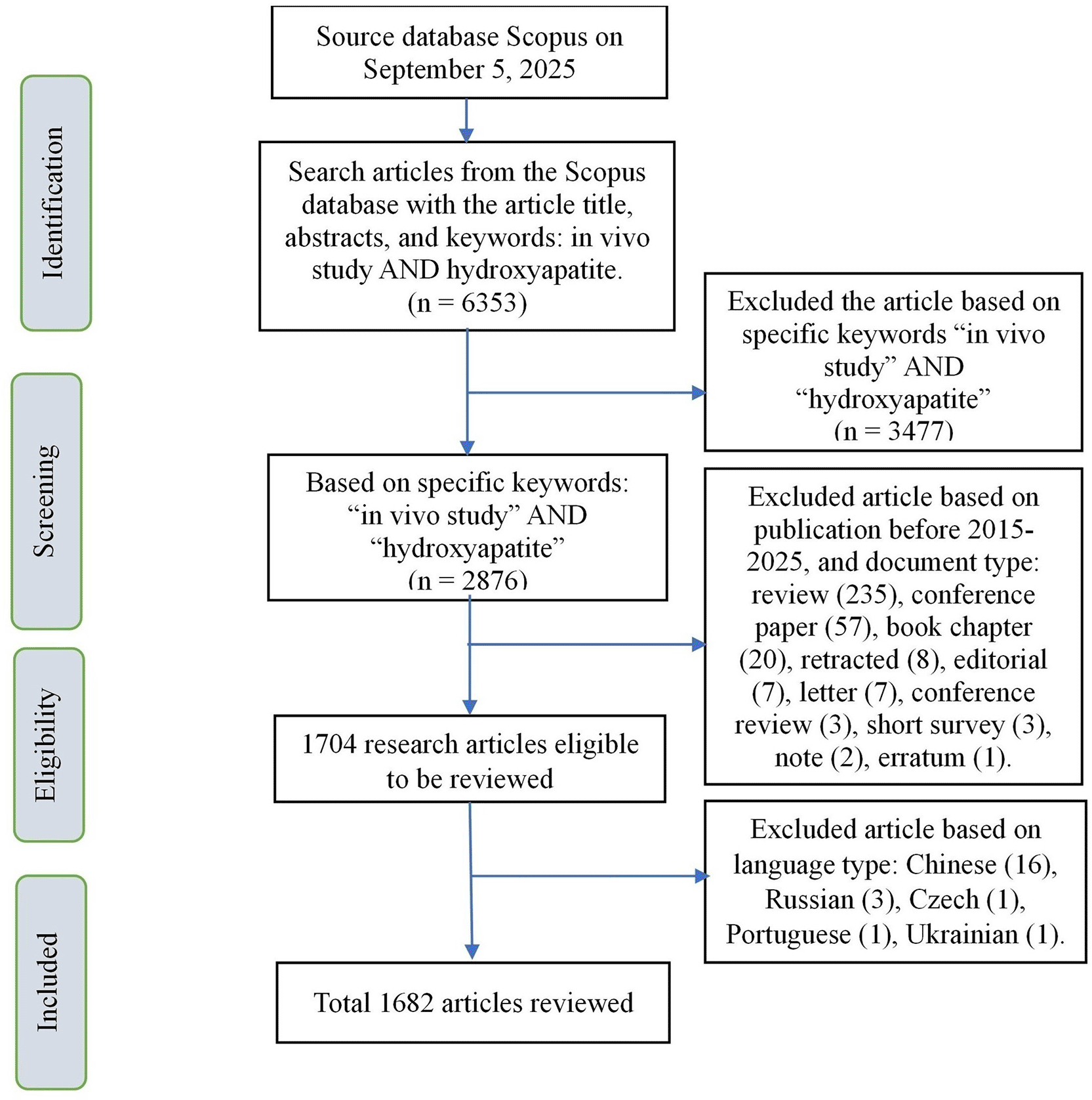
The initial phase entailed an identification process, during which an article search was performed in the Scopus database utilizing the keywords: in vivo study AND hydroxyapatite, focusing on the article title, abstract, and keyword section. The resultant documents totaled 6,353 records. Subsequently, article screening was conducted using the specific keywords: “in vivo study” AND “hydroxyapatite”. The specified keywords yielded 2,876 documents in total. The search for papers was subsequently restricted to those published between 2015 and 2025. Furthermore, document types including reviews, conference papers, book chapters, retracted editorials, letters, conference reviews, short surveys, notes, and errata were excluded. Consequently, 1704 eligible articles were retrieved. Additionally, articles in Chinese, Russian, Czech, Portuguese, and Ukrainian were excluded, resulting in the exclusive use of texts written in English. A total of 1,682 studies were included in the review.
In this study, a bibliometric analysis was performed using VOSviewer. Co-authorship analysis was applied to map collaborative networks among authors, institutions, and countries, highlighting the scientific partnerships that drive research development in this field. Co-citation analysis was conducted to reveal the intellectual structure of the domain by identifying journals and authors most frequently cited together, reflecting the theoretical foundations underpinning HAp research. Bibliographic coupling analysis was employed to assess similarities among documents, institutions, or countries based on shared references, which helped uncover research directions and thematic linkages across scientific entities. Finally, keyword co-occurrence analysis was used to map thematic clusters and emerging research trends, particularly in relation to scaffolds, osseointegration, ionic doping, and composite applications in in vivo HAp studies.18–22
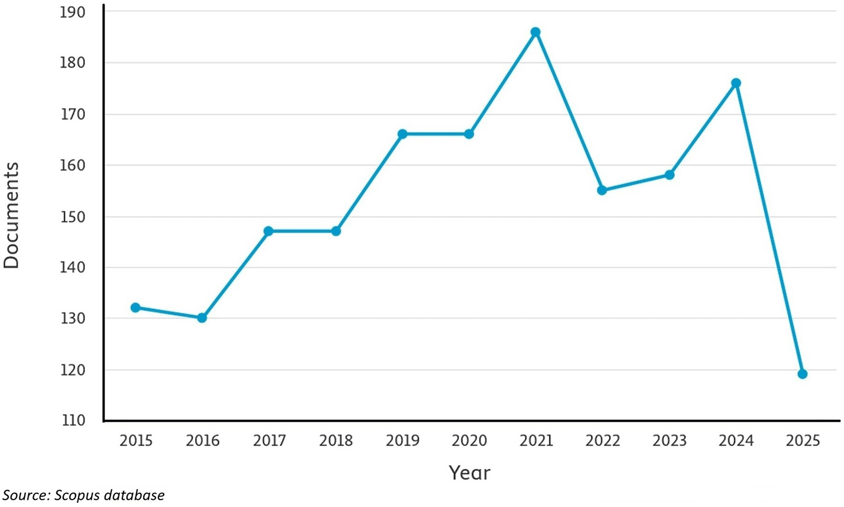
The conclusions of this study are based on the findings of 1,682 papers sourced from the Scopus database regarding in vivo investigations of HAp. Research from 2015 to 2025 indicated an upward trend until 2021 (186 documents), followed by a decline in 2022 (155 documents). Research escalated once more until 2024, yielding 176 documents, and then declined in 2025, resulting in 119 documents. This indicates that research on in vivo investigations of HAp remains limited, suggesting that it is a viable area for future studies ( Figure 2).
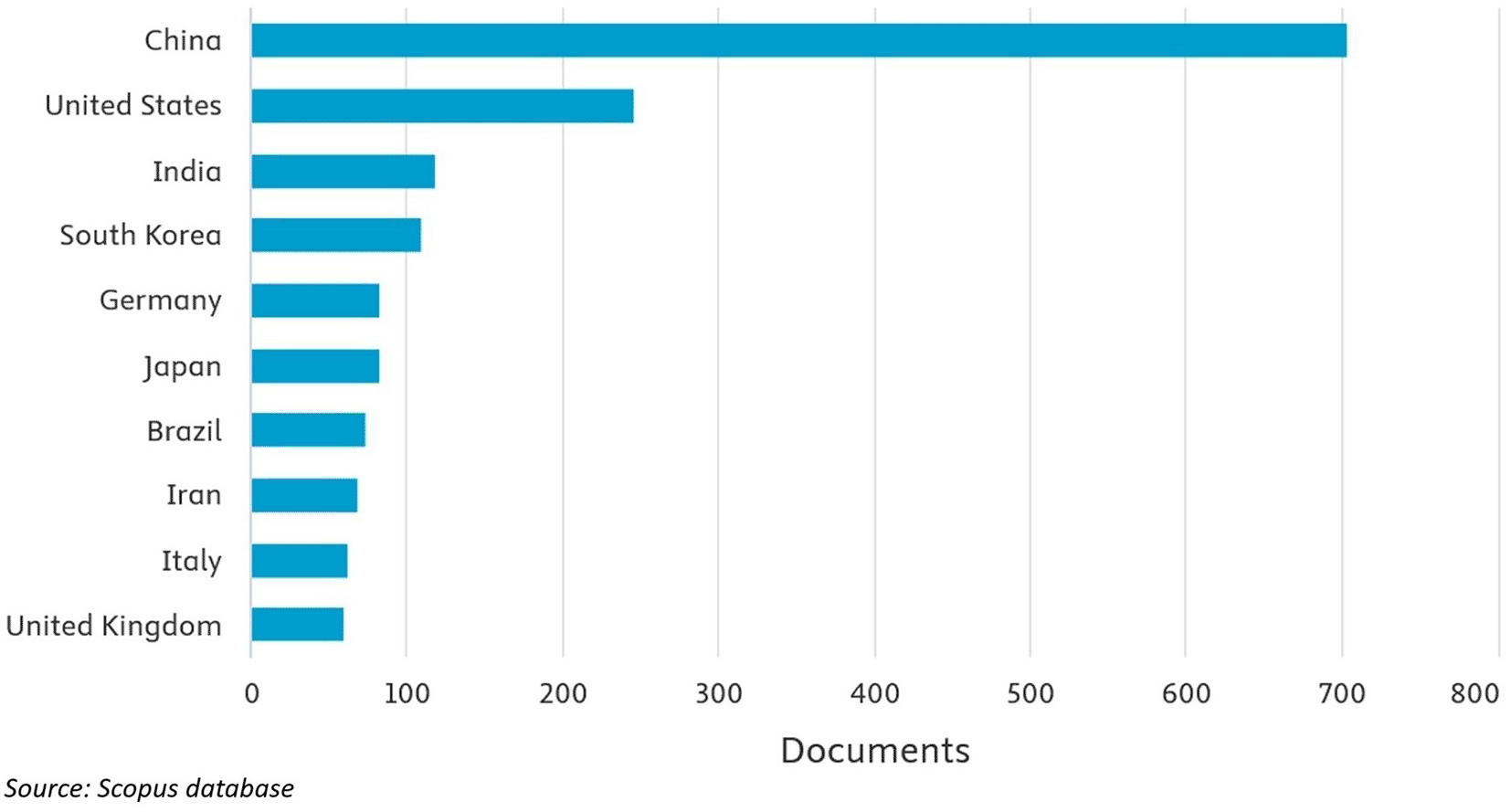
Analysis of international collaboration, visualized through VOSviewer using bibliographic coupling and focusing on countries, revealed that research publications were predominantly authored by China (704 documents), followed by the United States (245 documents), India (118 documents), South Korea (109 documents), Germany (83 documents), Japan (82 documents), Brazil (73 documents), Iran (68 documents), Italy (60 documents), and the United Kingdom (60 documents), as illustrated in Figure 3.
In recent years, research publications have been predominantly authored by the United Arab Emirates (2 documents) in 2025, Croatia (2 documents) in 2025, Iraq (3 documents) in 2023, Thailand (11 documents) in 2022, Colombia (5 documents) in 2022, Vietnam (4 documents) in 2022, and Tunisia (3 documents) in 2022. The findings indicate that the international research network focused on in vivo studies of HAp is expanding, resulting in new investigations in various nations. Research collaboration with China has yielded significant contributions from various countries, including the United Arab Emirates, Vietnam, Thailand, Iraq, Bosnia and Herzegovina, Ukraine, Colombia, and Croatia. Research collaboration with the United States yielded significant contributions from various countries, including Tunisia, Iraq, Vietnam, Thailand, Bosnia and Herzegovina, Croatia, and Colombia. Research collaboration with India has yielded significant contributions from several countries, including Vietnam, Thailand, and Colombia. Furthermore, this finding reinforces that research on in vivo HAp has emerged as a significant focus in numerous countries, as illustrated in Figure 4.
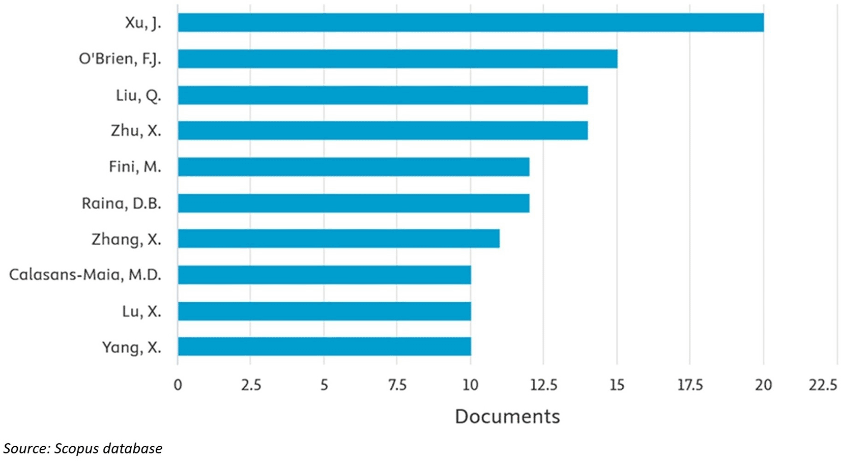
The top ten authors with the highest publication counts are Xu, J. (20 documents), O’Brien, F.J. (15 documents), Liu, Q. (14 documents), Zhu, X. (14 documents), Fini, M. (12 documents), Raina, D.B. (12 documents), Zhang, X. (11 documents), Calasans-Maia, M.D. (10 documents), Lu, X. (10 documents), and Yang, X. (10 documents). This is illustrated in Figure 5.
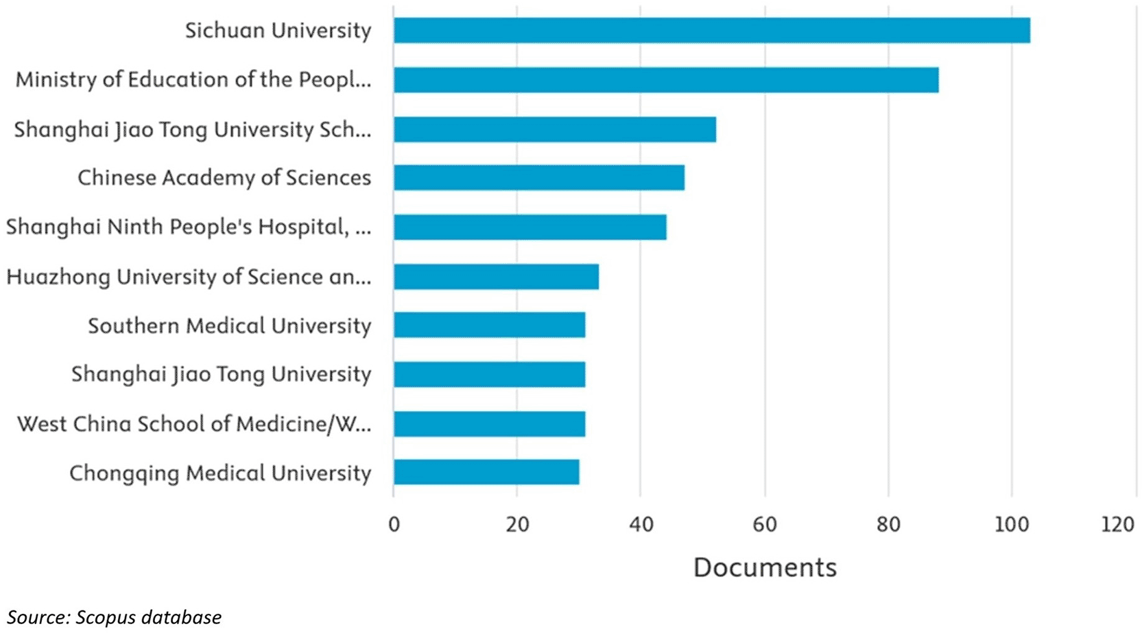
The in vivo research on HAp, based on institutional affiliation, reveals that the leading contributors in the top 10 positions are Sichuan University (103 documents), Ministry of Education of the People’s Republic of China (88 documents), Shanghai Jiao Tong University School of Medicine (52 documents), Chinese Academy of Sciences (47 documents), Shanghai Ninth People’s Hospital, Shanghai Jiao Tong (44 documents), University School of Medicine (44 documents), Huazhong University of Science and Technology (33 documents), Southern Medical University (31 documents), Shanghai Jiao Tong University (31 documents), West China School of Medicine/West China Hospital of Sichuan University (31 documents), and Chongqing Medical University (30 documents), as shown in Figure 6.
Based on Figure 7, in the realm of in vivo research, the term “hydroxyapatite” is intricately associated with “osseointegration,” “biomechanics,” “calcium phosphate,” and “bone remodeling.” This link suggests that several in vivo investigations have focused on assessing the capacity of HAp to facilitate bone-implant integration,23–28 enhance mechanical strength,29–34 and contribute to bone remodeling and regeneration.35–39 In addition, the term “in vivo” is located in proximity to “titanium implants,” “coating materials,” and “rabbit models.” This demonstrates that in vivo experiments frequently utilize animal models, particularly rabbits, focusing on the clinical application of titanium implants covered with HAp or its derivatives.
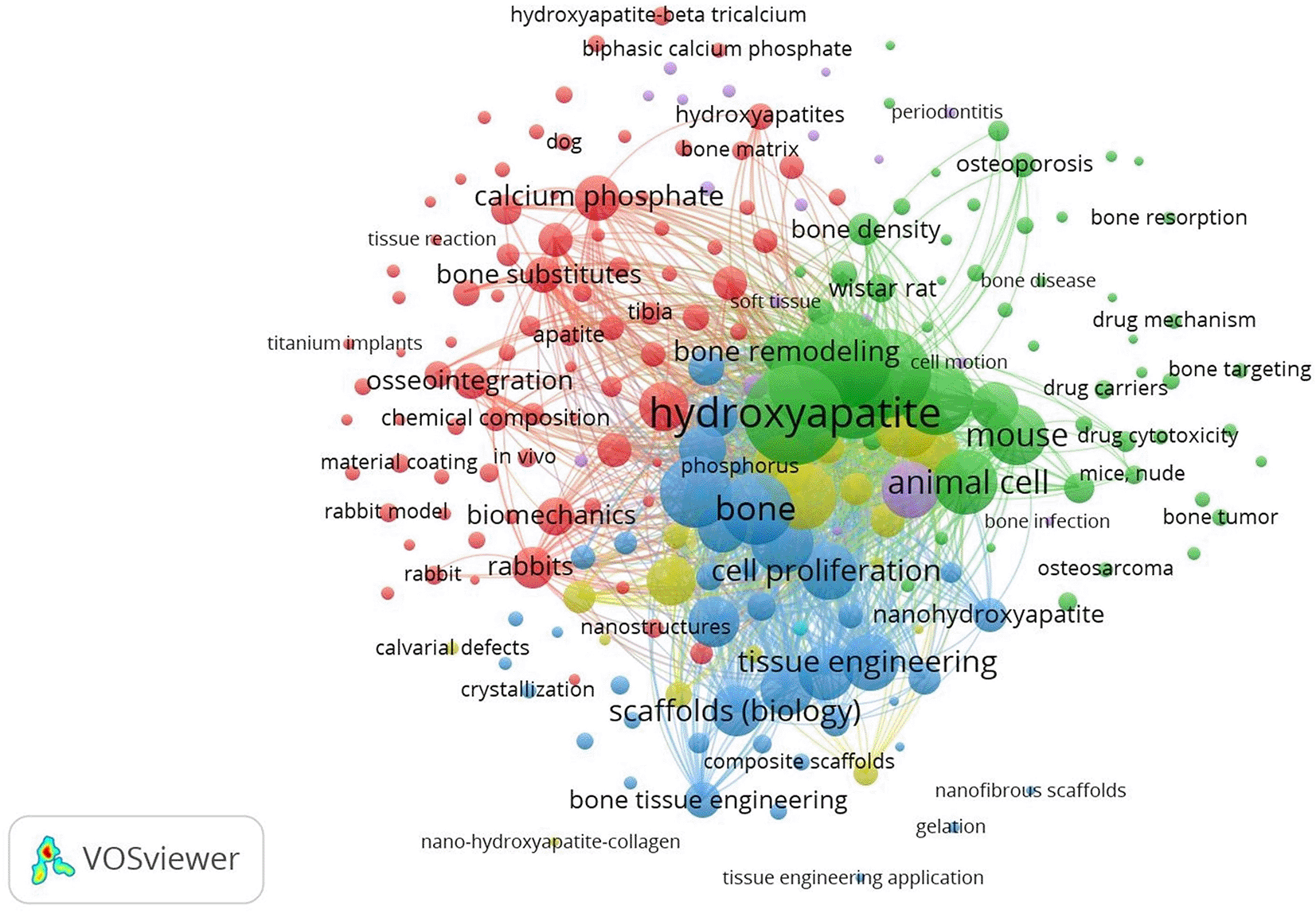
Next, the correlation between the terms “bone substitutes,” “tibia,” and “calvarial defects” indicates that in vivo research assesses both implants and the application of HAp as a bone replacement material in diverse anatomical sites.40–44 Furthermore, from a biological standpoint, the relationship between animal cells, cell proliferation, and nanohydroxyapatite (nHAp) indicates that in vivo studies should extend beyond macroscopic evaluations to examine the cellular biological response to the material, including at the nanoscale, which is thought to enhance biocompatibility.45–47
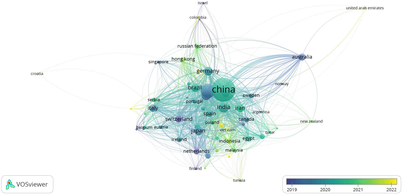
Figure 8 illustrates a highly centralized global collaboration network, predominantly led by the United States and China. The substantial node sizes and robust inter-country connections signify their preeminent role in the progression of HAp-based regenerative biomaterials, transitioning from conventional osteoconductive scaffolds to immunomodulatory platforms that facilitate bone regeneration, promote angiogenesis, and modulate macrophage polarization within the osteoimmune microenvironment.48–50
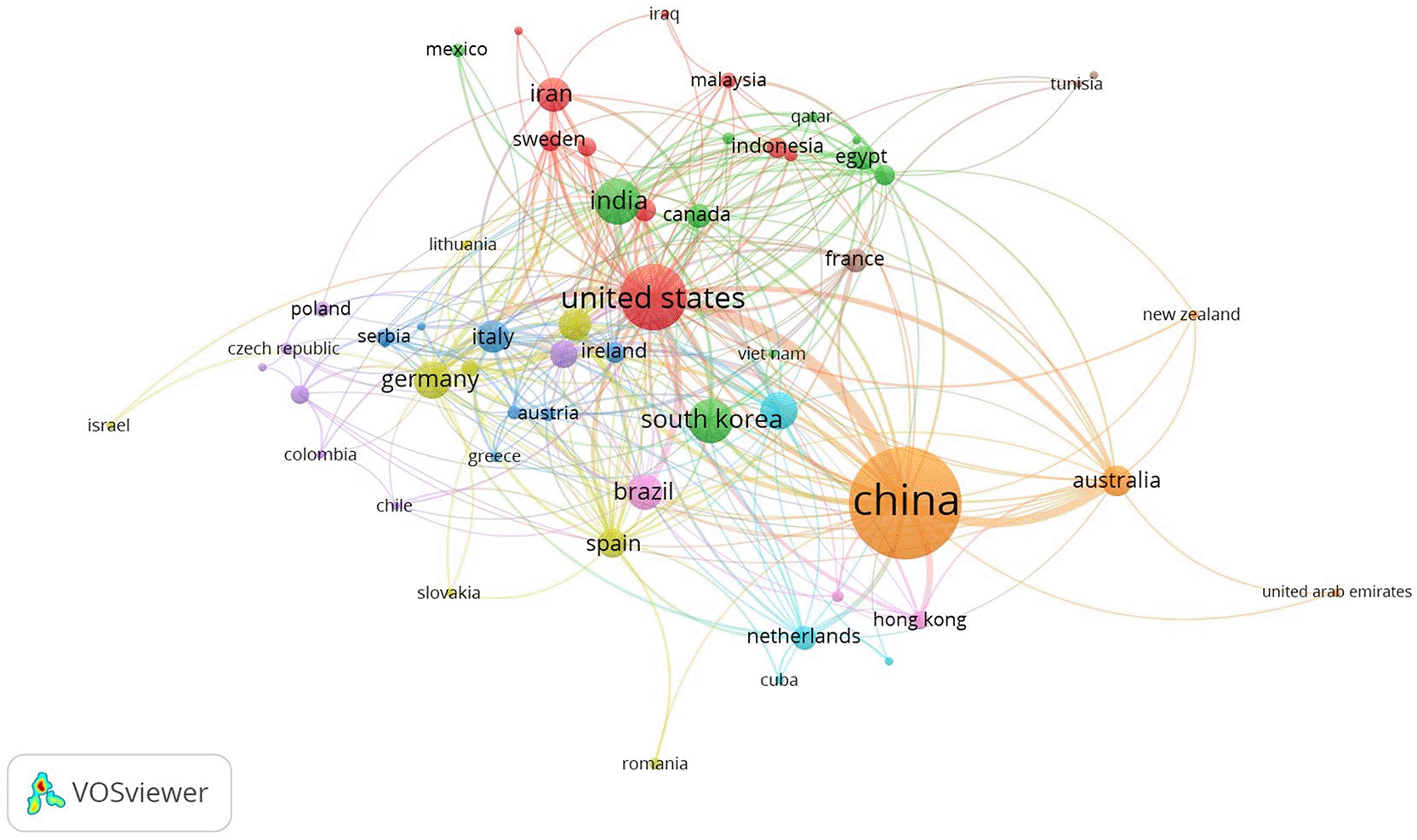
In Europe (Germany, Italy, France, and the Netherlands) and East Asia (China, South Korea, and Japan), dense regional clusters were also observed, suggesting developed research ecosystems with robust intra-regional collaboration. Because contemporary HAp biomaterials depend on interdisciplinary integration, including materials science, immunology, and bioengineering to regulate immune microenvironment remodelling and enhance implant stability and long-term regenerative outcomes, such coordinated collaborations are crucial.51
Crucially, emerging nations such as Egypt, Malaysia, Indonesia, Brazil, India, and others hold semi-peripheral positions, indicating their increasing yet still limited integration into global research networks with significant influence. This uneven allocation of collaboration capacity is crucial because new research shows that effective bone regeneration depends on precise control of immune signalling, macrophage phenotype, and ion-mediated microenvironment modulation in addition to scaffold structure. These processes necessitate sophisticated infrastructure and international cooperation.52
A small number of strongly connected innovation hubs dominate HAp research, according to the network topology overall, while new regions gradually become part of the global osteoimmunology research ecosystem. It will be crucial to improve clinical translation and expedite the development of next-generation immunomodulatory HAp biomaterials by fortifying international cooperation, especially with emerging nations.
According to journal sources that published the highest number of documents on in vivo HAp studies from 2015 to 2025, including Acta Biomaterialia, International Journal of Nanomedicine, ACS Biomaterials Science and Engineering, and Journal of Biomedical Materials Research - Part B Applied Biomaterials, International journal of biological macromolecules, Biomedical Materials (Bristol), Biomaterials, Colloids and Surfaces B: Biointerfaces, and Biomaterials Science. The top ten journals with the most substantial publications are listed in Table 1. The most frequently referenced journal was Acta Biomaterialia, with 4858 citations and an SJR of 2.007, classified in the first quartile (Q1).
| Rank | Source title | Number of documents | Number of citations | Pubisher | SJR 2024 | Scopus percentile (Q) |
|---|---|---|---|---|---|---|
| 1 | Acta Biomaterialia | 76 | 4858 | Acta Materialia Inc. | 2.007 | Q1 |
| 2 | International Journal of Nanomedicine | 57 | 1983 | Dove Medical Press Ltd. | 1.306 | Q1 |
| 3 | ACS Biomaterials Science and Engineering | 56 | 1506 | American Chemical Society | 1.105 | Q1 |
| 4 | Journal of Biomedical Materials Research - Part A | 49 | 1301 | John Wiley and Sons Inc. | 0.812 | Q1 |
| 5 | Journal of Biomedical Materials Research - Part B Applied Biomaterials | 49 | 1065 | John Wiley and Sons Inc. | 0.737 | Q2 |
| 6 | International journal of biological macromolecules | 45 | 953 | Elsevier B.V. | 1.285 | Q1 |
| 7 | Biomedical Materials (Bristol) | 43 | 904 | IOP Publishing Ltd. | 0.716 | Q2 |
| 8 | Biomaterials | 41 | 3220 | Elsevier Ltd. | 2.998 | Q1 |
| 9 | Colloids and Surfaces B: Biointerfaces | 41 | 1449 | Elsevier B.V. | 0.978 | Q1 |
| 10 | Biomaterials Science | 39 | 1087 | Taylor and Francis Ltd. | 1.215 | Q1 |
HAp is a calcium phosphate with a composition analogous to bone mineral,53 rendering it extremely biocompatible and bioactive,54–56 making it an optimal choice for bone graft and bone tissue engineering applications.4,5,57–59 in vivo investigations were conducted to evaluate HAp’s capacity to facilitate bone remodeling and regeneration by observing new bone formation, osseointegration, and mechanical integrity.60–65 in vivo studies on HAp and its applications are summarized in Table 2.
| Material type/modification of HAp | In Vivo model | Purpose/application | Main results | Reference |
|---|---|---|---|---|
| HAp-carbon nanotube (CNT) biocomposites | Bone defect of the rabbit femur | Bone implant | The biomechanical strength of the implant is increased. | 35 |
| Biomimetic bone composite hydrogel scaffold (BBCHS) | Rat cranial defect model | Bone repair | BBCHS exhibits strong biocompatibility and osteogenic properties. | 55 |
| Composite of poly (amino acid), HAp, and calcium sulfate (PAA/HAp/CS) | The ulna of sheep | Load-bearing bone substitutes | The composite exhibits good bioactivity, biocompatibility, and osteoconductivity in vivo. | 56 |
| Ultrathin PBAT, PBAT/nHAp, and PBAT/nHAp/GNR fibers | Critical tibia defects in rats | Bone grafts | The capability of scaffolds for bone regeneration. | 57 |
| PDLLA/VACNT-O:nHAp scaffolds | Mice | To evaluate the bone regeneration | The PDLLA/VACNT-O:nHAp scaffolds emulated immature bone and stimulated bone remodeling. | 63 |
| Beta-tricalcium phosphate and HAp with a unique unidirectional porous structure. | Animal experimentation and clinical uses | Bone graft and Clinical applications. | Both artificial bones, Affinos® and Regenos®, with a unidirectional porous structure, are appropriate for the majority of orthopedic surgical procedures. | 71 |
| Fluorinated porcine hydroxyapatite (FPHA) | Critical size calvarial defects in Sprague-Dawley rats | As a bone substitute | Consequences for the advancement of fluoride-modified immunometabolism-based bone regeneration biomaterials and the therapeutic utilization of FPHA or other fluoride-containing substances. | 72 |
| PCLLA-nanoHAp | Diabetic mice | Bone substitute | PCLLA-nanoHA promoting alveolar bone regeneration. | 73 |
| Ti-Zr and Ti-HA nanocomposite-coated Mg | Wistar rat model | Progressive bone regeneration | Ti-Zr and Ti-HA nanocomposite-coated Mg implants provide superior corrosion resistance, mechanical stability, and biocompatibility. | 74 |
| Polyamide 66/nano-hydroxyapatite (PAHA) scaffold | Rabbit femoral defects | Bone regeneration | The 3D-printed Gel/PTH@PAHA scaffold has superior mechanical capabilities and significant osteogenic activity, fulfilling the dual criteria for load-bearing applications and bone regeneration. | 75 |
| nd-CPC paste containing tetracalcium phosphate and anhydrous dicalcium phosphate | In rabbit femurs | To fill regeneration defects | nd-CPC paste did not elicit an inflammatory reaction post-implantation and exhibits enhanced remodeling activity. | 76 |
| L-pNIPAM co-DMAc loaded with HAPna and MSCs | Rat femur defect model | Bone regeneration for the management of minor osseous problems | The injectable hydrogel is biocompatible, capable of integrating with adjacent bone tissue and enhancing the deposition of early markers of bone development. | 77 |
Biologically, HAp demonstrates osteoconductivity, defined as its capacity to facilitate the migration of osteoprogenitor cells, angiogenesis, and deposition of the mineral matrix.6 The application of HAp coating on titanium implants enhances bone-to-implant contact (BIC) and pullout strength in a rabbit model.8,66 The amalgamation of HAp with bioactive agents, including bone morphogenetic proteins or ion doping (e.g., copper, gadolinium, and zinc), has been documented to enhance the expression of osteogenic and angiogenic markers, expediting bone healing.9,10
Furthermore, nHAp with interconnected porosity has demonstrated superior bone ingrowth and BV/TV ratios in critical defect models relative to traditional HAp.4,67 In addition, HAp–polymer composites (e.g., HAp-PLGA and HAp-chitosan) enhance mechanical characteristics and regulate degradation, thereby facilitating more stable bone regeneration.10 In addition, the use of HAp coating on metallic surfaces enhances osseointegration and expedites new bone growth surrounding implants.8,68 Biphasic calcium phosphate (BCP), comprising HAp and beta-tricalcium phosphate (β-TCP), achieves an equilibrium between resorption and new bone formation, which is crucial for sustained bone remodeling.12,69 Moreover, HAp loaded with medicinal compounds, such as statins or antimicrobial compounds, exhibits a dual function of promoting osteogenesis and mitigating infection risk.13,14,70
Next, in vivo investigations employ mice, rabbits, or lambs based on the translational objective.4,8 Evaluation is conducted using micro-computed tomography (micro-CT) to quantify bone volume, histomorphometry to evaluate BIC and the proportion of new bone, and biomechanical testing (push-out or pull-out test) to determine functional strength.6,8,12 Certain studies have employed immunohistochemistry to examine the expression of osteogenesis markers (RUNX2 and osteocalcin) and angiogenesis markers (VEGF).10,13
Research reviews consistently demonstrate that nHAp and HAp-polymer composites enhance bone bridging, elevate new tissue quality, and offer superior mechanical integration compared with controls lacking HAp.4,9,10 Ionic doping of HAp (e.g., Cu/Gd) leads to substantial enhancement of vascularization and osteoblast differentiation.9 Simultaneously, tailored BCPs yield a resorption rate that aligns with the rate of new bone production, thereby diminishing the likelihood of regeneration failure.12
HAp has been predominantly viewed as a passive osteoconductive material, functioning as a bone mineral analogue that promotes cell adhesion, matrix deposition, and new bone integration owing to its chemical compatibility and surface microarchitecture.40,78,79 Diverse HAp composite scaffolds and their modifications have shown the ability to enhance osteogenesis both in vitro and in vivo by optimizing mechanical properties, degradation rates, and bioactivity.56,80,81 Recent evidence indicates a substantial paradigm shift: HAp is not merely an osteoconductive substrate but functions as an active modulator of the immune response during the initial phase of bone healing. Furthermore, it was found that β-tricalcium phosphate particles can activate the NLRP3 inflammasome complex and enhance immune cell migration in vivo, indicating that calcium phosphate biomaterials directly interact with the innate immune pathway and influence inflammatory dynamics.82
The primary advancement is HAp’s capacity to systematically influence macrophage polarization (M1 → M2) via immunometabolic pathways. Furthermore, fluorinated HAp enhances a conducive osteo-immune milieu by inducing a metabolic transition in macrophages from glycolysis to oxidative phosphorylation, associated with the stabilization of the pro-regenerative M2 phenotype. This transition is not solely a phenotypic occurrence but offers insight into osteoblast differentiation and novel bone creation.72 Moreover, HAp-coated small intestinal submucosa membranes promote periodontal regeneration via immunomodulation and the activation of the BMP-2/Smad osteogenic pathway, thereby reinforcing the notion of osteoimmunomodulation as a cohesive mechanism linking immune regulation and bone formation.83
At the biological interface level, various HAp-based scaffold designs demonstrate that successful bone regeneration is significantly influenced by the quality of interaction at the immune–bone interface, rather than merely by the material’s intrinsic osteoconductive properties.84–86 Alterations to implants designed to avert infection and enhance osseointegration84 and HAp-based antimicrobial delivery systems87 demonstrate that controlling the local inflammatory burden is crucial for facilitating the shift to the regenerative phase. Thus, the immune response is currently considered not only a secondary consequence of implantation but also a controllable factor through the chemical and surface engineering of HAp.
These studies confirm a paradigm shift from HAp as a passive osteoconductive material to an osteoimmunoengineering platform that actively modulates inflammasome signaling, promotes the M1 to M2 macrophage transition, and orchestrates immune-bone interactions to improve regeneration. This conceptual shift positions HAp within regenerative immunology, where biomaterial design prioritizes the modulation of the immune microenvironment as a crucial element in successful bone reconstruction, rather than solely concentrating on the mechanics and bioactivity of minerals.
HAp is a pivotal biomimetic material for bone replacement applications owing to its mineral composition, which closely resembles the inorganic phase of human bone, and its capacity for bioactive interactions with bone tissue. Preclinical in vivo studies are essential for evaluating the efficacy of HAp as a bone substitute, specifically regarding its capacity to promote osteogenesis, achieve osseointegration with the host bone, and restore mechanical functionality in bone defects or reconstructions.4–6,88
In vivo investigations elucidating the fundamental mechanisms behind HAp’s function as a bone-replacement material encompass the following: First, Osteoconduction: HAp offers a porous scaffold that promotes the migration of osteoprogenitor cells and the deposition of the mineral matrix; micro-computed tomography (μCT) and histological analyses in several studies illustrate bone ingrowth from the scaffold’s periphery towards its core.5,88 Second, surface osseointegration: HAp coating on titanium or other substrates enhances bone-to-implant contact (BIC) and mechanical fixation (pull-out/push-out) in rabbit and sheep models.9,89 Third, biological modulation by modification: Ionic doping (e.g., Sr, Cu, Gd, Zn) and loading factors (BMPs, statins, antimicrobial drugs) can augment osteoinduction and angiogenesis when the release of biomolecules/ions is regulated.10,13,14
In vivo investigations have evaluated diverse HAp formulations, including nHAp/3D-printed porous HAp scaffolds. Scaffolds produced through 3D printing methods or biomimetic approaches exhibit accelerated bone bridging and enhanced BV/TV in crucial defects; the primary issue lies in achieving a balance between porosity (to facilitate vascularization) and mechanical strength.5,6,88–90 Second, HAp–polymer composites (PLGA/PLA/collagen/chitosan) mitigate the brittleness of ceramic HAp, provide controlled degradation, and frequently demonstrate superior in vivo outcomes (vascularization and matrix quality) compared to isolated HAp.91,92 Third, HAp coatings, specifically nanostructured HAp coatings, enhance bone-implant contact (BIC) and biomechanical properties in in vivo assessments, proving especially beneficial in low-density bone scenarios.9,89 Fourth, biphasic calcium phosphate (HAp + β-TCP) modulates the resorption rate to coordinate with new bone formation, thereby enhancing long-term remodeling.12,93 Fifth, HAp serves as a drug/therapeutic carrier: HAp is optimized for the localized administration of statins, antibiotics, or anticancer drugs (e.g., 5-fluorouracil), thereby integrating osteoconductive properties with localized treatments. In vivo, it has dual therapeutic advantages without considerable systemic effects when the release is regulated.13,14 in vivo studies on HAp and its applications are shown in Table 3.
| Material Type/modification of HAp | In Vivo model | Purpose/application | Main results | Reference |
|---|---|---|---|---|
| Biphasic calcium phosphate (β-TCP/HAp ratio optimally) | Rabbit (femur defects) | Bone replacement by hard tissue engineering. | In vivo investigations on rabbit femur lesions exhibited substantial bone repair, with bone-to-tissue volume ratios surpassing 50% within four weeks. | 12 |
| Substituted HAp and TCP (ZnMgSi-HAp and ZnMgSi-TCP) | Implanted into the rat’s skull bones | Osteogenesis promoting | The doping of HAp and TCP with silicon, zinc, or magnesium ions expedites the production of new bone tissue. | 44 |
| Microporous granular calcium phosphate (HAp/β-TCP) | in the sheep tibia | Evaluation of behavioral development in bone | Microporous HAp expedites bone growth and improves osseointegration. | 94 |
| HAp coating with ZnO, SiO2, Ag2O dopants (plasma spray) | Sprague-Dawley rats | orthopaedic or dental applications | Enhanced mechanical strength and antibacterial characteristics while maintaining bioactivity. | 95 |
| 3D printed HAp scaffolds | a rat tibial defect model | Utilization of 3D-printed HAp scaffolds for osseous regeneration | The 3D printed HAP scaffolds used in this study provide an appropriate matrix for facilitating patient-specific and defect-specific bone growth and require more testing for therapeutic applicability. | 96 |
| carbonate apatite (CO(3)Ap) block from DCPD | Rabbit (defects in the femur & tibia) | Bone defect reconstruction | CO3 Ap blocks possess significant potential as optimal artificial bone substitutes owing to their resorption and subsequent replacement by natural bone. | 97 |
| Composite scaffolds of porous HAp whisker-reinforced poly(L-lactide) (HAp-w/PLLA) with varying proportions of HAp and PLLA | male rats | Biodegradable bone scaffold | Porous HAp whisker-based biodegradable scaffolds for bone repair, replacement, and augmentation applications. | 98 |
| HAp and Ag@HAp scaffold (plasma spraying technique) | Rabbit (knee joint anterior cruciate ligament) | Rehabilitation and replacement of the anterior cruciate ligament of the knee joint. | The HAp and Ag@HAp scaffold inhibited bone resorption and enhanced the biomechanical integrity of the bone–joint complex, hence promoting osseointegration. | 99 |
| Crosslinked porous composite comprising collagen and HAp | New Zealand White Rabbit (NZWR) | Preparing biocompatible bone repair materials | The material demonstrated significant biocompatibility and effectively stimulated osteogenesis in vivo. It possesses significant commercialization potential and functions as an efficient bone-repairing material. | 100 |
| Fibrous PVA-HAp/Sr matrix composed of strontium (Sr)-substituted HAp. | Mouse | Bone tissue engineering scaffolds | Incorporating HAp or HAp/Sr nanoparticles into PVA and then electrospinning yields hybrid fibrous matrices of PVA-HAp or PVA-HAp/Sr with favorable characteristics. Also, the matrices containing HAp or HAp/Sr inclusion demonstrated significant bioactivity and enhanced cell survival and proliferation. | 101 |
| f-CDs-PE-Au@HAp biocomposite coatings; carbon dot-modified polyethylene (f-CDs-PE) and gold-doped hydroxyapatite (Au@HAp) bioceramic. | Rats | Shoulder joint replacement surgery | The f-CDs-PE-Au@HA biocomposite (BC2) exhibited significant bioactivity and cytocompatibility, facilitating the integration of collagen and the organization of ligament and hard tissue. The use of gamma-ray irradiation with f-CDs-PE-Au@HA biocomposite coating (BC2) at the tendon-hard tissue interface was demonstrated to enhance the healing of entheses and promote the growth of hard tissue and tendon. | 102 |
| Silver oxide-infused hydroxyapatite coating (Ag-HAp) | Hip osteoarthritis in an octogenarian female | The management of septic arthritis of the hip joint utilizing a cementless hip implant with antibacterial characteristics. | Ag-HAp implants demonstrated antibacterial activity in subcutaneous tissues and bone, exhibited osteoconductive characteristics, had no adverse effects, and showed no negative effects attributable to silver. | 103 |
| Triphasic nanostructured bioceramic (The CaNb2O6, PNb9O25 and Ca3(PO4)2 phases) | Calvaria of rats | Bone substitutes for applications requiring high and medium load-bearing capacity | The nanostructured biocomposite exhibited observed values for compressive strength (242 ± 29 MPa), Young’s modulus (19.63 ± 3.5 GPa), Poisson’s ratio (0.248 ± 0.016), and flexural strength (24 ± 5.9 GPa) comparable to some human bones. Histological analysis revealed osseointegration between the newly produced bone and the triphasic bioceramic merely 45 days post-implantation. | 104 |
| Composite coating on 316 L stainless steel using Direct Current Electrophoretic Deposition (EPD) | Rabbits (in the tibial bone) | Augment the osseointegration of 316 L stainless steel implants. | The enhanced osteoblast proliferation and mature bone creation, along with the biocompatibility and efficacy of the composite covering in stimulating bone growth, have demonstrated an improvement in the long-term performance of orthopedic implants by boosting bone formation and assuring stable fixation within the bone. | 105 |
| HAp-based solid cementitious material | rats | Evaluation of resorption of bioceramic grafts | Develop bone substitute materials exhibiting more reliable resorption characteristics. | 106 |
HAp has transitioned from a passive mineral carrier to a stimuli-responsive therapeutic platform that may convert problematic microenvironmental cues into programmed therapeutic responses. In pH-responsive release systems, an acidic inflammatory milieu serves as a trigger for regulated drug release, exemplified by HAp-based dual-responsive hydrogels for rheumatoid arthritis treatment, where microenvironmental circumstances govern local release kinetics.107 In the realm of infection-induced drug release, systems that respond to biofilms or reactive oxygen species (ROS) facilitate therapeutic activation solely upon an escalation of the infection burden, as evidenced by biofilm-targeting catalytic nanozymes108 and ROS-responsive antibacterial hydrogels for chronic wounds.109 This method diminishes premature release and enhances therapeutic specificity.
Moreover, in tumor microenvironments, the precise engineering of enzymes (e.g., MMP-2) facilitates tumor-specific release or activation of therapies, while concurrently altering the immunosuppressive microenvironment.110 Strategies based on pH and light activation illustrate the potential of utilizing acidic conditions for selective catalytic therapy.111 In the osteo-immune setting, HAp alteration has demonstrated the ability to create more regenerative microenvironments by metabolically regulating immune cells,72 hence validating the notion that HAp serves as a microenvironment-adaptive material. HAp in smart systems signifies a transition to precise biomaterials that not only administer medications but also modulate biological responses according to local disease dynamics.
A bibliometric analysis of worldwide in vivo HAp research shows that the field is still heavily focused on structural endpoints like implant coatings, bone remodeling, and osseointegration, with a preponderance of physicochemical optimization over immune-regenerative mechanisms.1 Since most in vivo studies still rely on static histological or micro-CT endpoints without longitudinal immune profiling, the mechanistic understanding of immune-bone dynamics is limited, despite growing osteoimmunology evidence that HAp can actively regulate macrophage polarization and immunometabolic reprogramming to promote bone regeneration.112,113
Despite evidence that translational-scale models are crucial for simulating human immune responses and remodeling kinetics, bibliometric mapping further emphasizes the dominance of small-animal models with comparatively little large-animal osteoimmunology data.4,88 Furthermore, in contrast with recognized structural measurements like bone-implant contact and BV/TV, the lack of standardized immunological markers makes it impossible to compare and synthesize mechanisms across investigations.112 Finally, bibliometric trends show that AI-guided biomaterial optimization is still largely unexplored, and scaffold design still relies on empirical methods rather than predictive modeling, despite the rapid growth in material modifications such as nanostructuring, ionic doping, and composite scaffolds.1,114
All of these experimental and bibliometric results point to a crucial translational bottleneck: long-term immune profiling, large-animal validation, standardized immune endpoints, and AI-assisted biomaterial design are necessary to move HAp from an empirically optimized osteoconductive scaffold toward a predictive osteoimmunomodulatory biomaterial.
Despite the positive results, obstacles, including diversity in animal models, discrepancies in defect size, and variations in follow-up duration, may influence inter-study comparisons. Future research should focus on optimizing scaffold porosity, integrating HAp with growth agents, and conducting long-term evaluations in large animal models before initiating human clinical trials. Consequently, in vivo investigations have substantiated the efficacy of HAp (particularly nHAp, HAp-polymer composites, and nanoscale HAp coatings) as a viable bone-replacement material, as HAp promotes bone ingrowth, osseointegration, and mechanical recovery in many animal models. Alterations (ionic doping, biphasic composition, and drug loading) may improve therapeutic efficacy and optimize resorption rates; however, clinical use necessitates long-term data from extensive models, nanoparticle safety evaluations, and standardization of in vivo study methodologies.
This bibliometric analysis confirms that HAp research remains predominantly focused in China and the US, with major trends focused on nHAp, polymer composites, and ion doping. Long-term in vivo studies using large-scale models are still needed before clinical translation.
This study is a bibliometric analysis using existing publication data from the Scopus database and does not involve the collection of new data from living subjects. Therefore, the section on Ethics Approval and Consent to Participation is not applicable because this study does not involve human or animal participants.
All authors who contributed to this study have read and approved the final version of the manuscript. All authors have agreed to the publication of this article and have declared no conflicts of interest. All data and materials generated or analyzed during this study are included in this paper.
| Views | Downloads | |
|---|---|---|
| F1000Research | - | - |
|
PubMed Central
Data from PMC are received and updated monthly.
|
- | - |
Provide sufficient details of any financial or non-financial competing interests to enable users to assess whether your comments might lead a reasonable person to question your impartiality. Consider the following examples, but note that this is not an exhaustive list:
Sign up for content alerts and receive a weekly or monthly email with all newly published articles
Already registered? Sign in
The email address should be the one you originally registered with F1000.
You registered with F1000 via Google, so we cannot reset your password.
To sign in, please click here.
If you still need help with your Google account password, please click here.
You registered with F1000 via Facebook, so we cannot reset your password.
To sign in, please click here.
If you still need help with your Facebook account password, please click here.
If your email address is registered with us, we will email you instructions to reset your password.
If you think you should have received this email but it has not arrived, please check your spam filters and/or contact for further assistance.
Comments on this article Comments (0)