Keywords
low bone mineral density, Indian women, nutrition, multi-site quantitative ultrasound.
low bone mineral density, Indian women, nutrition, multi-site quantitative ultrasound.
Osteoporosis is recognised as a disease of mainly postmenopausal women, but decreased bone mineral density (BMD) and rapid bone loss can also occur in young adulthood, a disease often referred to as premenopausal osteoporosis.1 Attainment of peak bone mass in early adulthood represents a key factor in subsequent fracture risk, and inadequate skeletal development at this critical period could be a risk factor for osteoporosis later in life.2–3 A constellation of biological and lifestyle factors, including poor nutrition, sedentary behaviour, endocrine dysregulation, menstrual irregularities, gestational influences, and inadequate sun exposure, has been implicated in the diminution of BMD among young women.4–7 In clinical practice, a Z-score of −2.0 or lower is generally considered low for pre-menopausal women, especially when used in combination with other risk determinants.7 Recent epidemiological studies show that low BMD is becoming more common in young women, especially in Asian populations; the prevalence of reduced BMD in young Asian women between the ages of 20–40 years may already reach 15–30 percent.8–9 These findings highlight the increasing public health concern of early skeletal health evaluation and the need for the identification of modifiable determinants during the peak bone building period.8 Quantitative ultrasound (QUS) has become a pragmatic, cost-effective and radiation-free modality for community-based skeletal screening, particularly in resource-constrained settings.10 Multisite instruments, for instance, the Omnisense, assess bone properties at peripheral skeletal sites and show reasonable concordance with dual-energy X-ray absorptiometry in identifying persons with decreased BMD.10,11 Although QUS is not a replacement for diagnostic imaging, it is sufficiently portable and feasible to be used for large-scale epidemiological studies and early risk identification.10,12 Moreover, community and cohort data from South Asia today show that women at reproductive age often have low BMD with the associated high rate of vitamin D insufficiency and inadequate calcium intake, which may impair peak bone mass and increase the risk of long-term fracture.13–15 Consequently, multisite QUS has been gradually used in field-based epidemiological screening, due to its portability, affordability, and acceptable concordance with conventional densitometric methods, but has been defined as a screening rather than a diagnostic tool.11 In spite of the growing body of evidence from around the world, community-based studies on low BMD in young Indian women are still limited, particularly those that incorporate nutritional, reproductive, lifestyle and psycho-social determinants in a coherent analytical framework.8,10,11 Existing research in India has mainly focused on postmenopausal cohorts or reference BMD databases, without comprehensive risk factor evaluations in younger women.15,16
Therefore, the present study was conducted to determine the prevalence of low bone mineral density and identify its associated risk factors among Indian women aged 20–40 years using multisite quantitative ultrasound in a community-based cross-sectional design.
The study was conducted at Guru Nanak Dev University, Amritsar, India. The current study design was population-based and cross-sectional in nature. The study started on October 15th, 2015, and was completed on July 17th, 2025. A cluster randomized sampling method was adopted, using the probabilities proportional to size (PPS) criteria to collect samples. The studied sample population comprised both rural and urban women of different socio-economic categories, belonging to the age group of 20–40 years.
The study was approved by the Institutional Ethics Committee, Faculty of Sports Medicine and Physiotherapy, Guru Nanak Dev University, Amritsar, India (No-1319/SMP). The protocol was carried out in accordance with the relevant guidelines and regulations from the “Declaration of Helsinki”. A written informed consent explaining the purpose and procedure of the study, translated both in Hindi and Punjabi, was obtained from each participant. The consent for the study was drafted as per the Indian Council of Medical Research (ICMR) guidelines. This study was registered in the Clinical Trial Registry of India (CTRI/2015/10/006240) on October 6th, 2015.
The sample size was calculated to estimate the prevalence of low bone mineral density (BMD) in young Indian women. Based on previous Indian studies indicating a prevalence of approximately 10%, and using a 95% confidence level with a relative precision of ±25%, the initial required sample was 540. Considering that data were collected across nearly 50 clusters, a design effect of 2.0 was applied to account for potential intra-cluster correlation, increasing the target to 1,080 participants. Ultimately, 1,176 women were recruited, exceeding the minimum requirement. This extended recruitment ensured adequate representation across age groups and geographic clusters, enhanced the precision of prevalence estimates, and strengthened the statistical power and generalizability of the findings.
Physically able women aged 20–40 years were included in the study. Pregnant and lactating women were excluded from the study. Women with a minimum gap of 6–9 months from the last delivery and lactation period were part of the study. Those who consented to participate were enrolled in the study.
The study methodology was precisely carried out as per the Strengthening the Reporting of Observational Studies in Epidemiology (STROBE) guidelines. Physical activity, a 24-hour dietary and self-designed questionnaire encompassing the anthropometric and socio-economic strata were collected. The self-designed questionnaire contained information related to factors that affect bone health.11 The questionnaire contained medical histories which included the questions on whether the participant was currently experiencing the following conditions: hyperparathyroidism, hypothyroidism, renal disease, diabetes mellitus, rheumatoid arthritis, eating disorders, medications that elevate bone density, history of back pain or not, and the question about whether the participant is or was undergoing glucocorticoids and/or thyroid hormone treatment. Another area explored by the interviewer was the obstetric and gynecological history of the participant, which included age at menarche and menopause (assuming it occurred), incidence of amenorrhea, number of offspring and pregnancies, length of breastfeeding, and whether the participant had undergone any surgical events or procedures, including oophorectomy or hysterectomy. There was also an investigation into the use of oral contraceptive pills and/or hormone replacement therapy. The next five questionnaires were used to collect information on socioeconomic status, family occupation, living expenditure, lifelong occupation, and educational attainment. Data on recreational activities and weight-bearing exercises were documented using a validated and translated Punjabi International Physical Activity Questionnaire (IPAQ). Perceived stress was measured using a local translation of a validated and translated Cohen Perceived Stress Scale (PSS-10) using the Punjabi local language. Each respondent was asked to give the time they spent under the sun every day, and a sun-exposure index was calculated according to the approach of Barger-Lux and Heaney (2002).3 The respondents also indicated their eating patterns through the 24-hour dietary recall technique.11
Bone health was assessed using a portable multisite quantitative ultrasound (QUS) device, the Sunlight Omnisense™ 7000S (BeamMed Ltd., Israel). Speed of Sound (SOS) measurements were taken at two peripheral skeletal sites, the distal one-third radius of the non-dominant arm and the mid-shaft tibia, after procedures to match those described by Shenoy et al. (2017).11 To ensure accuracy, measurements reproducible within a range of −2% of the expected calibration range were accepted daily to calibrate the device. The participants were sitting in a comfortable position as the palpation of the anatomical landmarks was carried out manually. Three successive readings were taken at each site, and the average of the three readings was taken to obtain the value of this mean or the average of the three values as near to the true value as possible. This multisite QUS protocol presented a viable and sound method of characterizing bone qualities in young adult women.11
Two physiotherapists, who had received structured training in the presence of an experienced supervisor, were used to carry out all QUS measurements. Their competence was first put to a pilot sample of 30 people. Inter-operator reliability was high, and the coefficient of variation was 1.8% at the radius and 2.1% at the tibia. Only those operators who passed a training stage and received more than 95 percent agreement in the training phase could carry out measurements during data collection to ensure consistency and accuracy.
The device automatically produced Z-scores based on sex- and age-specific reference data based on several populations of Asians. In line with the recommendations of the International Society of Clinical Densitometry (ISCD), a Z-score less than −2.0 was considered as low bone mineral density, whereas a Z-score below −3.0 was regarded as very low bone mineral density. The reason behind the choice of these cut-offs is to enhance the sensitivity of screening in women of the target age group of the present study, i.e., between 20 and 40 years.
Data was collected with a pre-established quality-control protocol that included daily checks of calibration, monitoring of signal quality indicators, and verification of the correct positioning of the anatomy during each scan. The entire data was entered in a two-entry procedure to reduce transcription errors. An automated check was used to find outliers, incomplete entries, or inconsistent responses. Records where anthropometric, QUS, or questionnaire data were missing were categorized as missing and were not included in statistical analyses.
The descriptive statistics in the study have been evaluated using the exact binomial method. For the multinomial logistic regression analysis, bone mineral density (BMD) was divided into three categories based on z-score values: (1) normal BMD (z-score ≥ −2.0), (2) low BMD (z-score between −2.0 and − 3.0), and (3) very low BMD (z-score ≤ −3.0). These thresholds are aligned with the recommendations of the International Society of Clinical Densitometry (ISCD) for premenopausal women and have been widely used in population-based studies to assess skeletal risk in younger adults. Categorizing BMD in this way allowed us to capture the gradual decline in bone health while ensuring sufficient sample sizes in each group to produce robust and meaningful statistical results. To analyze the relationship of categorical variables (PSS score, obstetrics and gynecological, categories of socio-economic status, physical activity level, nutrition variables, medical history, and anthropometric characteristics) with low bone mineral density, the chi-square test with Bonferroni’s correction was used. Bonferroni’s correction, p-value <0.025, was considered significant. To evaluate the baseline characteristics among different groups, one-way ANOVA with Tukey’s post hoc adjustment was used. Additionally, one-way ANOVA was used to draw a comparative analysis of the average mean of all nutrient variables among three groups. The Levene’s test was applied to check the homogeneity of variances for all variables across groups; when the test was significant, the Welch correction was used. Post-hoc comparisons were conducted using Tukey’s test when variances were equal, and Dunnett T3 when they were unequal. In addition, a multinomial logistic regression analysis using the force-entry method was performed to examine the independent association of various factors with reduced bone mineral density, with the normal BMD group serving as the reference category. To minimize model overfitting, the predictors entered into the model were selected based on biological relevance, bivariate correlations with p < 0.20, and prior evidence from young female cohorts. The final model included age at menarche, gravidity, hypothyroid status, perceived stress, socioeconomic status, daily sun exposure, and dietary calcium intake. Model fit was evaluated using the Hosmer–Lemeshow test, and multicollinearity was assessed through variance inflation factors (VIF < 2.5 considered acceptable). To reduce the likelihood of type I error, multiple comparisons were adjusted using the Bonferroni correction. Adjusted odds ratios (ORs) with 95% confidence intervals were reported, and a p-value <0.05 was considered statistically significant. All analyses were carried out using IBM SPSS Statistics version 26.0.
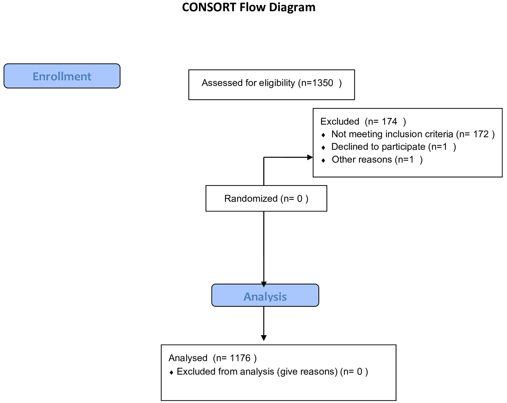
A total of 1,350 women were screened, of whom 1,176 participants met the inclusion criteria and were retained in the final analysis to find the prevalence of low bone mineral density conditions in the age group of 20–40-year-old women in the population of Punjab ( Figure 1).
For statistical analysis, the participants were categorized into three groups: normal (z score ≥ −2.0), z score < −2.0 and ≥ −3.0, and z score < −3.0.
Table 1 shows the baseline characteristics of the women participants. Both z-score categories had similar mean ages (~33 years). The mean values of anthropometric covariates (weight, waist circumference, waist: hip ratio) were higher in women having a z-score < −3.0. Further, in terms of obstetrics and gynecological covariates, the average menarche age was almost similar (~13 years) in all three groups. Significantly greater number of pregnancies (3.94), children (2.39), and prolonged duration of breastfeeding (14.41 months) was reported from women belonging to the z-score below −3.0 category in comparison to the normal group. In addition, significantly low mean physical activity level (488.41 METS-min/week), low sun exposure index (0.83), and high perceived stress (PSS) (25.67) were reported from women in the category of z score below −3.0 (p < 0.025). Moreover, women in the category of z score below −3.0 had significantly low mean SOS values of radius (3743.65 m/s) and tibia (3903.18 m/s), and low SES when compared to the mean values of the normal group (p < 0.025).
The results in Table 2 revealed that 83.9% of the participants had high waist circumference. 32.48% of the women from the total sample population had a complaint of low back pain. Out of 1176 women studied, 13 women had undergone the oophorectomy procedure; of them, 53.84% of the women had a z-score below −3.0. No woman had reported having undergone hormonal therapy. Only 0.5% of the women mentioned the history of osteoporosis as their medical history. 7.57% of the women reported rheumatoid arthritis as their medical history; of them, 71.15% of the women had z scores less than −2.0. 27.72% of the total sample population reported having more than 3 children, of which 41.71% of the women had a z-score < −2.0. Further, 9.27% of the women surveyed reported having consumed calcium supplements (minimum 2 months continuously); of them, 77.98% were in the normal category (Table-2).
The crude prevalence of low bone mineral density conditions: z score below −2.0) and z score below-3.0 was found to be 22.6% (95% CI, 20.3–25.1), and 8.1% (95% CI, 6.6–9.8), respectively.
Table 3 presents the distribution of bone mineral density (BMD) categories among the 1,176 women who participated in the study. Most women (69.30%) were classified as having normal BMD, indicating generally healthy bone status in this age group. Even so, a considerable number showed reduced bone density. About 22.61% fell into the low BMD range (Z-score − 2 to −3), suggesting early deterioration in bone strength. An additional 8.07% had very low BMD (Z-score < −3), placing them at the greatest risk for osteoporosis-related issues.
Table 4 highlights noticeable differences in dietary intake across the three BMD groups. Women with normal BMD were much more likely to fulfil their daily calcium requirements, and this relationship was statistically significant. This suggests that sufficient calcium intake plays an important role in maintaining healthier bone density. A similar trend was seen for energy intake, where women with very low BMD were less likely to meet the recommended calorie levels.
For several other nutrients, such as protein, magnesium, folic acid, iron, and vitamin C the proportions were relatively similar across all groups, indicating that these deficits are widespread in the population rather than specific to any BMD category. On the other hand, nutrients like niacin and riboflavin showed meaningful differences, with adequate intake observed more frequently among women with normal BMD.
In summary, the findings point to insufficient calcium intake as the most influential dietary factor associated with reduced BMD, while shortages of other micronutrients appear to be common across all women, regardless of their bone density status.
Table 5 presents the multinomial regression analysis results after adjusting for the confounding variables BMI, SES, exercise, history of osteoporosis, and stress) showed that more than 3 pregnancies and late menarche age were found to be significant risk factors of low bone mineral density (p < 0.001). The presence of hypothyroidism was positively associated with low bone mineral density conditions. Further, lifestyle variables (high perceived stress, low SES, low sun exposure) were also positively correlated with low z-score categories. In addition, it was found that an increase of every 10 unit increase in physical activity levels was found to be a significant protective factor in delaying the onset of low bone mineral density (p < 0.001).
| Very low BMD (3) (z score < −3) (n = 95) | Low BMD () (z score < −2 to - ≥ 3) (n = 266) | Over-all p-value | |||||
|---|---|---|---|---|---|---|---|
| Risk factor | N | OR (95% CI) | P-value | N | OR (95% CI) | p-value | |
| BMI (kg/m 2) | |||||||
| >23 | 53 | 1.38 (0.54–3.53) | 0.499 | 129 | 1.12 (0.61–2.01) | 0.727 | 0.792 |
| ≤23 | 42 | 1.0 | 137 | 1.0 | |||
| Number of pregnancies | |||||||
| ≤3 | 27 | 1.0 | 110 | 1.0 | |||
| >3 | 68 | 5.22 (2.16–12.63) | <0.001 | 156 | 1.89 (1.02–3.50) | 0.042 | 0.001 * |
| Hysterectomy | |||||||
| Absent (1) | 28 | 1.0 | 224 | 1.0 | |||
| Present (0) | 67 | 1.60 (0.20–12.89) | 0.661 | 42 | 1.46 (0.22–9.36) | 0.694 | 0.905 |
| Menarche age (years) | |||||||
| ≤14 | 50 | 1.0 | <0.001 | 86 | 1.0 | 0.074 | 0.001 * |
| >14 | 45 | 4.73 (2.06–10.82) | 180 | 1.71 (0.95–3.08) | |||
| Hypothyroid | |||||||
| Absent (1) | 80 | 1.0 | 248 | 1.0 | |||
| Present (0) | 15 | 3.84 (0.89–16.49) | 0.070 | 18 | 1.41 (0.47–4.22) | 0.545 | 0.130 |
| Hyperthyroid | |||||||
| Absent (1) | 77 | 1.0 | 239 | 1.0 | |||
| Present (0) | 18 | 4.48 (1.12–17.97) | 0.034 | 27 | 1.59 (0.52–4.86) | 0.416 | 0.048 |
| Diabetes | |||||||
| Absent (1) | 88 | 1.0 | 258 | 1.0 | |||
| Present (0) | 7 | 0.49 (0.05–5.23) | 0.554 | 8 | 0.78 (0.12–5.00) | 0.796 | 0.807 |
| Hypertension | |||||||
| Absent (1) | 69 | 1.0 | 225 | 1.0 | |||
| Present (0) | 26 | 3.05 (0.97–9.52) | 0.055 | 41 | 1.43 (0.62–3.29) | 0.398 | 0.134 |
| Perceived Stress | |||||||
| Upto High | 91 | 1.0 | 129 | 1.0 | |||
| Very high | 4 | 7.87 (2.26–27.36) | 0.001 | 78 | 5.30 (2.63–10.67) | <0.001 | <0.001 * |
| Sun Exposure | |||||||
| Low | 69 | 6.43 (2.81–14.72) | <0.001 | 127 | 2.87 (1.61–5.12) | <0.001 | <0.001 * |
| Optimum +high | 26 | 1.0 | 139 | 1.0 | |||
| Physical-Activity (METS) | |||||||
| Per 10-unit increase | 0.40 (0.34–0.46) | <0.001 | 0.52 (0.46–0.58) | <0.001 | <0.001 * | ||
Multiple regression analysis was used to find the dietary risk factors related to low bone mineral density. After adjusting for some confounding variables for low bone mineral density categories, the results revealed that low calcium intake was found to be independently associated with low z-score categories (OR: 7.09, 95% CI: 2.89–17.22; OR: 6.19, 95% CI: 1.52–25.24) (p < 0.01), respectively. All other factors were non-significant (p > 0.05).
The current study revealed that low and very low bone mineral density (BMD) is a significant concern in young Indian women, underscoring the importance of early skeletal health assessment. Consistent with previous studies, later age at menarche, limited sun exposure, low dietary calcium intake, parity, and hypothyroid status were significantly associated with low BMD. Given the cross-sectional design of this research, these associations cannot be interpreted as causal; however, they align with biological plausibility and existing evidence. Similar prevalence trends have been reported in other Asian populations, with low BMD observed in 20–26% of Vietnamese premenopausal women14 and 18–22% of Chinese cohorts.17 In the present study, nearly one-third of the participants demonstrated compromised bone health, emphasizing the need for routine screening and preventive measures in young adult women.18,19 European studies reported lower rates, possibly due to higher dietary calcium intake and more structured physical activity.20–22 These findings contribute to the growing consensus that low BMD is not confined to postmenopausal women but is increasingly relevant in younger populations.
Since the sample size was large and to simplify the data grouping for statistical computation, the data was divided into two categories: z scores below −2.0,23 as having low bone mineral density, and z score below-3.0 has very low bone mineral density, for women who had not attained their menopausal status were used using QUS. The prevalence of low bone mineral density, categorized as z-score below −2.0 and z- score below −3.0, was found to be 22.6% and 8.1%, respectively. Our study results showed a higher prevalence (8.1%) of low bone mineral density (z score below −3.0), whereas a large sample-sized QUS study based on Vietnamese women of a similar age group showed a prevalence of osteoporosis as 3.9%.20 The current study results also suggest that, on average, every third woman surveyed had low bone mineral density.
Numerous studies have suggested various factors are responsible for low bone mineral density conditions using the QUS technique.19,20,24 In the current study, analysis of the adjusted regression model revealed that an increased number of pregnancies (>3) and late menarche (>14 years) appeared as significant risk factors for low bone mineral density conditions. The results showed 45.3% of the women surveyed had a history of more than 3 pregnancies, of which 86.65% had low SOS values. Further, 63.77% of the women had a late onset of menarche (>14 years), and 35.6% of women had breastfed for more than a year. It is known that late menarche, multiple pregnancies, and prolonged lactation period in the absence of adequate calcium intake result in decreased BMD levels.25–28 Further, it is to be noted that no woman participant was pregnant nor breast-feeding at the time of the study. All women surveyed had recorded their last lactation period 6–9 months before at the time of investigation, which is sufficient time for the bone loss to recover post-weaning.29 Our findings are consistent with recent studies reporting rising prevalence of low BMD among younger women in South and East Asia, emphasizing the importance of early screening in this population.16
A notable finding in this study was the presence of several young women who had undergone hysterectomy or oophorectomy at an unusually early age. Although these procedures were not significant predictors in the regression analysis, their occurrence raises concerns about the reproductive health status of young women. Most affected participants belonged to rural communities and had undergone these surgeries following pregnancy-related complications. This pattern aligns with national reports showing that early marriage, poor nutrition, and limited access to quality maternal healthcare contribute to higher gynaecological morbidity in rural India.13 Prior studies also highlight that many women undergo hysterectomy or sterilization without adequate counselling and may not even be aware of whether their ovaries were retained.18 These observations underscore the need for better reproductive health awareness, improved counselling, and access to conservative treatment options in rural regions.
Further, it was observed that 69.04% had high perceived stress. Increased stress levels are known to be related to osteoporosis.30 Increased monetary expenses owing to rapid economic growth, poor spousal relations causing physical abuse, especially among rural women, were cited as possible causes for their increased stress levels. Thus, high perceived stress scores were found to be a significant risk factor for low bone mineral density status of women.
Another notable observation found in the study was the increased prevalence of central obesity (WC > 80 cm) in the studied women. The results showed that 86.39% of the women had high WC, of which 50.39% of the women had low bone mineral density scores. High fat intake could be one of the possible reasons for this obesity, as 98.99% of the women surveyed had excess of fat intake in their diet.31 These figures suggest that the main form of meeting the calorie demands is through increased fat intake. Rapid urbanization and a changing economy might have stimulated these habits, as a high-fat and high-sugar diet is cheaper than healthy foods.32 In addition, we also found increased mobility and physical activity levels reflected as a significant protective factor against low bone mineral density conditions (p < 0.001). It was observed that 55.95% of the women surveyed were physically active, that is, they expended 600 METS/min-wk. But no women were reported to be in the “Active” category (meeting the minimum criteria of expending 3000 METS-min/wk).33 Moreover, the majority (75.09%) of the women surveyed had optimal sun exposure, suggesting that in spite of social and cultural limitations posed on Indian women, the traditional habit of sitting in the sunlight still exists. The results of the current research correspond to the existing literature that records bone-related issues at the early ages in young women living in Asian and low-middle-income countries. Emerging factors in low BMD in younger women younger than 40 years have been pointed out by modern research due to nutritional deficiencies, lifestyle habits, and late reproductive maturation.34,35 These similarities support the utilization of bone health assessment as a priority area of concern in reproductive-age populations.
An additional goal of the study was to observe the role of micro and macronutrients in bone health. It is known that a sufficient amount of calcium and other nutrients play a vital role in maintaining good bone health status.31 In the current study, analysis of nutrient intake through the 24-hour dietary recall method showed low calcium levels posed as an independent risk factor for low bone mineral density conditions among women(p < 0.001). Further, all major and micro nutrients (protein, folic acid, niacin, riboflavin, β-carotene, vitamin B12, and zinc) assessed did not meet the recommended dietary levels, which may lead to several multiple nutritional deficiencies. Also, deficiency of vitamin A and iron, which is viewed as a well-recognized problem in Indian women (WHO, 2010) was prevalent in the current study as well.32
The present study has several limitations that should be considered. First, the cross-sectional design precludes any causal inferences between risk factors and bone mineral density. Second, although multi-site quantitative ultrasound (QUS) is a practical and widely used screening tool for assessing bone health in large population studies, it cannot replace dual-energy X-ray absorptiometry (DXA), the gold standard for BMD measurement. Additionally, reference ranges of z-scores for young Indian women using multi-site QUS have not yet been firmly established, which may affect interpretation. Important variables such as serum vitamin D levels, genetic factors, biochemical markers related to bone metabolism, and detailed quantification of physical activity were not assessed. Finally, the study sample was drawn from northern India, limiting the generalizability of the findings to other regions. Despite these limitations, the results emphasize the public health importance of early bone health screening and lifestyle interventions in young Indian women, particularly in settings where access to DXA is limited.
The study has several major strengths. It is the first large-scale Indian study to estimate the prevalence of low bone mineral density in young women using QUS technology. The accuracy and reliability of bone measurements were ensured, as all densitometer assessments were performed by a single trained operator. Nutritional assessments were consistently conducted by a qualified dietician, and all relevant factors related to low bone mineral density were included in the study. Additionally, the questionnaires were translated and validated in Punjabi, and the multisite Omnisense QUS device was validated for use in the Punjabi population for epidemiological purposes. Importantly, the final analytical sample (n = 1,176) exceeded the minimum required size, reflecting higher-than-expected participation and improving statistical precision, without introducing methodological bias, as the same inclusion criteria and ethical protocols were applied throughout.
The present study presents the prevalence of low bone mineral density status in young women of Punjab using a validated multisite QUS technique. Our results show that there is an increased prevalence of low bone mineral density in these young women. A multiple logistic regression model assessed associations between various factors and low SOS. Our study strongly advocates the need to propagate effective preventive measures (increased physical activity and healthy dietary habits) and educate women to improve bone health on a large scale, as most of the women are unaware of the importance of these healthy practices in building healthy bone status.
The study was conducted in line with the Declaration of Helsinki for human participants and approved by the Institutional Ethics Committee, Faculty of Sports Medicine and Physiotherapy, Guru Nanak Dev University, Amritsar (No-1319/SMP). This study is registered with the Clinical Trial Registry of India (CTRI/2015/10/006240).
| Views | Downloads | |
|---|---|---|
| F1000Research | - | - |
|
PubMed Central
Data from PMC are received and updated monthly.
|
- | - |
Provide sufficient details of any financial or non-financial competing interests to enable users to assess whether your comments might lead a reasonable person to question your impartiality. Consider the following examples, but note that this is not an exhaustive list:
Sign up for content alerts and receive a weekly or monthly email with all newly published articles
Already registered? Sign in
The email address should be the one you originally registered with F1000.
You registered with F1000 via Google, so we cannot reset your password.
To sign in, please click here.
If you still need help with your Google account password, please click here.
You registered with F1000 via Facebook, so we cannot reset your password.
To sign in, please click here.
If you still need help with your Facebook account password, please click here.
If your email address is registered with us, we will email you instructions to reset your password.
If you think you should have received this email but it has not arrived, please check your spam filters and/or contact for further assistance.
Comments on this article Comments (0)