Keywords
Dermatofibroma, Benign fibrous histiocytoma, CD34, D2-40, S100
Dermatofibroma, Benign fibrous histiocytoma, CD34, D2-40, S100
Dermatofibroma or benign fibrous histiocytoma represents one of the most common cutaneous soft tissue tumors, typically arising in the lower extremities of the young to middle-aged adults.1 The exact pathogenesis of dermatofibroma remains a subject of debate, with evidence supporting both a reactive and a neoplastic origin. The reactive hypothesis suggests that these lesions may arise following minor trauma, such as insect bites or folliculitis, triggering a localized proliferation of fibroblasts and histiocytes.2 Conversely, the neoplastic perspective is supported by the presence of clonal cytogenetic abnormalities and the ability to establish proliferating cell lines in vitro.3–5 Immunohistochemical analysis has further elucidated the histogenesis, indicating that dermatofibroma derives from dermal dendrocytes, such as factor XIIIa-positive, CD34 -negative mesenchymal cells that serve as cutaneous antigen-presenting cells.3,5
While generally benign, certain histologic variants of dermatofibroma present significant diagnostic challenges, particularly when differentiating them from more aggressive spindle cell lesions such as DFSP and atypical fibrous histiocytoma.3 Although the classic variant has a low recurrence (2–3%), the cellular variant demonstrates a markedly higher propensity for local recurrence, with rates reported between 10% and 26%.6 Research indicates that recurrent cellular dermatofibromas are often significantly larger at initial presentation (diameter ˃1 cm) compared to non-recurrent lesions.6 Notably, recurrence risk appears independent of the anatomical site, emphasizing the clinical importance of attaining clear surgical margins and definitive histologic confirmation.6
Immunohistochemistry (IHC) is indispensable for the definitive diagnosis and differentiation of dermatofibroma (DF) from its mimics.7 A scoping review encompassing 63 studies and 99 unique IHC markers confirmed that CD34 and Factor XIIIa (FXIIIa) remain the primary diagnostic standards, despite significant interstudy variability.3 While CD34 expression is observed in majority of DFSP cases, it is not entirely exclusive. A small percentage of DFs, particularly those with variant histology, exhibit focal or weak CD34 positivity.3,8,9 This overlap underscores that CD34 expression alone is insufficient for a definitive diagnosis, necessitating a multi-marker approach.
In contrast, FXIIIa demonstrates positivity in majority of the DFs while remaining negative in majority of the DFSP cases, serving as a robust countervailing marker.3 Furthermore, novel biomarkers such as Wilms tumor 1 (WT1), connexin 43 (Cx43) and lymphocyte-specific protein 1 (LSP-1) have demonstrated significant expression disparities between these entities, though their routine clinical utility awaits further large-scale validation.3
We present a classic dermatofibroma, managed by complete surgical excision and established through immunohistochemical confirmation. Following the case description, we provide a discussion of the differential diagnosis, synthesized from current literature and established diagnostic algorithms. This report emphasizes the clinicopathological correlation required to distinguish common fibrohistiocytic variants from more aggressive mesenchymal neoplasms.
A 36-year-old female, with an unremarkable medical history presented with a slowly progressive, painless cutaneous mass on the medial aspect of the right thigh. The lesion had been present for approximately two years, with a notable increase in size over the preceding months. The patient denied any preceding trauma or prior dermatological conditions in the affected area. Physical examination revealed an afebrile, hemodynamically stable patient. Localized examination identified a firm, non-tender, palpable cutaneous formation. Radiography of the femur confirmed a 23 × 16 mm cutaneous mass without osseous involvement. The lesion was managed via wide local excisional biopsy under general anesthesia. Following a fusiform incision and primary closure, the specimen was submitted for histopathological evaluation.
Gross examination revealed a rhomboid skin fragment measuring 3.5 × 2 cm, featuring a central, slightly elevated, gray-white firm lesion. The specimen included a subcutaneous tissue depth of 1.5 cm. On sectioning, the tumor displayed a variegated cut surface.
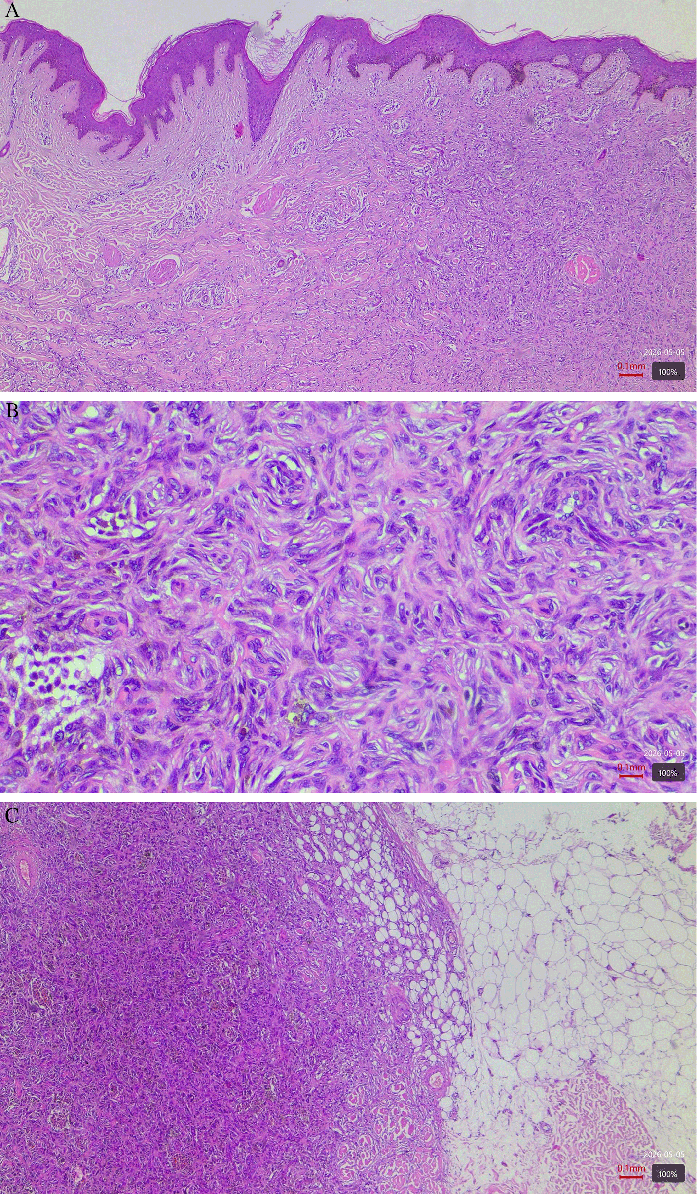
Histopathological analysis demonstrated a relatively well-circumscribed, dermal based neoplastic proliferation composed of spindle and epithelioid cells which were separated by a narrow, uninvolved dermal band – a distinct ‘Grenz zone” (Figure 1A). A storiform pattern was also well seen (Figure 1B) within an abundant collagenous stroma. The overlying epidermis displayed basal cell hyperpigmentation. The tumor lesion focally exhibited infiltration into the underlying subcutaneous adipose tissue (Figure 1C). Other findings included some lymphocytic infiltration and hemosiderin pigmentation.
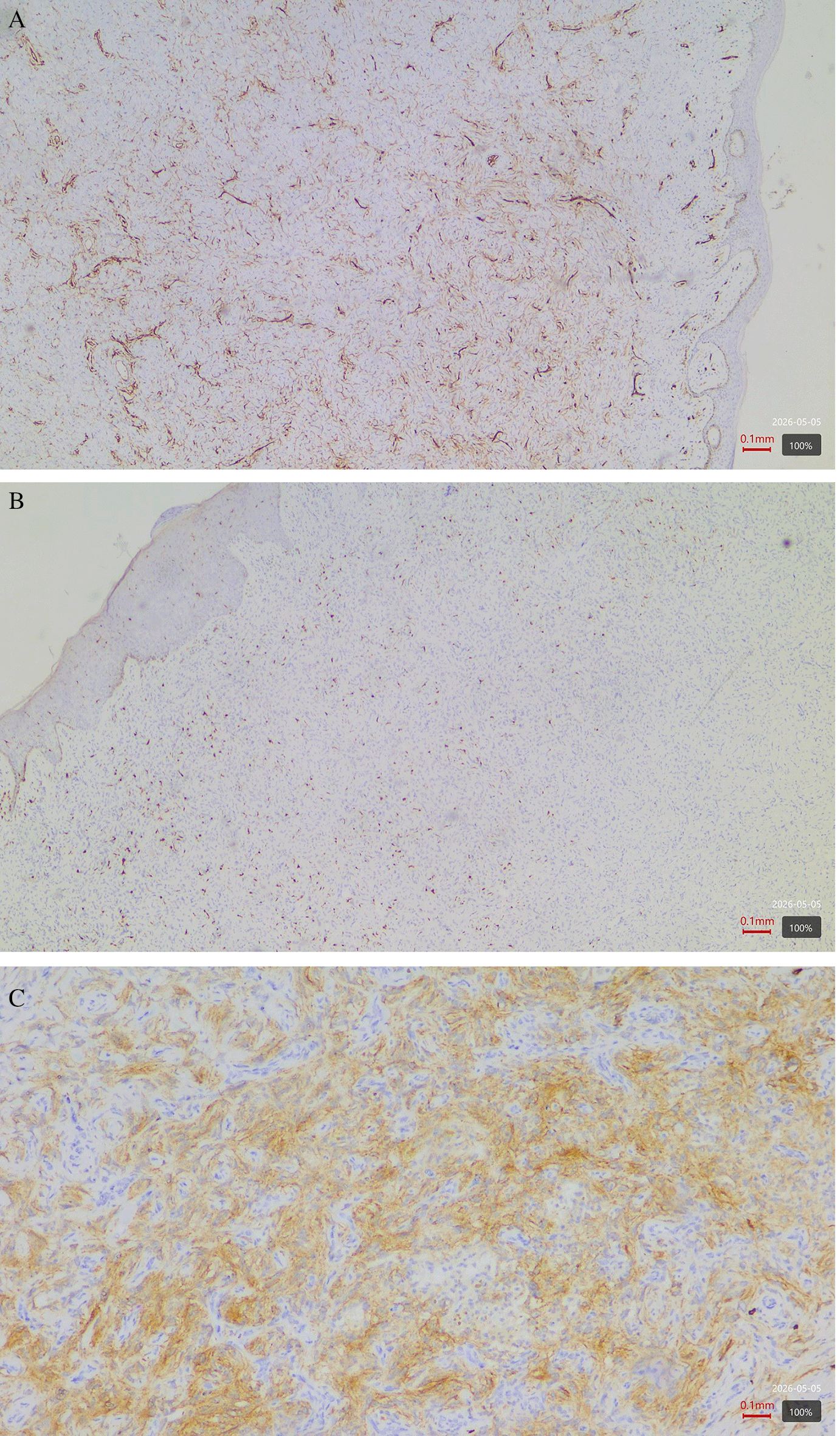
To exclude morphologically similar spindle cell neoplasms, immunohistochemical analysis was performed. The latter revealed a weak expression of CD34 localized primarily at the tumor periphery (Figure 2A), a pattern that contrasts with the diffuse, strong positivity typically observed in dermatofibrosarcoma protuberans. S100 protein staining was limited to focal expression within isolated cells (Figure 2B), suggesting melanocytic colonization or the presence of dendritic cells rather than a neural origin. Additionally, D2–40 expression was noted at the tumor periphery (Figure 2C), indicating non-specific lymphatic differentiation, supported the diagnosis of dermatofibroma over more aggressive mimics such as DFSP.
The morphological and immunohistochemical profiles were collectively diagnostic of dermatofibroma (benign fibrous histiocytoma).
The postoperative course was unremarkable, with the patient remaining afebrile and hemodynamically stable. Postoperative pain was effectively managed with a regimen of metamizole (Omalgin) and tramadol (Tramalgin). Standard prophylactic measures included the administration of cefazolin and low-molecular-weight heparin (Nadroparin, 0.4 ml). The patient was discharged in improved condition.
The current case demonstrated the classic histopathological and immunohistochemical hallmarks of dermatofibroma (benign fibrous histiocytoma). Features such as storiform architecture, collagen deposition and siderophages align with established criteria, while the prominent “Grenz zone” remains a hallmark though not a universal diagnostic feature.11
The immunohistochemical profile is critical for excluding aggressive mimics. Our findings of weak, peripheral CD34 expression distinguishes this lesion from the diffuse positivity of DFSP, a distinction vital for management given the metastatic potential and wide margin requirements.12,13 These findings are further supported by recent reviews that confirm CD34 and Factor XIIIa as the most reliable diagnostic markers despite intra-study variability.3 Furthermore, focal S100 positivity likely reflects melanocytic colonization rather than neural differentiation10 and peripheral D2–40 expression likely represents reactive stromal changes.7 While this case is a classic variant with a low recurrence risk, it must be distinguished from cellular variants which exhibit significantly higher recurrence rates and larger initial sizes.6 This case also emphasizes the importance of recognizing subtle histopathological and immunohistochemical distinctions in the diagnostic gray zones of spindle cell tumors. While dermatofibroma is a common entity, its diverse histological spectrum, ranging from classic to cellular variants, can pose significant diagnostic challenges.
In our case, DFSP was considered in the differential diagnosis because of the spindle cell morphology and CD34 expression and since it has a potential for high local recurrence risk. However, the lesion lacked the diffuse, strong CD34 immunoreactivity which is typically associated with it. In addition, DFSP, presents with a characteristic ‘honeycomb’ infiltration in the subcutaneous fat, which was absent here and therefore arguing against this diagnosis.13
Atypical fibrous histiocytoma is also considered in the differential diagnosis due to its characteristic histologic features. It presents usually with a marked cellular pleomorphism and atypical mitotic figures and in some cases, possible areas of tumor necrosis.14 The diffuse expression of S100 is also seen spindle cell melanoma along with significant atypia and also a high expression of HMB-45 and Melan-A and in some cases junctional activity.15 These features together can aid in excluding them from the differential. Since our case showed only S100 positivity with absence of any other melanocytic markers this diagnosis was also deemed unlikely. S100 positivity is also seen in Neurofibromas. Although, it also presents with spindle cells with a characteristic wavy or comma-shaped nuclei and it usually lacks a storiform growth pattern.16 In contrast, the lesion in this case presented a prominent storiform architecture which argued against a neurofibroma diagnosis.
The integration of classical morphological features with a modern multi-marker IHC panel, specifically addressing the nuances of focal CD34, S100 and D2–40 expression serves as a high-fidelity verification model to avoid a misdiagnosis ( Table 1). By contrasting these findings with recent reviews, we underscore the necessity of a standardized diagnostic approach to prevent the significant surgical morbidity associated with misdiagnosing benign lesions as aggressive sarcomas.
| Marker | Expression Pattern | Diagnostic Interpretation |
|---|---|---|
| CD34 | Weak, focal expression restricted to the tumor periphery. | Consistent with dermatofibroma; distinguishes the lesion from the diffuse, strong positivity characteristic of DFSP.3 |
| S100 | Focal expression in isolated, scattered cells. | Likely represents melanocytic colonization or entrapped dendritic cells; effectively excludes neural tumors10 . |
| D2–40 | Expression localized to the tumor periphery. | Non-specific finding; potentially indicating lymphatic differentiation or peripheral stromal remodelling.7 |
A key morphological highlight in this case was the identification of the “Grenz zone”. While this is a well-documented histopathological characteristic of dermatofibroma,17,18 its presence is not universal and it remains a critical diagnostic anchor when observed. Furthermore, the sparse infiltration of the tumor into the superficial subcutaneous fat was interpreted as a benign architectural variation rather than an indicator of malignancy. Complete surgical excision with negative histological margins remains the curative standard. When such margins are achieved, recurrence rates for classic variants are exceptionally low. Consequently, this case offers an evidence-based framework for achieving definitive diagnosis and ensuring a favorable prognosis through conservative but complete local excision.
The diagnostic evaluation of spindle cell neoplasms requires a meticulous integration of histomorphology and immunohistochemical profiling. As demonstrated in this case, classical architectural features, such as the storiform growth pattern and the Grenz zone, provide a strong diagnostic foundation. However, since dermatofibroma can exhibit focal subcutaneous involvement or variable marker expression, IHC remains an indispensable tool for excluding aggressive mimics.
The differentiation from DFSP is of paramount clinical importance. Our findings confirm that weak, peripheral CD34 immunoreactivity, contrasted with the diffuse and strong positivity characteristic of DFSP, is a reliable diagnostic anchor. Furthermore, the absence of marked cellular pleomorphism and the localized expression of secondary markers like S100 and D2–40 further validate the benign nature of the lesion.
In conclusion, complete surgical excision with negative histological margins remains the curative gold standard for classic dermatofibroma, ensuring an excellent prognosis with minimal risk of recurrence. This report reinforces the value of standardized diagnostic algorithms and multi-marker IHC panels in navigating complex differential diagnoses, ultimately safeguarding patients from unnecessary surgical radicality while ensuring definitive therapeutic outcomes.
This study was conducted in accordance with institutional ethical standards. Ethical approval was not required for this single case report in line with local institutional policy.
Written informed consent for publication of the patient’s clinical details and histopathological images was obtained from the patient. No identifiable patient information is included in the manuscript or images.
| Views | Downloads | |
|---|---|---|
| F1000Research | - | - |
|
PubMed Central
Data from PMC are received and updated monthly.
|
- | - |
Provide sufficient details of any financial or non-financial competing interests to enable users to assess whether your comments might lead a reasonable person to question your impartiality. Consider the following examples, but note that this is not an exhaustive list:
Sign up for content alerts and receive a weekly or monthly email with all newly published articles
Already registered? Sign in
The email address should be the one you originally registered with F1000.
You registered with F1000 via Google, so we cannot reset your password.
To sign in, please click here.
If you still need help with your Google account password, please click here.
You registered with F1000 via Facebook, so we cannot reset your password.
To sign in, please click here.
If you still need help with your Facebook account password, please click here.
If your email address is registered with us, we will email you instructions to reset your password.
If you think you should have received this email but it has not arrived, please check your spam filters and/or contact for further assistance.
Comments on this article Comments (0)