Keywords
Pulmonary embolism, Syncope, Low risk
Pulmonary embolism, Syncope, Low risk
Pulmonary embolism (PE) is a medical emergency that can lead to sudden death. This condition is usually associated with factors that may increase the risk of its occurrence, such as age, high tendency for intravascular coagulation and the presence of certain concomitant conditions such as deep venous thrombosis (DVT) and malignancy1. In this case, we have a presentation of a young, low-risk patient with only recurrent syncope and dyspnea that was eventually diagnosed as a massive PE. It is an uncommon case of pulmonary embolism of unknown cause that deserves attention. Such a common presentation could have led to misdiagnosis of the condition which could have ended with the sudden death of the patient.
A 30 year old male patient, a farmer from Upper Egypt, attended the cardiology outpatient clinic at Assiut University Hospitals, Egypt, on June 2012 with repeated attacks of syncope. They took place at irregular intervals, about two or three times per month.
The attacks of syncope had started eighteen months earlier, with each one lasting about one minute. There was progressive grade III dyspnea between the initial syncope symptoms and presentation at our clinic, but no evidence of orthopnea, paroxysmal nocturnal dyspnea or lower limb swellings.
General examination found that the patient had a body mass index of 28.4 kg/m2. The pulse was 110 beats/min, the respiratory rate was 24 breaths/min and the arterial blood pressure was 110/70 mmHg. There was a raised jugular venous pressure line about 6 centimeters above the level of the sternal angle. There were multiple, soft, painless subcutaneous masses over both thighs and the back of the patient with ill-defined edges. Cardiac examination showed that there was a pansystolic murmur over the tricuspid area which increased with inspiration, ejection systolic murmur over the pulmonary area with accentuated pulmonary component of the second heart sound. Electrocardiography showed sinus tachycardia with inverted T wave in leads II, III, aVF and V1-V6. Chest X-Ray revealed cardiomegaly. 2-D transthoracic echocardiography showed a dilated right side of the heart with a 5.7 cm × 1.4 cm mass in the right atrium that protruded through the tricuspid valve towards the right ventricle. Valvular morphology was normal with moderate tricuspid regurgitation and moderate pulmonary hypertension (Figure 1). Liver and kidney function tests were normal. According to this data, the patient was given a Wells score of 1.5, which gave an indication to a low risk of PE.

2-D transthoracic echocardiography apical 4 chamber view shows a mass in the right atrium that protrudes through the tricuspid valve (arrow).
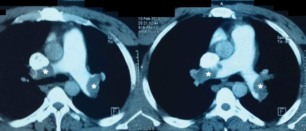
The patient was diagnosed initially with a cardiac tumor in the right atrium with suspected positional obstruction of right ventricular inflow track causing syncopal attacks, which was managed accordingly with diuretics, aspirin 150 mg/day and bisoprolol 10 mg/day to control heart rate. He underwent a duplex scan of the deep venous system of both lower limbs, which was found to be normal. While the medical team investigated the patient in hospital, a sudden attack of syncope occurred, with a sudden severe hypotension down to 90/60 mmHg. The Wells score of the patient has sharply increased from the initial 1.5 to 8, which indicated a very high risk of the presence of PE. Urgent Multi-slice CT pulmonary angiography found a massive bilateral pulmonary embolism involving the left and the right pulmonary arteries and most of their segmental branches (Figure 2). A diagnosis of high-risk pulmonary embolism was made.

Multi-slice CT pulmonary angiography shows massive bilateral pulmonary embolism involving the left and the right pulmonary arteries (asterisks).
In order to save the patient’s life, we put the patient under thrombolytic therapy. A central venous line was set up, and streptokinase was given initially as a loading dose of 250,000 IU over 30 minutes, followed by a dose of 100,000 IU/hour over 24 hours. After the recovery of the patient from the acute stage, anticoagulant therapy was given, initially in the form of enoxaparin sodium at a dose of 90 mg/kg for seven days. In conjunction, warfarin was given at a daily dose of 5 mg until the international normalized ratio (INR) reached 2.8.
The patient fully recovered from the high-risk pulmonary embolism, and an echocardiogram made 24 hours after recovery showed complete dissolution of the intra-atrial mass with no evidence of any remnants left, suggesting that it was a massive ball thrombus that had a protrusion across the tricuspid valve (thrombus in transit). However, right atrial and ventricular dilation persisted. The patient was discharged after a short period of uneventful monitoring. Right before the discharge of the patient, surgical excisional biopsy of one subcutaneous mass was performed, and pathological assessment revealed superficial subcutaneous lipomas. His brothers also have these lipomas, so we believe that it is of familial origin. Upon taking further history from the patient, it was found that the patient and his brothers had a history of familial hyperlipidemia. Tests for the patient’s hypercoagulable state as assessed by measuring levels of protein C and S and antithrombin III appeared normal. The patient was followed up for 6 months and showed no more syncopal attacks. We do not have details of any further investigations that may lead to a cause of this condition.
PE is a serious medical emergency condition, in which obstruction of the pulmonary trunk or any of its branches takes place. It could be partial or complete, which causes sudden obstruction of the blood flow to the lungs, resulting in dyspnea, hypoxia, chest pain and insufficient blood flow to the left side of the heart. PE can be caused by any cause of embolism, including thrombosis, air, fat, talc or amniotic fluid1. About 1–2 people per 1000 may suffer from pulmonary embolism while 1 in every 100 people over the age of 80 may suffer from this condition2.
Signs and symptoms of pulmonary embolism occur suddenly. Dyspnea, tachypnea, chest pain, cough, and hemoptysis take place. In more severe cases, cyanosis, syncope and circulatory instability occur, and sometimes peripheral edema may be present. Sudden death can occur in the most severe cases1. About 25% of PE cases present as sudden death while 15% of all cases of sudden death are attributable to PE2.
The criteria for diagnosis of PE are put into clinical prediction systems, such as the Wells score, which was initially proposed by Wells et al. in 1995 (Table 1)3. Additional prediction systems to determine the probability of getting a PE are available, such as the Geneva rule, which has a similar manner of prediction, but with some modifications4.
Adapted from Neff MJ (2003)16. DVT: deep vein thrombosis, PE: pulmonary embolism.
Syncope, as a presenting symptom in PE, is not uncommon. While it can have multiple causes, syncope is still an important sign of PE, especially if it is accompanied by dyspnea. A study conducted by Thames et al. in 1977 found that 13% of patients with PE present initially with syncope5. Another study conducted by Calvo-Romero et al. in 2004, in Spain, showed that 9.1% of patients with PE presented with syncope. However, the authors concluded that it was impossible to tell whether syncope could determine the prognosis in such cases6. Apart from these two studies, in 1988, two patients in Israel were reported to have suddenly died of massive PE after having a hip surgery 3 weeks earlier. Several syncopal episodes started in the first week after surgery, and they were considered as a sign of fatal outcome7.
The mechanisms that cause syncope in PE are variable. One mechanism is that the presence of PE decreases blood flow to the lungs, thus leading to hypoxemia and cerebral hypoxia. Another theory suggests that a vasovagal attack is triggered with the occurrence of PE. A third mechanism suggests that syncope occurs due to the arrhythmias that result from overloading of the right ventricle. A fourth mechanism suggests that PE leads to circulatory obstruction and decreased left ventricular filling, thus leading to decreased cardiac output and ultimately diminished cerebral blood flow8.
A determinant factor in the diagnosis of the cause of syncope is the composition of arterial blood gases. Hypoxemia can indicate the presence of a condition that obstructs normal cardiovascular or respiratory functions. If there is no airway obstruction, PE is highly suspected. Advanced imaging techniques, such as multi-slice CT pulmonary angiography, can give an accurate image of the condition within the pulmonary vascular tree8.
Lipomas are common benign tumors formed of adipose tissue. They are the most common soft tissue tumor9. The most common lipoma is the superficial subcutaneous lipoma10. They can be found wherever fat is present under the skin surface. The tendency to develop them is not exclusively hereditary11. Nevertheless, there are familial conditions that may include the presence of lipomas, such as familial multiple lipomatosis12. In 1989, it was reported in Israel that there could be an association between familial combined hyperlipodemia and the presence of nonsymmetric subcutaneous lipomatosis in a family, which is similar to this case13. A growing body of evidence suggests the presence of a relationship between hyperlipidemia and the occurrence of DVT14. Therefore, we feel this co-occurrence is worth noting.
In this case, the ball thrombus (thrombus in transit), which was present in the right atrium, caused transient obstruction to the tricuspid valve, thus causing a transient obstruction to the outflow of the right atrium. That obstruction caused a transient obstruction to the left ventricular filling that brought about decreased cerebral blood flow. The ball thrombus probably got displaced as the position of the patient changed so that blood flow was restored and the syncopal episode ended. Eventually, the ball thrombus caused further thrombosis and released emboli that were large enough to occlude the pulmonary trunk and its main branches. Fortunately for the patient, the medical team successfully managed to initiate thrombolytic therapy in time, and the ball thrombus, along with the other emboli, were lysed. A similar case in Turkey was reported in 2009, but it was different in that the patient had a high risk due to long-term occupational immobilization and the presence of DVT8. Also a similar case series was reported in 1998 by Wolfe and Allen. They reported three cases that initially presented with syncope and later diagnosed with PE, and one of these cases ended up fatally15. An interesting aspect of the case presented here is that the medical team initially evaluated the patient as low risk according to the Wells score. Duplex scans and tests for hypercoagulable state were normal, unlike the results usually found in most patients with PE. That leaves us with a question as to what could have caused that thrombus to occur. Additional investigations are necessary to find a cause of this condition, such as an undiagnosed malignancy or a familial cause.
Pulmonary embolism is a medical emergency that can be presented by a common symptom such as syncope. Physicians should pay attention to this presentation and check for pulmonary embolism in cases of syncope with dyspnea. Such cases can be easily missed, which may end up with sudden death if not diagnosed and treated promptly.
Written informed consent was obtained from the patient for publication of this case report and any accompanying images.
AO was a major contributor to the writing of the manuscript. He also gathered and analyzed the patient’s data about syncope and pulmonary embolism. AT performed the acute treatment to the patient and was a major contributor in writing the manuscript. AH performed and supervised the treatment of the patient and the manuscript writing process. All of the three authors have read and approved the final manuscript.
We would like to express our sincere and deepest gratitude towards the Medical Sector Reform Group of Egypt for their continuous support throughout the case report preparation process as a part of the Mentor-Student Research Project. Our special thanks go to Dr. Noha A. Moussa and Omnia M. Omar for their tremendous efforts as coordinators of this project. We are also grateful to Dr. Sameh Nashat for performing echocardiography to the patient and providing us with the results.
| Views | Downloads | |
|---|---|---|
| F1000Research | - | - |
|
PubMed Central
Data from PMC are received and updated monthly.
|
- | - |
Competing Interests: No competing interests were disclosed.
Competing Interests: No competing interests were disclosed.
Alongside their report, reviewers assign a status to the article:
| Invited Reviewers | ||
|---|---|---|
| 1 | 2 | |
|
Version 1 25 Nov 13 |
read | read |
Provide sufficient details of any financial or non-financial competing interests to enable users to assess whether your comments might lead a reasonable person to question your impartiality. Consider the following examples, but note that this is not an exhaustive list:
Sign up for content alerts and receive a weekly or monthly email with all newly published articles
Already registered? Sign in
The email address should be the one you originally registered with F1000.
You registered with F1000 via Google, so we cannot reset your password.
To sign in, please click here.
If you still need help with your Google account password, please click here.
You registered with F1000 via Facebook, so we cannot reset your password.
To sign in, please click here.
If you still need help with your Facebook account password, please click here.
If your email address is registered with us, we will email you instructions to reset your password.
If you think you should have received this email but it has not arrived, please check your spam filters and/or contact for further assistance.
Comments on this article Comments (0)