Case presentation
A 67 year old Hindu female presented to us in May 2010 with history of right flank pain, fever and vomiting. She had raised total leukocyte count: 16600/μL and deranged renal function (serum creatinine: 3.1mg/dL). A non-contrast CT (NCCT) scan revealed moderate hydronephrosis, right upper ureteric calculus and a well circumscribed lesion on the medial aspect of the kidney. A percutaneous nephrostomy was performed on account of the deranged renal function. Subsequently, the patient underwent a percutaneous nephrolithotomy (PCNL).
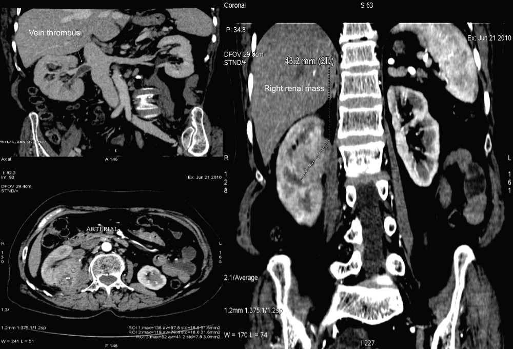
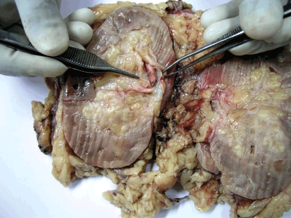
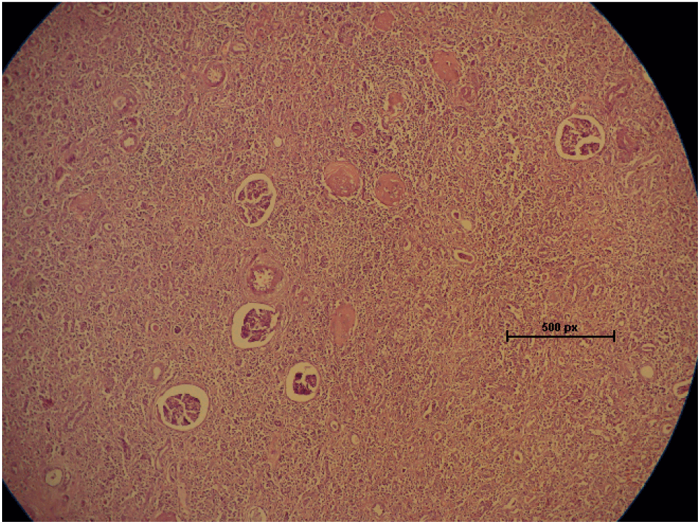
At one month from presentation and after the serum creatinine improved to 1.47mg/dL, a contrast CT revealed an enhancing mass (enhancement from 33 to 118 Hounsfield units) on the medial aspect of the kidney (Figure 1; a contrast CT not done at initial presentation due to deranged renal function) with evidence of renal vein thrombosis and multiple paracaval lymph nodes. A provisional diagnosis of renal cell carcinoma with renal vein thrombus was made. The clinical stage was T3aN2M0. A laparoscopic radical nephrectomy was done. The gross specimen revealed evidence of renal vein thrombus and Xanthogranulomatous pyelonephritis (XGPN) (Figure 2). On H & E (Hematoxylin & Eosin) microscopic examination, it was composed of foamy macrophages admixed with inflammatory infiltrate (Figure 3). There was no evidence of malignancy. The patient recovered well and was discharged in stable condition after 4 days with a serum creatinine of 1.16mg/dL.

Figure 1. Well defined soft tissue density mass of right kidney measuring 49 × 35 × 43 mm enhancing from 33 HU to 118 HU with non-enhancing areas of necrosis.

Figure 2. Gross specimen showing thrombus in renal vein.

Figure 3. Microscopic examination at 100X magnification showing collection of foamy macrophages and inflammatory infiltrate diffusely infiltrating the renal parenchyma.
Discussion
XGPN is an uncommon, severe, chronic suppurative renal parenchymal infection characteristically leading to renal destruction. The majority of cases are unilateral and result in a nonfunctioning, massively enlarged kidney associated with obstructive uropathy secondary to urolithiasis. XGPN has been described as a great imitator or a masquerading tumor in adults and pediatric age groups1,2. The etiological factor in this case was the renal calculus with chronic infection. The imaging findings in this case showed a significantly enhancing mass, lymph nodes and a renal vein thrombus. The mass was seen closely abutting the psoas as well. The CT findings mimicked a case of T3N2Mx renal cell carcinoma. Localised XGPN is amenable to partial nephrectomy if diagnosed preoperatively. XGPN has been found to be associated with renal cell carcinoma, papillary transitional cell carcinoma and squamous cell carcinoma and hence nephrectomy should be performed when malignancy cannot be excluded. This case highlights the need to keep XGPN as a differential diagnosis of a renal mass especially in presence of urolithiasis.
Consent
Written informed consent for publication of clinical details and clinical images was obtained from the patient.
Author contributions
Arvind Ganpule and Jitendra Jagtap drafted the manuscript and carried out the literature search. Sanika Ganpule, Amit Bhattu and Shailesh Soni prepared the illustrations and helped to draft the manuscript. Ravindra Sabnis and Mahesh Desai revised the manuscript and did the final proofreading of the manuscript. All authors approved the final manuscript for publication.
Competing interests
No competing interests were disclosed.
Grant information
The author(s) declared that no grants were involved in supporting this work.
Faculty Opinions recommendedReferences
- 1.
Zorzos I, Moutzouris V, Petraki C, et al.:
Xanthogranulomatous pyelonephritis--the "great imitator" justifies its name.
Scand J Urol Nephrol.
2002; 36(1): 74–6. PubMed Abstract
| Publisher Full Text
- 2.
Gerber WL, Catalona WJ, Fair WR, et al.:
Xanthogranulomatous pyelonephritis masquerading as occult malignancy.
Urology.
1978; 11(5): 466–71. PubMed Abstract
| Publisher Full Text


Comments on this article Comments (0)