Keywords
PET, CT, Basal cell Carcinoma (BCC), Metastatic, albinism, vismodegib
PET, CT, Basal cell Carcinoma (BCC), Metastatic, albinism, vismodegib
Basal cell carcinoma (BCC) is the most common human malignancy worldwide, yet it is typically indolent and rarely possesses metastatic potential1. Reported rates of metastases range from 0.0028 to 0.5%1. Despite the high incidence of BCC, there have been only 257 cases of metastatic BCC (MBCC) reported in the English medical literature between 1894 and 1991, 82 of which demonstrated metastases to the lung2–5.
In this article, we review the clinical, radiological, and histopathological presentation of a patient with a history of multiple non-head and neck BCC with subsequent numerous metastases to the bilateral lungs. We also briefly review the literature, and discuss the epidemiology, risk factors, TNM staging, therapeutic modalities, and prognosis for patients with MBCC.
A 62-year-old Caucasian male with oculocutaneous albinism (Fitzpatrick type I skin) had been followed extensively by both the dermatology and the general surgery services at the University of Arkansas for Medical Sciences. His past medical history was significant for multiple BCCs, the most recent of which (2012) involved the back and flank, requiring adjuvant radiation therapy and split thickness skin grafting. No other significant medical history was noted aside from shortness of breath.
Additionally, four months prior to these excisions, the patient underwent excisions of morpheaform (infiltrative) BCCs of the right arm and back, as well as nodular BCCs of the left cheek and temple. In 2009, he had an initial large wide excision for BCC on his back and flank which demonstrated positive deep margins. The most recent re-excision in 2012 demonstrated all negative margins. Moreover, in 2012 he had a singular squamous cell carcinoma of the right upper extremity that was less than 1.0 mm to the nearest margin, and measured 4.0 mm in maximum depth of invasion.
A routine chest X-ray in 2009 was effectively within normal limits, displaying no mass or tumor. A diagnostic CT scan was ordered in 2012 for surveillance due to the extensive nature of his BCCs. It demonstrated numerous solid and sub-solid nodules measuring up to 2.0 cm, in multiple stages of cavitary change, in both lungs. It was considered possible that the nodules were metastases from the squamous cell carcinoma of the right arm but further evaluation was recommended to confirm this.
Follow-up F-18 fluoro-deoxy-glucose PET/CT scan (15.14 mCi, 69-minutes of uptake time, and a fasting blood glucose of 104 mg/dL) was performed from the base of the orbits through the mid-thigh with 3-axis reconstructions, and attenuation correction with a non-diagnostic CT scan. It demonstrated no less than thirty non-specific foci with significant hypermetabolic activity (>3-times background), most of which were associated with nodules in multiple stages of cavitary change (Figure 1). In light of the patient's history of multiple malignancies, in addition to an inflammatory/infectious etiology, the possibility of metastasis, although less likely, was also considered. Clinical and pathologic correlations were recommended, as well as a repeat PET/CT of the vertex through the feet, for definitive evaluation of the dermis.
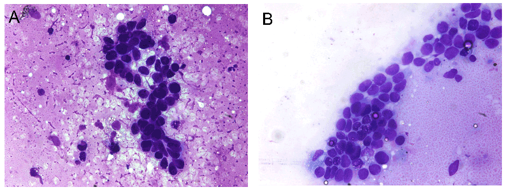
CT-guided fine needle aspiration and subsequent core biopsy from one of the lung nodules from the right upper lobe were interpreted as positive for malignant cells, basal cell carcinoma. The right upper lobe core biopsy showed small cohesive nests and cords of basaloid cells with scant cytoplasm. Artifactual clefts containing mucin were present around the periphery of many of the nests. The cells demonstrated hyperchromatic chromatin without nucleoli and displayed some nuclear overlap and molding. The tumor cells were negative for synaptophysin, chromogranin, and cytokeratin 20 by immunohistochemistry, findings which argue against the possibility of primary or metastatic neuroendocrine carcinoma (Figure 2).

Fine needle aspiration (Diff-Quick preparation, original magnification ×400) from a lung nodule demonstrates a) Cells with a high nuclear to cytoplasmic ratio and mildly enlarged nuclei compared to surrounding inflammatory cells in the background, b) Enlarged, fairly uniform cells forming sheets.
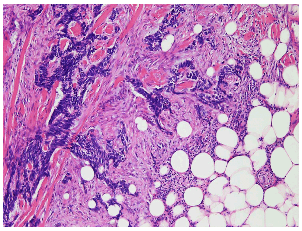
A right flank wide local re-excision from 2012, several months prior to the discovery of lung metastases, demonstrated infiltrative cords and strands of basaloid cells characteristic of the infiltrative (morpheaform) type of basal cell carcinoma. Multiple tumor nodules were present in this specimen and the tumor invaded into the subcutis. Margins were negative. This right flank basal cell carcinoma specimen was re-examined and immunohistochemical stains were performed following discovery of the lung metastases; the tumor cells were negative for synaptophysin, chromogranin, and cytokeratin 20 by immunohistochemistry, findings which argue against the possibility of a cutaneous neuroendocrine carcinoma (Merkel cell carcinoma), (Figure 3).

Biopsies and excisions from the scalp, left temple, right arm, and back performed several years previously were also reviewed and all were found to show characteristic histologic features of basal cell carcinoma.
The histologic features in the lung biopsy strongly resembled the histologic features of the multiple infiltrative basal cell carcinomas that this patient has had previously. The similarity between the tumors, the history of multiple aggressive and deeply invasive BCCs, and the exclusion by immunohistochemistry of the histologic mimics are all features that suggest that the pulmonary nodules represent a very rare example of metastatic basal cell carcinoma.
The patient was started on vismodegib, the cyclopamine-competitive antagonist of the smoothened receptor, at a dosage of 150 mg by mouth each day. After approximately two months of therapy, he began to have an improvement in his ability to breathe, and he was able to self-taper off of his home supplemental oxygen requirement. A follow up CT scan at this time showed disease improvement, and the vismodegib was discontinued. Most recently, a follow up CT, seven months after initial diagnosis of pulmonary metastases, showed interval worsening of the metastatic burden in the lungs. He is scheduled to restart vismodegib and is now being followed jointly by the palliative care and medical oncology services.
In 1894, Beadles described a singular case of “rodent ulcer” deposition within the lymphatic gland of a deceased 46-year-old male, who had a previous history of “rodent ulcer” of the cheek. At autopsy, caseating foci were found within his lungs3. Although the historical medical terminology precludes absolute certainty, this is thought to represent the first reported case of metastatic basal cell carcinoma (MBCC).
Lattes and Kessler used three criteria to define MBCC in their 1951 report of two cases. First, they required that neither the primary nor the metastases could be squamous in type. Second, tumors could not be considered new primaries or a result of direct extension. Third, the tumors could not be mucoid or salivary in origin5.
In total, there have been only 257 cases of MBCC reported in the literature between 1894 and 1991 (based on a PubMed search using the term “metastatic+basal+cell+carcinoma” and adding the number of cases presented in each article)”.
Metastases are thought to arise from both lymphatic and hematogenous pathways1. The most frequent reported sites of metastases were regional lymph nodes (60–65%), lung (32.3–40%), bone (19–25.8%), and skin (10–19%)2,4.
In light of the rarity of MBCC, it is unclear which histological or clinical features might be correlated with the risk of metastasis. With regards to histological features, there is a correlation with increased risk of local recurrence in specific subtypes like morpheaform or infiltrating types, but no correlation has been found with an increased risk of metastases among these more locally aggressive subtypes6. Of the 257 cases reported between 1894 and 1991, only five cases of MBCC were in black patients -the remainder were all in patients with a light complexion4. Disorders such as Basal Cell Nevus Syndrome or Xeroderma Pigmentosum have been shown to predispose patients to BCC. However, none have been shown to have a higher rate of metastases7,8. Patients with specific genodermatoses such as oculocutaneous albinism, in which there is a disorder of the melanin biosynthesis, have been shown to have a much less specific association with BCC. The significance of this association is unknown at this time8. However, a history of radiation therapy and history of local tumor recurrence have both been implicated in higher rates of MBCC8. In 170 of the reported cases of MBCC, the most frequent primary sites were head (67.6%) and trunk (16.5%), anatomic locations which are not significantly different than the most frequent sites of typical non-metastatic BCC1. In a review of 41 publications by Snow et al., BCCs larger than 4.0 cm had a 1.9% chance of metastases5. However, primary BCCs as small as 1.1 cm have been reported to metastasize9.
Survival time after the development of distant metastases in MBCC has been reported at 8–10 months2,4. A median interval between discovery of the primary focus of BCC and the discovery of metastasis has been reported as 9 years2, much longer than the interval seen in many other types of carcinoma.
For surveillance of patients with only local BCC, close follow up with a thorough skin examination, loco-regional lymph node evaluation, periodic Roentgenographic evaluation, liver function tests and alkaline phosphatase tests have been suggested to evaluate for occurrence of distant metastases10,1. However, given the extreme rarity of metastasis from BCC, these suggestions may not be feasible or reasonable for most patients. Further studies to elucidate risk factors for metastasis in BCC would be useful in determining which patients should receive a higher level of follow up screening.
As with many rare diseases, there is no established standard of care for management of metastatic foci in MBCC. A variety of therapies have been reported in the literature including local excision, radiation therapy, and chemotherapy. Given the aforementioned short survival times, large BCCs (>5 cm) and multiple metastatic sites pose a difficult dilemma for treatment, surgery may result in functional and cosmetic impairment, and radiation therapy is poor at providing local control12.
For metastatic disease, debulking prior to local surgery, and for a failure of local treatment, cis-platinum containing regimens had been a commonly accepted therapeutic approach12. However, preliminary results from NCT00833417 [a study evaluating the efficacy and safety of vismodegib (GDC-0449, Hedgehog pathway inhibitor) in patients with advanced basal cell carcinoma] prompted the USFDA to approve vismodegib as a treatment for “adults with BCC, that has spread to other parts of the body, or that has come back after surgery, or that their healthcare provider decides cannot be treated with surgery or radiation”, on January 30, 2012.
In the present patient, with history of oculocutaneous albinism, there were in excess of 30 metastatic foci within the lungs, whose histologic features strongly resembled those of the patient’s multiple other infiltrative BCCs. The primary tumor as well as the metastases were not squamous in type. Also, the squamous cell carcinoma of the arm was well differentiated and did not demonstrate basaloid features. Because primary basal cell carcinomas do not arise in the lungs and there was no evidence of direct extension from the back and flank into the lung tissue, the lung lesions are considered to be true metastases, as evidenced by the multiple lung nodules. Lastly, none of the primary or metastatic lesions of BCC demonstrated any mucoid or salivary features. Given the immunohistochemical exclusion of histologic mimics and history of multiple deeply invasive and aggressive BCCs it is considered most likely that the lung based cavitary foci were MBCC. Neuroendocrine primary tumors of the lung were systematically ruled out by immunohistochemical stains and the previous skin resections were ruled out for Merkel cell involvement.
Because of the tumor burden involving the lungs, surgery was not considered an option. He was treated with a two month course of vismodegib, to which he initially responded well. Surveillance CT scans and clinical examinations are currently being used to determine the need for resumption of vismodegib. Secondary to a lack of prospective studies or large sample sizes of previous studies, it remains unclear if the oculocutaneous albinism of the present patient was simply an association or a predisposing factor.
In conclusion, an unusual case of MBCC is presented that is presumed to arise from a non-facial primary BCC, with metastasis to the lung, detected by radiographic interrogation. It is important for providers to be aware that BCC may rarely metastasize, and that the risk for metastasis may be higher in patients with a primary BCC that is >4 cm or that has been previously irradiated. Imaging specialists should be sure to keep MBCC in the differential diagnosis when faced with a lung, lymph node, or cutaneous focus that is hypermetabolic and there is a history of BCC.
No consent was obtained from the patient. The case report is fully anonymized and HIPAA-compliant, contains no patient identifiers in either the text or the figures, and was performed as an IRB exempt study.
Mickaila Johnston: Manuscript preparation. Whitney Winham: Manuscript preparation. Nicole Massoll: Manuscript review, data generation. Jerad M. Gardner: Manuscript preparation, project supervision, data generation. All authors critically revised the manuscript and agreed to its publication.
This project was not funded by any commercial vendor or by any grant. Costs were covered by the Department of Pathology at the University of Arkansas for Medical Sciences.
The views expressed in this presentation are those of the author and do not necessarily reflect the official policy or position of the Department of the Navy, Department of Defense, or the United States government. “I am a military service member (or employee of the U.S. Government). This work was prepared as part of my official duties. Title 17, USC, §105 provides that ‘Copyright protection under this title is not available for any work of the U.S. Government’. Title 17, USC, §101 defines a U.S. Government work as a work prepared by a military service MBR/employee of the U.S. Government as part of that person's official duties”.
| Views | Downloads | |
|---|---|---|
| F1000Research | - | - |
|
PubMed Central
Data from PMC are received and updated monthly.
|
- | - |
Competing Interests: No competing interests were disclosed.
Competing Interests: No competing interests were disclosed.
Competing Interests: No competing interests were disclosed.
Competing Interests: No competing interests were disclosed.
Alongside their report, reviewers assign a status to the article:
| Invited Reviewers | ||||
|---|---|---|---|---|
| 1 | 2 | 3 | 4 | |
|
Version 1 15 Jan 14 |
read | read | read | read |
Provide sufficient details of any financial or non-financial competing interests to enable users to assess whether your comments might lead a reasonable person to question your impartiality. Consider the following examples, but note that this is not an exhaustive list:
Sign up for content alerts and receive a weekly or monthly email with all newly published articles
Already registered? Sign in
The email address should be the one you originally registered with F1000.
You registered with F1000 via Google, so we cannot reset your password.
To sign in, please click here.
If you still need help with your Google account password, please click here.
You registered with F1000 via Facebook, so we cannot reset your password.
To sign in, please click here.
If you still need help with your Facebook account password, please click here.
If your email address is registered with us, we will email you instructions to reset your password.
If you think you should have received this email but it has not arrived, please check your spam filters and/or contact for further assistance.
Comments on this article Comments (0)