ALL Metrics
-
Views
-
Downloads
Get PDF
Get XML
Cite
Export
Track
Case Report

Case Report: Large nested variant urothelial carcinoma –invasive malignancy masquerading as low grade disease

[version 1; peer review: 3 approved]
PUBLISHED 23 Dec 2014
Author details Author details
OPEN PEER REVIEW
REVIEWER STATUS

Abstract

Introduction
The large nested variant of urothelial carcinoma (LNVUC) is a newly described and rare subtype of urothelial carcinoma. It is characterised by bland cytological features and a large nested architecture similar in appearance to low grade urothelial carcinoma with an inverted growth pattern. To date only 23 cases in a single series have been described.
 
Case Report
We describe the case of a 59 year old male with LNVUC whose tumour was initially misdiagnosed as a non-invasive low grade urothelial carcinoma. At a subsequent re-resection, his tumour was correctly re-classified as LNVUC with extensive invasion of the muscularis propria. Radical cystectomy and formation of an ileal conduit was performed. His operative specimen revealed invasion of prostatic stroma and perivesical fat, with all surgical margins clear. He is currently free from clinical recurrence 12 months after his cystectomy.
 
Conclusion
LNVUC is a newly described and rare urothelial carcinoma subtype. It characteristically possesses bland cytological features and may mimic low grade urothelial cancer. Despite its bland appearance it behaves aggressively with invasion, metastasis and death being common.

Keywords

LNVUC, Urothelial carcinoma

Introduction

The large nested variant of urothelial carcinoma (LNVUC) is a newly described variant of urothelial cancer (UC), with a single series of 23 cases being the only examples reported thus far1. This aggressive UC variant has deceptively bland cytological features, which may confound correct tumour classification. We present the case of a 59 year old male with a large bladder tumour who was initially diagnosed histologically as non-invasive low grade UC on initial resection. At re-resection the tumour was correctly identified as LNVUC.

Case report

A 59 year old Caucasian male was transferred to our unit from a regional hospital with a two week history of macroscopic haematuria. He sought medical attention only after he developed clot retention. He denied any previous history of haematuria or urinary problems prior to the two week period immediately before his hospital admission.

His medical history was unremarkable other than extensive carcinogen exposure, with both a 40 pack-year smoking history and significant occupational exposure, working as a fly-in, fly-out diesel fitter on a mine site.

On admission he required placement of an indwelling urinary catheter and continuous bladder irrigations. His initial serum creatinine was elevated, but soon normalised following catherisation. He was transferred to our secondary referral centre following failure of conservative therapies to control his persistent haematuria.

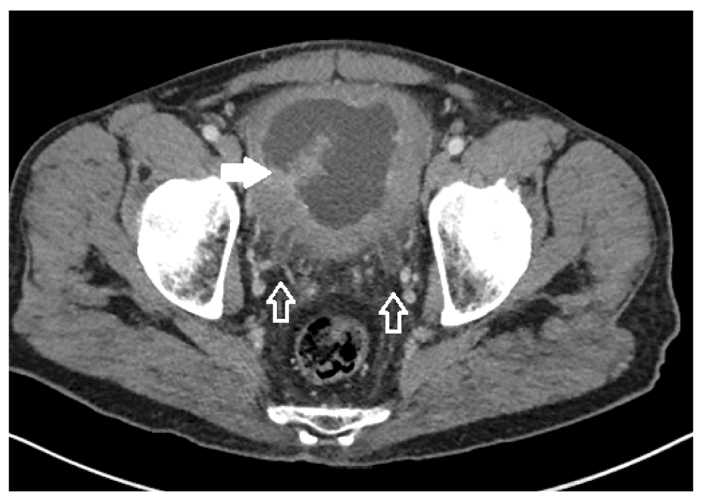
On his arrival to our facility we arranged Computerised Tomography (CT) to assess his bladder and upper renal tracts. CT demonstrated a grossly thick walled bladder with a large enhancing intra-vesical mass, and bilateral hydroureteronephrosis (Figure 1). His haematuria continued and he became transfusion dependant. He was taken to the operating theatre two days after his arrival for cystoscopic assessment.
5818a65e-d8c1-4373-af84-a094bdbc29e0_figure1.gif

Figure 1. Axial contrast enhanced CT demonstrating bilateral ureteric dilation (hollow arrows) and a large enhancing intravesical mass (solid arrow).

At cystoscopy, there was a large papillary tumour involving the prostatic urethra, the trigone, and both lateral walls of the bladder. (Figure 2) Neither ureteric orifice was identifiable. The tumour was macroscopically resected after an extensive procedure.

5818a65e-d8c1-4373-af84-a094bdbc29e0_figure2.gif

Figure 2. The bland appearance of the tumour at the bladder neck at his initial cystoscopy.

Histologically the tumour was classified as a low grade urothelial carcinoma with no evidence of superficial or muscle invasion. We found this finding inconsistent with the operative and radiologic findings and repeated a cystoscopy four weeks later.

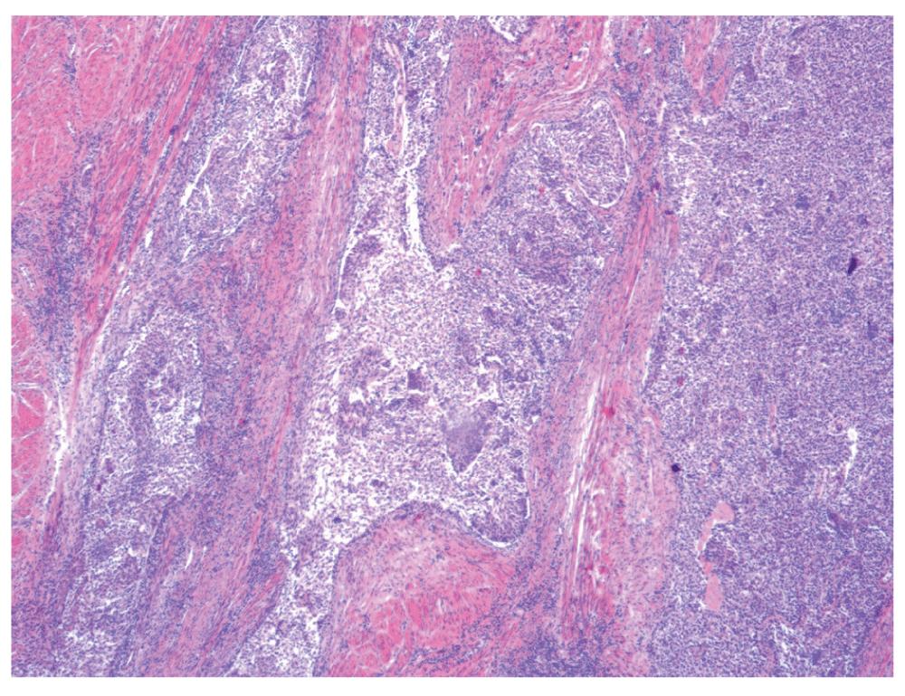
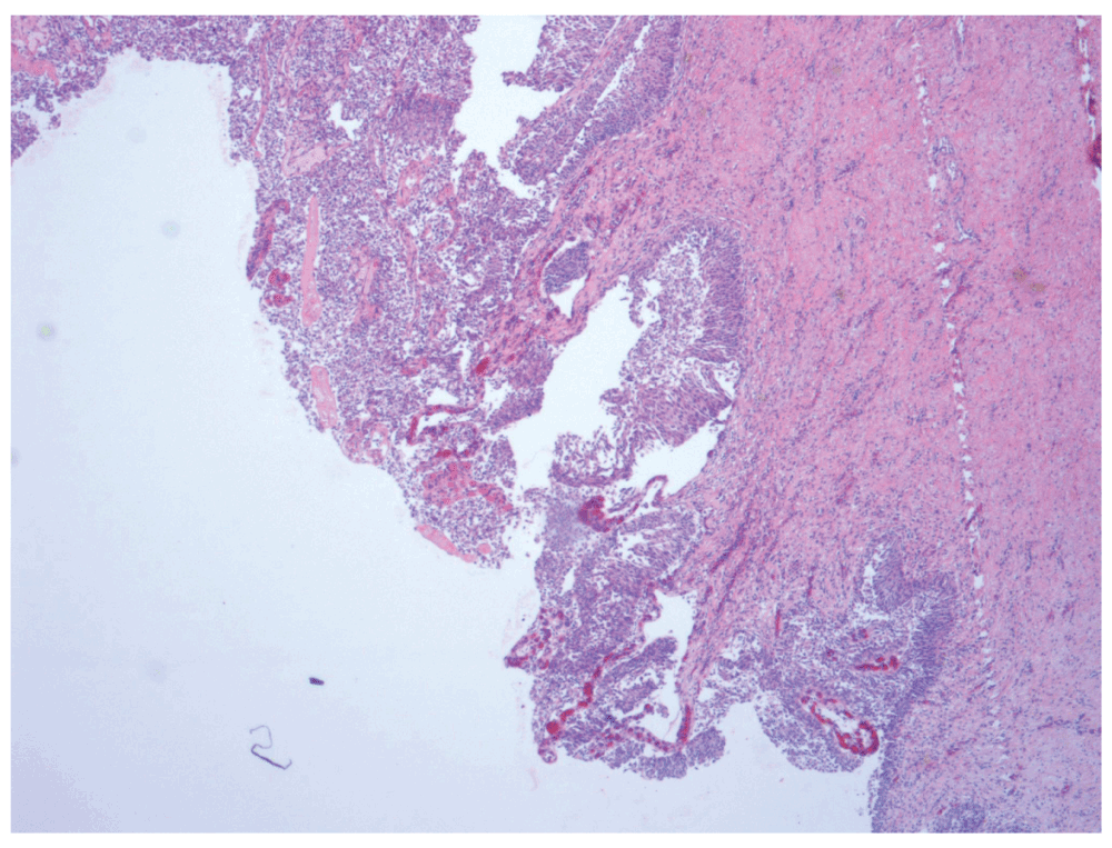
At repeat cystoscopy large volume tumour regrowth had occurred and a further 90 minute resection was performed. Tumour histology this time demonstrated invasion into the muscularis propia by a large nested variant of UC (Figure 3) with an adjacent superficial component of low grade papillary UC (Figure 4).
5818a65e-d8c1-4373-af84-a094bdbc29e0_figure3.gif

Figure 3. Low power view (×4 objective lens) of Hematoxylin and Eosin (H and E) stained bladder resection specimen showing the large nests of the invasive component of urothelial carcinoma with abundant interspersed stromal tissue.

5818a65e-d8c1-4373-af84-a094bdbc29e0_figure4.gif

Figure 4. Low power view (×4 objective lens) of Hematoxylin and Eosin (H and E) stained bladder resection specimen showing the superficial component of LNVUC.

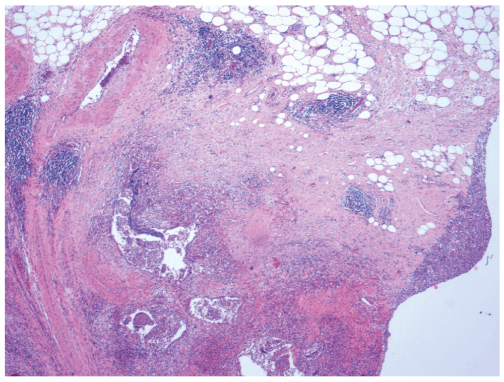
A staging Positron Emission Tomography (PET) CT was negative for metastatic disease and a cysto-prostatectomy and formation of an ileal conduit was performed. The operative specimen histology again revealed the large nested variant of UC with focal invasion into peri-vesical fat (Figure 5) and the prostatic stroma (Figure 6). A component of low grade UC was also present superfically. The tumour was clear of all operative margins. All lymph nodes sampled were negative for metastatic deposits.
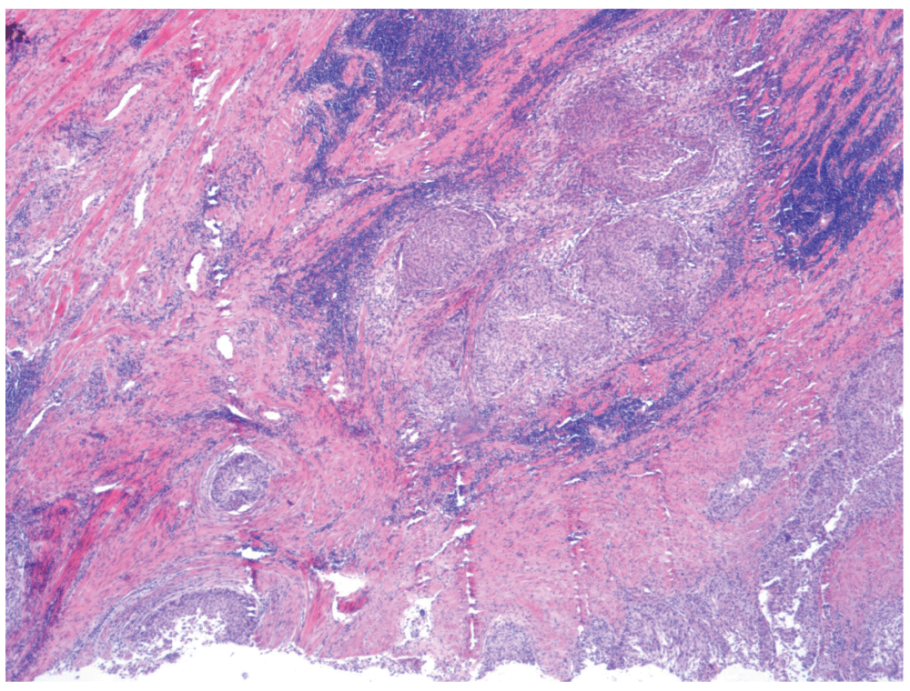
5818a65e-d8c1-4373-af84-a094bdbc29e0_figure5.gif

Figure 5. Low power view (×4 objective lens) of Hematoxylin and Eosin (H and E) stained bladder resection specimen showing focal invasion of tumour into peri-vesical fat.

5818a65e-d8c1-4373-af84-a094bdbc29e0_figure6.gif

Figure 6. Low power view (×4 objective lens) of Hematoxylin and Eosin (H and E) stained bladder resection specimen showing tumour nests invading prostatic stromal tissue.

The patient’s post-operative period was unremarkable and he made a swift recovery. He was discharged from hospital one week post-operatively. He was referred to medical oncology for consideration of adjuvant chemotherapy, however after discussion with oncologists the patient declined any additional treatment. He is presently twelve months post cysto-prostatectomy and he remains clinically well and free from clinical disease recurrence. We will continue to closely monitor this patient.

Discussion

The large nested variant is a newly described subtype of UC. The first and to date only case series was published in 2011 by Cox and Epstein and describes 23 cases1. They describe tumours with universally bland histologic appearances but with invasion of large nests resembling von Brunns nests into the underlying stroma. In contrast to the normal nested variant of UC, a surface papillary component is present and there is abundant fibrous stroma between individual tumour nests1,2. LNVUC is most commonly mistaken for low grade urothelial cancer with an inverted growth pattern2.

LNVUC behaves aggressively, of the 17 cases with adequate follow-up in Cox and Epstein’s series, 3 had died of their disease and another two were alive but had developed metastatic spread of their cancer1.

Conclusion

The large nested variant is an extremely rare, newly described variant of UC. Our case is only the 24th described in the literature, and the first case reported since the condition was first classified in 2011. LNVUC can confound accurate diagnosis by masquerading as Von Brunn’s nests or, in our case, low grade non-invasive UC. Despite the bland macroscopic and histologic appearance of LNVUC it behaves in an aggressive manner, and should be treated the same as any invasive urothelial malignancy.

Consent

Written informed consent for publication of their clinical details and/or clinical images was obtained from the patient.

Comments on this article Comments (0)

Version 1
VERSION 1 PUBLISHED 23 Dec 2014
Comment
Author details Author details
Competing interests
Grant information
Copyright
Download
 
Export To
metrics
Views Downloads
F1000Research - -
PubMed Central
Data from PMC are received and updated monthly.
- -
Citations
CITE
how to cite this article
Keller A, Lim AJ and Ali A. Case Report: Large nested variant urothelial carcinoma –invasive malignancy masquerading as low grade disease [version 1; peer review: 3 approved]. F1000Research 2014, 3:314 (https://doi.org/10.12688/f1000research.5966.1)
NOTE: If applicable, it is important to ensure the information in square brackets after the title is included in all citations of this article.
track
receive updates on this article
Track an article to receive email alerts on any updates to this article.

Open Peer Review

Current Reviewer Status: ?
Key to Reviewer Statuses VIEW
ApprovedThe paper is scientifically sound in its current form and only minor, if any, improvements are suggested
Approved with reservations A number of small changes, sometimes more significant revisions are required to address specific details and improve the papers academic merit.
Not approvedFundamental flaws in the paper seriously undermine the findings and conclusions
Version 1
VERSION 1
PUBLISHED 23 Dec 2014
Views
15
Cite
Reviewer Report 02 Feb 2015
Daron Smith, Institute of Urology, University College London Hospitals NHS Trust, London, UK 
Paula Pal, University College Hospital, London, UK 
Approved
VIEWS 15
This case report does an excellent job of highlighting the deceptive nature of LNVUC and the clinical importance of early detection. This aggressive and rare subtype of urothelial cancer can very easily be mis-diagnosed as a low-grade urothelial tumour. The article identifies ... Continue reading
CITE
CITE
HOW TO CITE THIS REPORT
Smith D and Pal P. Reviewer Report For: Case Report: Large nested variant urothelial carcinoma –invasive malignancy masquerading as low grade disease [version 1; peer review: 3 approved]. F1000Research 2014, 3:314 (https://doi.org/10.5256/f1000research.6383.r7541)
NOTE: it is important to ensure the information in square brackets after the title is included in all citations of this article.
Views
12
Cite
Reviewer Report 02 Feb 2015
Levent Turkeri, Department of Urology, Marmara University School of Medicine, Istanbul, Turkey 
Approved
VIEWS 12
This is a timely report on large nested variant of urothelial carcinoma. It may pose a diagnostic confusion since histological appearance may resemble more indolent forms of the disease although it certainly has a high invasive potential and may be ... Continue reading
CITE
CITE
HOW TO CITE THIS REPORT
Turkeri L. Reviewer Report For: Case Report: Large nested variant urothelial carcinoma –invasive malignancy masquerading as low grade disease [version 1; peer review: 3 approved]. F1000Research 2014, 3:314 (https://doi.org/10.5256/f1000research.6383.r7138)
NOTE: it is important to ensure the information in square brackets after the title is included in all citations of this article.
Views
17
Cite
Reviewer Report 07 Jan 2015
M. Hammad Ather, Department of Surgery, Aga Khan University, Karachi, 74800, Pakistan 
Approved
VIEWS 17
Nested variant of UC is indeed rarely reported. It is however, not certain whether the actual incidence is low or it is less frequently recognized. The estimated incidence is 0.3% (Hong et al., 2007).

Authors have correctly recognized the disparity in clinical ... Continue reading
CITE
CITE
HOW TO CITE THIS REPORT
Ather MH. Reviewer Report For: Case Report: Large nested variant urothelial carcinoma –invasive malignancy masquerading as low grade disease [version 1; peer review: 3 approved]. F1000Research 2014, 3:314 (https://doi.org/10.5256/f1000research.6383.r7134)
NOTE: it is important to ensure the information in square brackets after the title is included in all citations of this article.
  • Author Response 07 Jan 2015
    Andrew Keller, Department of Urology, Ipswich General Hospital, Ipswich, QLD, 4305, Australia
    07 Jan 2015
    Author Response
    Thanks for your review.

    The described pathology in this case report is actually "large nested UC" which is a distinct clinical entity to "nested UC" and one only recently described. I ... Continue reading
COMMENTS ON THIS REPORT
  • Author Response 07 Jan 2015
    Andrew Keller, Department of Urology, Ipswich General Hospital, Ipswich, QLD, 4305, Australia
    07 Jan 2015
    Author Response
    Thanks for your review.

    The described pathology in this case report is actually "large nested UC" which is a distinct clinical entity to "nested UC" and one only recently described. I ... Continue reading

Comments on this article Comments (0)

Version 1
VERSION 1 PUBLISHED 23 Dec 2014
Comment
Alongside their report, reviewers assign a status to the article:
Approved - the paper is scientifically sound in its current form and only minor, if any, improvements are suggested
Approved with reservations - A number of small changes, sometimes more significant revisions are required to address specific details and improve the papers academic merit.
Not approved - fundamental flaws in the paper seriously undermine the findings and conclusions
Sign In
If you've forgotten your password, please enter your email address below and we'll send you instructions on how to reset your password.

The email address should be the one you originally registered with F1000.

Email address not valid, please try again

You registered with F1000 via Google, so we cannot reset your password.

To sign in, please click here.

If you still need help with your Google account password, please click here.

You registered with F1000 via Facebook, so we cannot reset your password.

To sign in, please click here.

If you still need help with your Facebook account password, please click here.

Code not correct, please try again
Email us for further assistance.
Server error, please try again.