Keywords
error evaluation, ventricular contraction
error evaluation, ventricular contraction
A cardiac strain pattern explains how a part of the ventricular wall is shortening or lengthening during the cardiac cycle. In ventricles of healthy subjects, all parts of the ventricular wall shorten fairly uniformly during systole1,2. This is different in patients whose patterns of myocardial electrical activation and/or regional distribution of myocardial contractility are irregular3,4.
In these patients, the contraction is discoordinate and the uniformity of strain shortening is replaced by a complex interplay of simultaneous shortening and stretching in different regions. The regions that are activated earlier shorten, and those that are activated later and have not developed enough tension, stretch.
In studies evaluating ventricular mechanics, the accurate identification of local mechanics from all regions is essential. The existing measurement techniques such as magnetic resonance (MR)-tagging and ultrasound speckle tracking only measure a limited number of regions within the left ventricular wall5–7. Some parts of the ventricular wall that are important in determining the global ventricular mechanics may be excluded from the regions measured8–10. Therefore, the resulting average strain may appear non-physiological, for example, as lengthening during systole, which indicates cardiac filling. But we know that ventricle is contracting during ejection and therefore the average strain must always have negative slope during this period. We suggest that this characteristic could be used to evaluate the quality of the measurements.
In this study, a reference strain was determined from a series of measurements by averaging. Separate reference strains were determined for patients with discoordinate contraction and healthy controls.
The study included 10 patients (age 67±8) with idiopathic dilated cardiomyopathy (DCM) and left bundle branch block (LBBB) who were selected for cardiac resynchronization therapy (DCM+LBBB group) and 9 healthy volunteers (control group). The ethics committee of the Catharina Hospital approved the study protocol, and all subjects gave informed consent.
The methods for acquiring the MR-tagged images, the measurement parameters, the technique of strain extraction from the recorded MR-tagged images and the partial data analyses of the measured data have been described previously11,12. Briefly, images were acquired using a conventional 1.5-T scanner (Philips Gyroscan T5-II, Philips Medical Systems, Best, The Netherlands). A 500-ms acquisition period was started 20 ms after the electrocardiogram trigger, and the time interval between two consecutive images was 20 ms. MR-tagged images were recorded in five parallel short-axis cross-sections, each slice was further divided into 32 sectors. For each of 160 sectors, we determined the pattern of circumferential mid-wall strain (εcc).
A global strain was obtained as an average of 160 strain measurements. A reference strain was obtained as an average of global strains separately for patients and controls. The ejection phase based on the reference strain was determined as described previously11. Briefly, we located the peak negative slope during systole on the strain curve and fitted a linear curve to it. The beginning and the end of the ejection phase were determined where the linear curve crossed the zero strain and maximum strain points, respectively.
Figures 1A and 1B show the measured strain patterns from 160 regions of a patient with DCM+LBBB and a healthy control, respectively. The patient with DCM+LBBB showed an exchange of negative and positive slopes during the initial systole phase in the average strain pattern. In contrast, the control subject showed an increasingly negative slope during the same period. The strain patterns in the patient with DCM+LBBB were highly heterogeneous compared to the control subject, and the curve band patterns were similar. The peak shortening for the average strain was -0.06 and -0.17 for the patient and control, respectively.
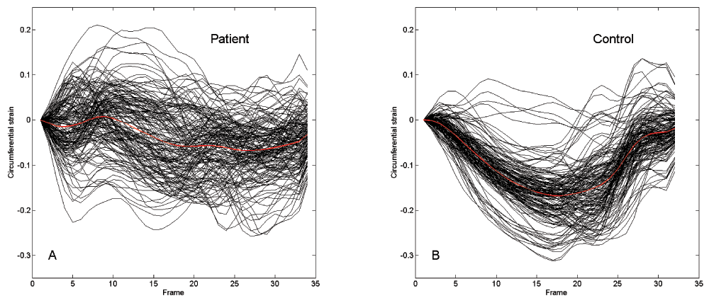
Left ventricular mid-wall circumferential strain patterns from 160 regions in (A) a patient with idiopathic dilated cardiomyopathy (DCM) and left bundle branch block (LBBB) and (B) a healthy control. Note the large variability seen in the patient and relative uniformity in healthy control. The average value of the cardiac strains (global strain) is represented by a red line.
The average strains for the patients with DCM+LBBB and the controls are shown in Figures 2A and 2B, respectively. The average strain patterns in five cases showed a period of positive slope during the first half of systole. The average strains were more homogenous in the controls, where they decreased smoothly during the entire early systole phase. The obtained reference strain decreased in patients and controls during the entire initial systole phase. The peak shortening for the reference strain was -0.034 and -0.15 for the patient and control, respectively (red lines, Figures 2A and 2B).
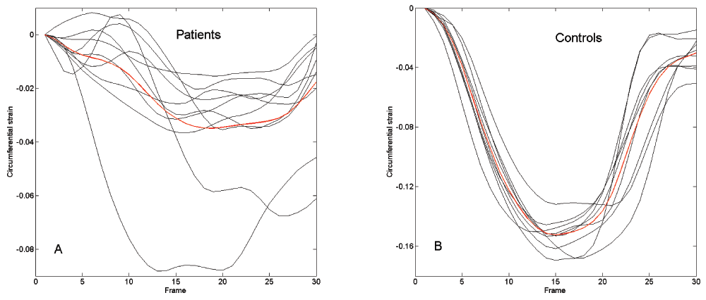
The global strain from 10 patients with idiopathic dilated cardiomyopathy (DCM) and left bundle branch block (LBBB) (A) and from 9 healthy controls (B). The group average, that is, the reference average strain, is identified by a red line.
On the basis of the reference strain, the ejection phase was from 1 to 16 of the MR-tagged images in patients with DCM+LBBB and from frames 1 to 14 in controls.
We constructed a reference strain for patients with cardiac activation disorder and healthy controls. As expected, the resulting reference strain presented a negative slope during the entire early systole phase. A large difference between the measured global strain of each subject and the reference strain is indicative of insufficient measurements.
The shortcomings of this measurement technique are reflected in patient individual average strains. The first measured point in the strain pattern is during systole, that is, 20 ms after the electrocardiogram QRS trigger has started the measurements. In five of the patients assessed, the average strain pattern (Figure 2A) had a positive slope during the first half of systole. A positive slope on the curve indicates ventricular filling, but ventricular filling does not occur after the onset of tension generation in the ventricular wall. This measurement technique does not assess the mechanics of the entire ventricular wall, just a part of it. It relies on the coincidence of individual cardiac mechanics and selection of regions, which are measured irrespective of whether this artifact is pronounced. Because of the stochastic nature of this event, the averaging of cardiac strains throughout the group yields a better estimate of the actual cardiac strain than individual measurements.
A standard procedure to determine the ejection phase on the basis of the average cardiac strain is based on identifying the peak negative slope during systole, fitting a linear curve to it, and identifying the time points where the linear curve crosses the zero strain and maximum strain points, which represent the beginning and end of the ejection phases, respectively. Without considering if the patients had positive slopes during systole, this procedure would be associated with several errors in case of our measured strains. The reference strain averages out the measurement errors in patients and thus facilitates more accurate detection of cardiac phases.
Currently, ventricular function is frequently evaluated on the basis of patterns of a regional strain. A simple test of global ventricular strain could additionally verify whether the measurements included all the relevant regions of the ventricle.
figshare: Data of ventricular mechanistic measurements evaluation. http://dx.doi.org/10.6084/m9.figshare.94097313
Written informed consent for publication of clinical details and clinical images was obtained from all the participants involved.
| Views | Downloads | |
|---|---|---|
| F1000Research | - | - |
|
PubMed Central
Data from PMC are received and updated monthly.
|
- | - |
Provide sufficient details of any financial or non-financial competing interests to enable users to assess whether your comments might lead a reasonable person to question your impartiality. Consider the following examples, but note that this is not an exhaustive list:
Sign up for content alerts and receive a weekly or monthly email with all newly published articles
Already registered? Sign in
The email address should be the one you originally registered with F1000.
You registered with F1000 via Google, so we cannot reset your password.
To sign in, please click here.
If you still need help with your Google account password, please click here.
You registered with F1000 via Facebook, so we cannot reset your password.
To sign in, please click here.
If you still need help with your Facebook account password, please click here.
If your email address is registered with us, we will email you instructions to reset your password.
If you think you should have received this email but it has not arrived, please check your spam filters and/or contact for further assistance.
Comments on this article Comments (0)