Keywords
arteriovenous malformation, AVM, grading, management
arteriovenous malformation, AVM, grading, management
In this revised version, we have included two new figures
To read any peer review reports and author responses for this article, follow the "read" links in the Open Peer Review table.
As of now, many tenets exist regarding management of high grade cerebral arterio-venous malformation (AVM) management, making a rigid algorithm impossible to create. In experienced hands, microsurgery proved to have better results, compared to other treatments1,2. Herein, we report a microsurgical management of a grade 5 arteriovenous malformation (AVM) in a young patient with a high predicted risk for rebleeding.
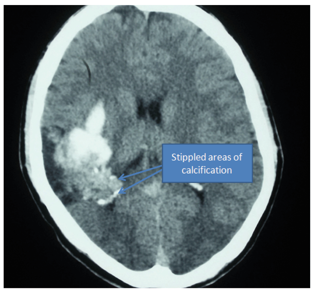
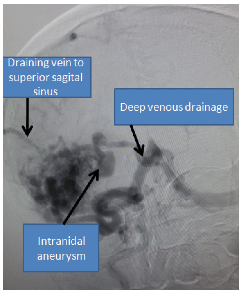
A 22-year-old Brahmin male from Khaireni, a remote village in Nepal, presented to our emergency room with a sudden-onset severe headaches and left-sided weakness over the last 24 hours. Physical examination revealed a Glasgow coma scale (GCS) of 14/15 with left-sided hemiparesis of 3+/5(Medical research council grading). Medical history was significant for a few episodes of paroxysmal headaches since last couple of years, which improved after taking 500 mg Paracetamol tablet on an ‘as needed’ basis. The frequency and intensity of the headache had worsened in the last few months. There was no significant family history. An urgent head computerized tomogram (CT) revealed evidence of a hyperdense lesion with peripheral stippled calcification on the right side in the territory of posterior limb of internal capsule and the retro-thalamic region (Figure 1). There was also coating of vessel along the middle cerebral artery (MCA) territory (Figure 2) and hyperdensity along the deep venous territory. A four-vessel diagnostic carotid angiography revealed Grade 5 Spetzler-Martin AVM in the right sub corticol region with feeders from lenticulostiates of the middle cerebral artery (Figure 3). Drainage was to the deep draining veins and also to the superior sagittal sinus (Figure 4).


Multiple factors such as young age at presentation, the fact that the lesion had bled, presentation of patient with deficits associated with the lesion on the non-dominant side, presence of deep venous drainage and intra-nidal aneurysm led to a high calculated risk for rebleeding in the patient. We therefore decided on surgical management, despite the high grade of the lesion. After explanation of the risks of the treatment and role of adjuvants in the form of radiosurgery and embolisation the patient was taken up for microsurgical excision. Since the facility of radiosurgery is not available in the country, we only had the option of embolisation of the feeders prior to the surgical excision of the lesion. However, since the lesion had only low velocity feeders from the lenticulostriate vessels, we opted for direct microsurgical management. After a liberal craniotomy, basal cisterns were opened to gain access to the M1 branch of the MCA. We identified the major deep draining vein that was looping over the MCA bifurcation with the help of Indocyanine Green (ICG) venography. We placed a temporary clip on M1, then made a minimal corticostomy over the parietal cortex and continued our dissection over the gliotic tissue surrounding the AVM taking care of the minimal bleeders with the help of bipolar cauterization and avoiding inadvertent entry to the nidus. Lastly a clip was applied to the draining vein after completely dissecting the AVM nidus. The lesion was finally excised (Figure 5). Complete hemostasis was confirmed.
Postoperatively his blood pressure was rigorously monitored so as not to overshoot the mean arterial pressure above 100 mm of mercury so as to prevent breakthrough perfusion rebleeding. Patient was started on Sodium Valproate (1 gm stat followed by 300 mg IV 8 hourly) and Nimodipine (60 mg 4 hourly via nasogastric tube) for seizure and vasospasm prophylaxis, respectively. Repeat head CT scan the following morning revealed no cavity hematoma or any evidence of vasospasm (Figure 6). Patient was extubated uneventfully. He had hemiparesis of 3+ in upper limbs and 3 in lower limbs. Patient was started on physiotherapy and finally discharged home on the 7TH post-operative day after removal of sutures. Patient came for follow-up 2 weeks later walking on his own with left upper limb weakness of grade 3+/5. The Nimodipine was tapered off in the subsequent three weeks. The patient was advised to continue Na Valproate 300 mg orally three times a day for at least a year. Post operative angiography revealed complete excision of the AVM with no remaining feeders (Figure 7). The patient followed up in the outpatient clinic 6 months later with minimal pronator drift on the left arm and grade 2 spasticity on his left leg.
Bleeding within the AVM is considered a significant predictor of rebleeding. Other important factors moderating risk of rebleeding include deep venous drainage3. Studies have verified that the risk of rebleeding under these circumstances is as high as 34.4% compared to just 0.9% per year in patients without these risk factors4,5. Another important factor to be considered while calculating the risk of rebleeding is the presence of concurrent aneurysm within the AVM (6.93% with aneurysm Vs 3.99% without aneurysm)3.
Up to 40% of cases with AVM manifest neurological deficits6, mostly attributable to hemorrhage. A minority of only 5% to 15% of such deficits are related to factors such as coronary steal phenomenon and venous hypertension7–9.
The Spetzler-Martin Scale is used to estimate the risk of surgical resection of an AVM with higher grades being associated with greater surgical morbidity and mortality10. Multivariate studies have shown this grading system to reliably predict permanent major morbidity or mortality at the following levels: Grade I (4%), Grade II (10%), Grade III (18%), Grade IV (31%), and Grade V (37%)11. This data has been further validated prospectively, and this grading system remains the most widely used among neurosurgeons and neurointerventionalists12.
Han et al. reported the management of 73 grade 4 and 5 lesions and found the annual hemorrhage rate for untreated lesions to be only 1.5% versus 10.4% for partially treated lesions13. Grade IV or V lesions are only treated in circumstances of progressive neurological deterioration from hemorrhage, vascular steal, or seizure as seen in our case, which had a high risk of rebleeding because of presentation at young age with hemorrhagic episode, large size of the nidus, deep venous drainage pattern and associated aneurysm within the AVM.
There is time-lag of about two years following radiosurgery for complete obliteration of the nidus in the lesion. The risk of hemorrhage in this time period is around 4.8% per year14 which parallels the natural history of the lesion after bleeding. However there is a risk of inadvertent radiation injury to the adjacent eloquent brain area15 and also a risk of symptomatic radiation necrosis in around 9% of cases15,16.
The main indication for other embolisation options in such a high grade of AVM is in order to downgrade the lesion and to minimise the intraoperative blood loss so as to make the lesion amenable for microsurgical excision, which bears an acceptable complication rate of around just 6.5%17. One study has shown that the deep venous drainage, higher grade of the lesions and the periprocedural hemorrhage are predictors of post procedural complications following the embolisation treatment17.
In our case there were only few feeders from the lenticulostriate branches from MCA: not ideal for embolization. Partial embolisation of the lesion will not reduce the risk of hemorrhage to zero3. Partial embolisation of the high grade lesions are only justified in few circumstances, such as in vascular steal phenomenon or an AVM with associated aneurysm18.
In a few selected cases who have a high calculated risk of rebleeding, microsurgical excision remains a therapeutic option even for a high grade AVM especially in centers with limited resources for intervention and radiosurgery. However, all the patients should be well counseled about the available alternative mode of intervention and the associated risks. The management plan in each patient should be tailored addressing factors such as age of the patient, mode of presentation, grade of the lesion, treatment modalities and expertise availability etc.
Written, informed consent was sought and attained from the father of the patient as per medical protocol in Nepal.
Dr Sunil prepared the manuscript and obtained the pictures. Dr Binod and Dr Cherian revised and confirmed the final manuscript. All authors have seen and agreed to the final content of the manuscript.
| Views | Downloads | |
|---|---|---|
| F1000Research | - | - |
|
PubMed Central
Data from PMC are received and updated monthly.
|
- | - |
Competing Interests: No competing interests were disclosed.
Competing Interests: No competing interests were disclosed.
Competing Interests: No competing interests were disclosed.
Competing Interests: No competing interests were disclosed.
Competing Interests: No competing interests were disclosed.
Alongside their report, reviewers assign a status to the article:
| Invited Reviewers | |||
|---|---|---|---|
| 1 | 2 | 3 | |
|
Version 2 (revision) 04 Jul 16 |
read | read | read |
|
Version 1 02 Nov 15 |
read | read | |
Provide sufficient details of any financial or non-financial competing interests to enable users to assess whether your comments might lead a reasonable person to question your impartiality. Consider the following examples, but note that this is not an exhaustive list:
Sign up for content alerts and receive a weekly or monthly email with all newly published articles
Already registered? Sign in
The email address should be the one you originally registered with F1000.
You registered with F1000 via Google, so we cannot reset your password.
To sign in, please click here.
If you still need help with your Google account password, please click here.
You registered with F1000 via Facebook, so we cannot reset your password.
To sign in, please click here.
If you still need help with your Facebook account password, please click here.
If your email address is registered with us, we will email you instructions to reset your password.
If you think you should have received this email but it has not arrived, please check your spam filters and/or contact for further assistance.
Comments on this article Comments (0)