Keywords
Magnetic Resonance Imaging, Prostate cancer, biopsy, diagnostic test accuracy
Magnetic Resonance Imaging, Prostate cancer, biopsy, diagnostic test accuracy
Current practice mandates a prostate biopsy for histological confirmation of prostate cancer prior to a radical prostatectomy. Prostate biopsy, whether performed trans-rectally or trans-perineally, is an invasive procedure which usually requires an anaesthetic and has the inherent risks of urosepsis, urinary retention and haematoma1,2.
Post-biopsy inflammatory changes can also obliterate natural tissue planes there by potentially compromising the quality of a nerve sparing procedure and increase positive margin rates3. For this reason prostatectomy is usually delayed by at least 6 weeks to allow for a reduction in the peri-prostatic inflammatory change that follows any biopsy procedure4.
The use of 3T mpMRI-p is gaining increasing acceptance for both the diagnosis and localisation of prostate cancer. In experienced centres, the positive predictive value of Prostate Imaging-Reporting and Data System (PI-RADS) 5 lesions has a specificity of 97–100% in biopsy naïve patients5–8. We describe two case reports of radical prostatectomy without tissue diagnosis. The patients involved both had objections to confirmative biopsy, PI-RADS 5 lesions on mpMRI-p and high pre-test probabilities of prostatic malignancy. We believe this report will be of interest for urologists dealing with the dilemma of patients with a high risk of prostatic malignancy, positive mpMRI-p and patient refusal of biopsy procedures.
A 56 year old man was referred to our service with elevated Prostate Specific Antigen (PSA) titres, which had risen progressively from 1.7 ng/mL to 4.6 ng/mL over a five year period. He had a positive family history of prostate cancer (PC), with his brother having undergone radical prostatectomy 5 years earlier. His digital rectal exam (DRE) showed a malignant nodule in his right lobe and a firm contralateral lobe.
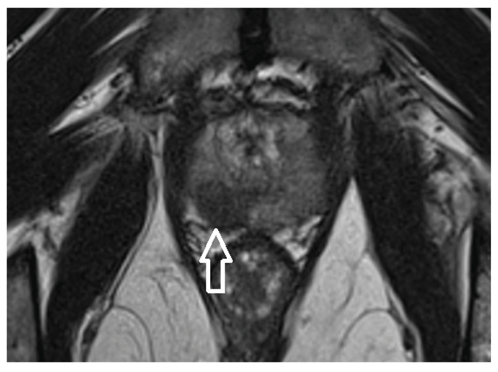
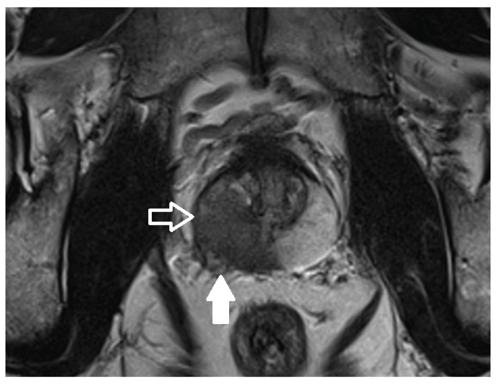
We requested a 3-Tesla Multi-parametric Magnetic Resonance Imaging of the prostate (mpMRI-p) which identified a Prostate Imaging-Reporting and Data System (PI-RADS) 5 lesion in the right lobe of the prostate with probable extra-prostatic extension (Figure 1). A smaller lesion was present in the left mid peripheral zone (Figure 2) with possible left external iliac node involvement. Staging Computerised Tomography (CT) of the abdomen and pelvis and Tc-99m bone scan showed no radiological evidence of metastatic spread other than the previously mentioned borderline enlarged left external iliac nodes.
The results of the investigations (PSA titres, DRE) and the chance of a false positive mpMRI-p result of approximately 5% was explained to the patient. The patient was adamantly against undergoing confirmatory prostate biopsy as he was concerned about biopsy related sepsis. He also reported pre-existing anxiety regarding PC since his brother had been diagnosed and treated.
The patient subsequently underwent a bilateral incremental nerve sparing Robot Assisted Laparoscopic Radical Prostatectomy (RALRP) with left sided extended pelvic lymph node dissection.
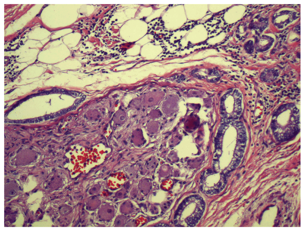
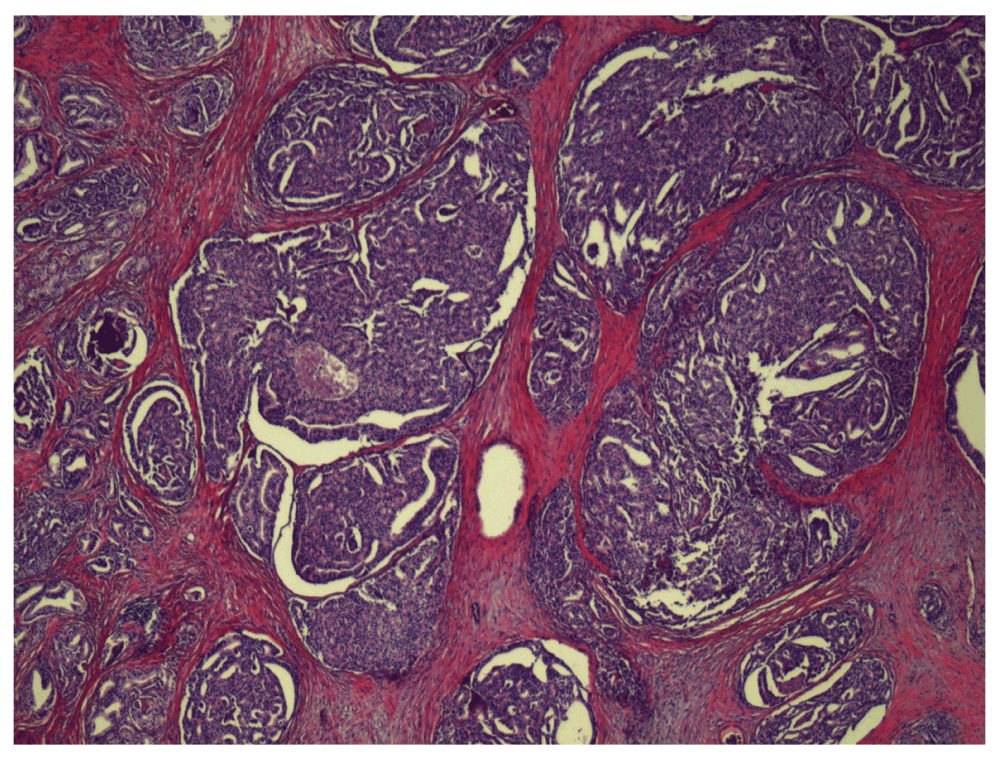
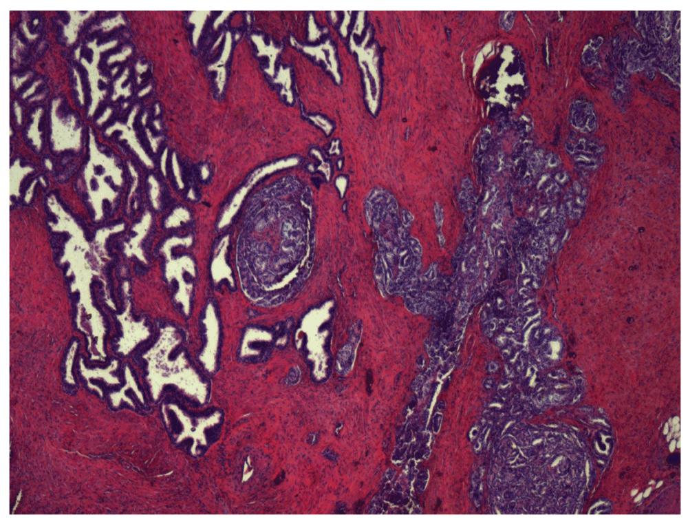
The Operative specimen’s histology demonstrated Gleason 4+3=7 primary tumour with tertiary pattern 5 present (Figure 3) with extra-prostatic extension (EPE) present. Peri-neural invasion was present (Figure 4). A focally positive margin at region of EPE was present over a 0.5 mm base. The specimen’s histological stage was T3a (AJCC 7th Edition 2010).
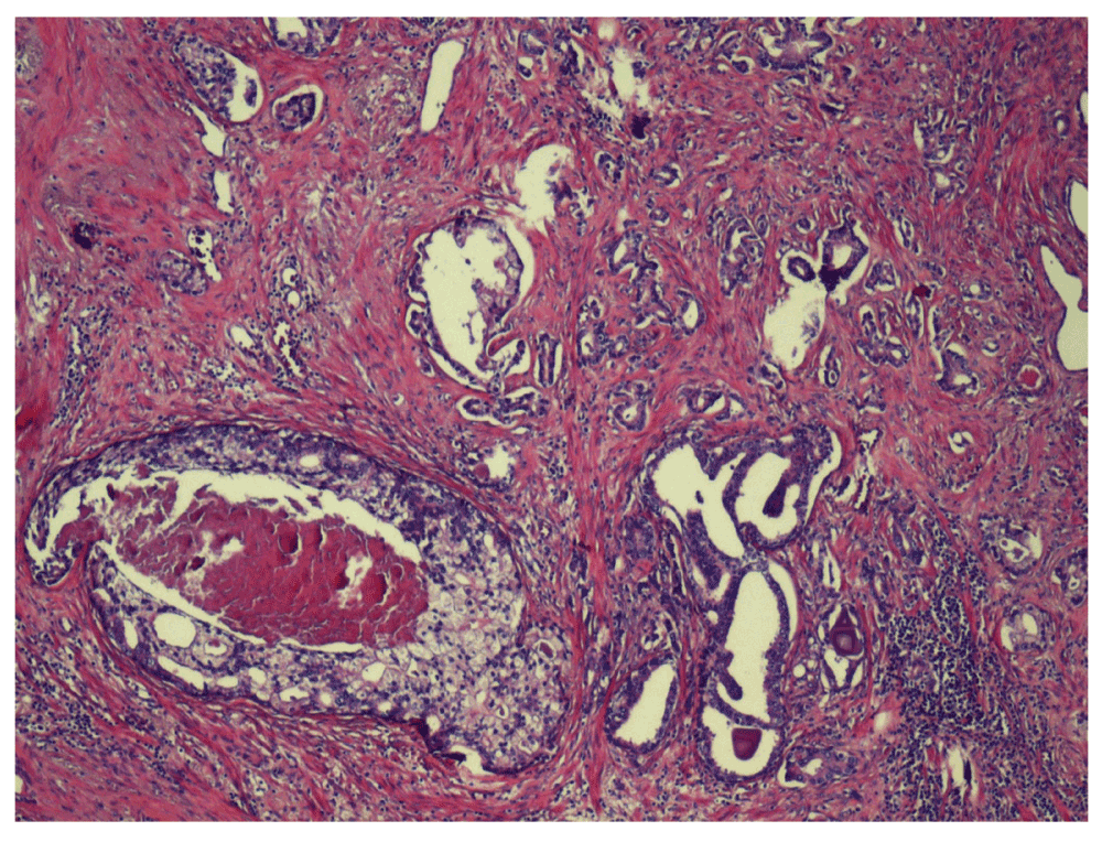
Cribiform glands with comedonecrosis represent Gleason pattern 5.
The patient made an uneventful recovery from surgery. Post-operative incontinence was mild at 6 weeks, using a single safety pad during the day only. He is trialling sildenafil for his post-operative erectile dysfunction. His PSA at 5 weeks was low at 0.033 ng/mL. His follow-up is ongoing.
A 64 year old with no family history of prostate cancer and an elevated serum PSA of 9 ng/mL was refereed to our service. His DRE revealed a right sided palpable prostate nodule with extension into the ipsilateral seminal vesicle.
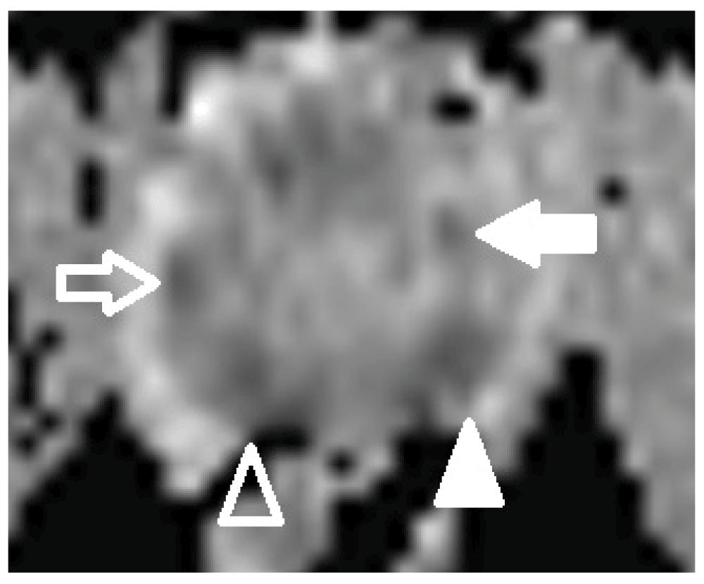
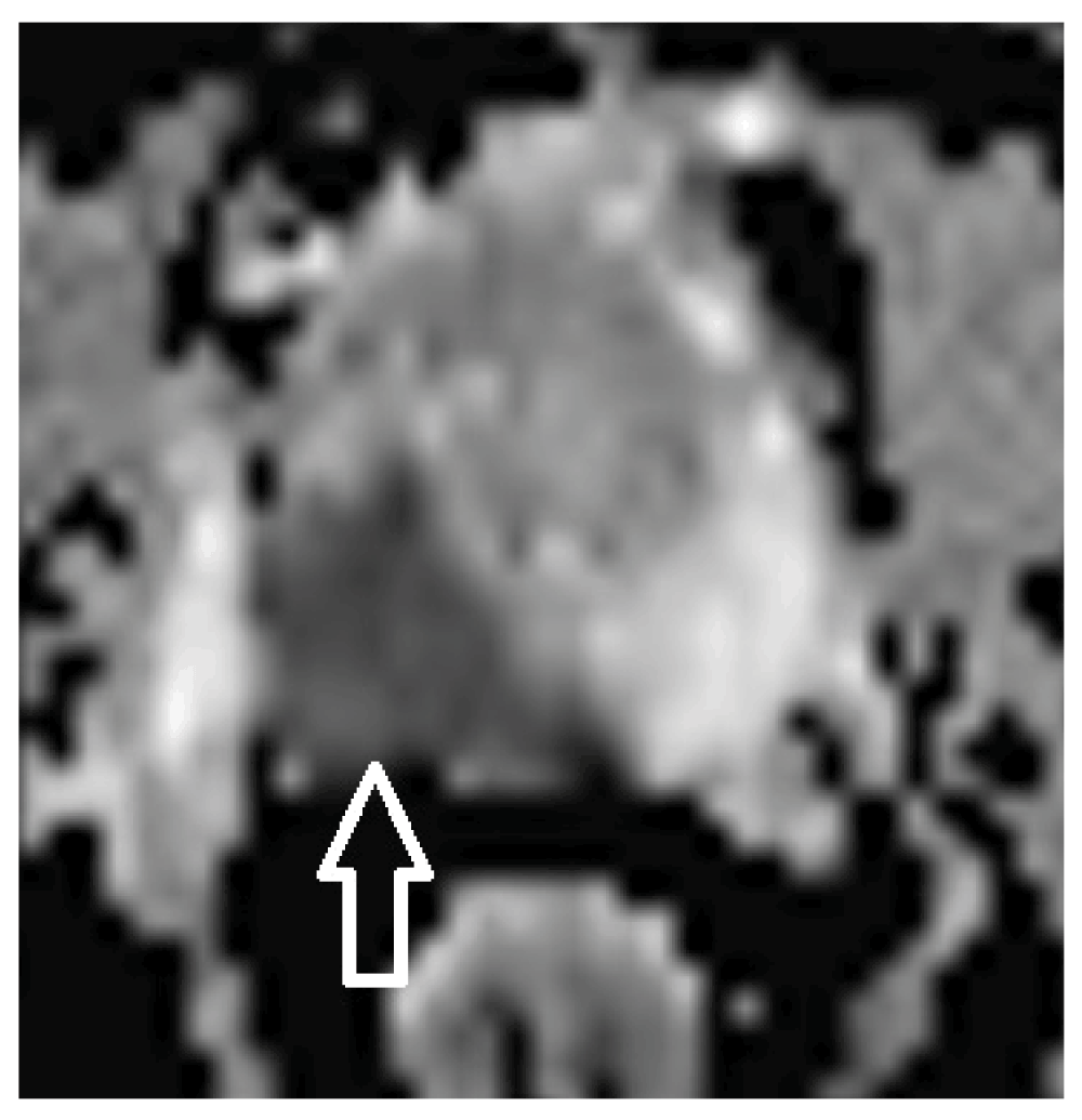
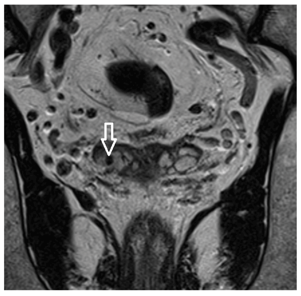
mpMRI-p was requested and confirmed a PI-RADS 5 lesion in the right base and mid-zone of the gland (Figure 5). The lesion extended across midline into the left lobe. Diffusion restriction was present on ADC map (Figure 6). Right-sided seminal vesicle invasion was also demonstrated radiologically (Figure 7).
The patient was advised to undergo confirmatory prostate biopsy but declined, being satisfied with his diagnosis based on PSA, DRE and Mp-MRI-p findings. CT and Tc-99m bone scans were negative for metastatic spread.
The patient underwent RALRP. Due to his seminal vesicle invasion a wide non-nerve sparing approach was taken on the right side, with the contralateral nerve bundle spared due to the patient being sexually active. A right sided obturator node dissection was performed concurrently.
Histology demonstrated Gleason 4+4 with tertiary pattern 5 (Figure 8). Tumour volume was 14.92cc. Right neurovascular bundle invasion and bilateral seminal vesicle invasion was present (Figure 9). All resection margins were uninvolved and 0/3 resected nodes were infiltrated by malignancy. His tumours histologic stage was T3b (AJCC 7th Edition 2010).
The patient made a good recovery following his surgery. Post-operatively his early urinary incontinence was very mild, using a single safety pad per day. His erectile dysfunction is successfully managed with sildenafil.
Initially, at 2 months post-operatively his PSA was undetectable, however, at 5 months it had risen to 0.03 ng/mL, and by 9 months post-operatively it had risen further to 0.14 ng/mL. He was subsequently referred to radiation oncology for salvage radiotherapy for biochemical failure.
Both patients in our small case series were diagnosed and their tumours correctly localised by mpMRI-p. Whilst we strongly recommended both patients proceeded to confirmative prostate biopsies, they both declined further investigation. Both patients had high pre-test probabilities of malignancy even prior to their positive mpMRI-p findings and were satisfied that their diagnoses were correct, and had concerns about the risks of prostate biopsy. They were both informed of and willing to accept the small risk of a false positive diagnosis.
It is important to note that radical prostatectomy without tissue diagnosis is not recommended as standard practice by the authors, even with a PI-RADS 5 lesion on mp-MRI-p. The case studies presented here are exceptional cases where both patients had strong opposition to biopsy despite counselling otherwise. Whilst mpMRI-p has demonstrated great sensitivity and specificity for identification of high risk prostate cancer, there remains the possibility of over diagnosis, and consequent overtreatment if a confirmatory biopsy is not also obtained pre-operatively. With further development of this imaging technology it is plausible that in the future, high risk patients with PI-RADS 5 lesions on mpMRI-p could undergo a radical prostatectomy without the need for a prostate biopsy. While we have successfully performed two prostatectomies without pre-operative biopsy we do not advocate this as a standard approach.
In exceptional circumstances and with appropriately counselled patients who have a high pre-test probability of prostate cancer (rising and elevated PSA, malignant nodule on DRE and a corresponding PIRADS 5 lesion on mpMRI-P), it may be appropriate to proceed to a radical prostatectomy without a prostate biopsy. Further advancements in mpMRI-p imaging techniques may negate the need for routine biopsies in high risk lesions prior to prostatectomy, but this cannot be recommended as routine practice with currently available imaging protocols.
Written informed consent for publication of their clinical details and clinical images was obtained from the patients.
| Views | Downloads | |
|---|---|---|
| F1000Research | - | - |
|
PubMed Central
Data from PMC are received and updated monthly.
|
- | - |
Provide sufficient details of any financial or non-financial competing interests to enable users to assess whether your comments might lead a reasonable person to question your impartiality. Consider the following examples, but note that this is not an exhaustive list:
Sign up for content alerts and receive a weekly or monthly email with all newly published articles
Already registered? Sign in
The email address should be the one you originally registered with F1000.
You registered with F1000 via Google, so we cannot reset your password.
To sign in, please click here.
If you still need help with your Google account password, please click here.
You registered with F1000 via Facebook, so we cannot reset your password.
To sign in, please click here.
If you still need help with your Facebook account password, please click here.
If your email address is registered with us, we will email you instructions to reset your password.
If you think you should have received this email but it has not arrived, please check your spam filters and/or contact for further assistance.
Comments on this article Comments (0)